
Outgoing and incoming channels of cavernous sinus
Reading File
The cavernous sinus receives blood from the ophthalmic veins, sphenoparietal sinus, and superficial middle cerebral vein (incoming), and drains via the superior and inferior petrosal sinuses into the transverse sinus and internal jugular vein respectively (outgoing).
Searching sources...
Finding Sources
Finding Sources
Good results. Let me read the detailed anatomy sections from Gray's Anatomy and Scott-Brown's for the complete tributary/drainage information.
Reading File
Reading File
Let me fetch the coronal section diagram of the cavernous sinus and get more detailed tributary info from Gray's Anatomy veins section.
Loading Image
Reading File
Now let me also get the sphenoparietal sinus and intercavernous connections info for completeness.
Finding Sources
Now let me read the detailed Gray's Anatomy section on cavernous sinuses for the full tributary/drainage table.
Reading File
Let me also fetch the dural venous sinuses diagram for a visual, and read the detailed cavernous sinus section from Gray's.
Loading Image
Reading File
Excellent - I now have all the information needed for a complete, well-sourced answer. Here is the full breakdown:
Cavernous Sinus - Incoming and Outgoing Channels
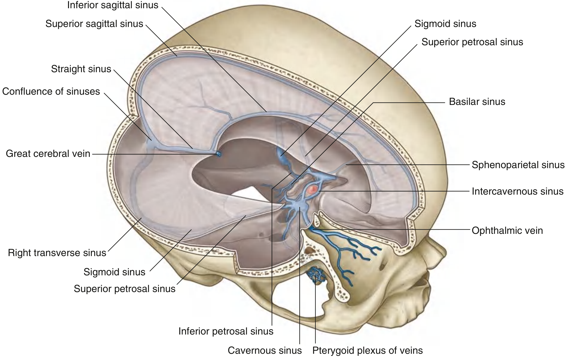
Fig. 8.46 - Veins, Meninges, and Dural Venous Sinuses (Gray's Anatomy for Students)
Incoming Channels (Tributaries)
These channels bring blood into the cavernous sinus:
| Channel | Source/Notes |
|---|---|
| Superior ophthalmic vein | Begins in the anterior orbit from the supra-orbital and angular veins; passes through the superior orbital fissure to enter the cavernous sinus. Principal orbital tributary. |
| Inferior ophthalmic vein | Smaller; may drain into the cavernous sinus directly through the superior orbital fissure, or join the superior ophthalmic vein first, or drain into the pterygoid plexus via the inferior orbital fissure. |
| Superficial middle cerebral vein | Drains the temporal lobe cortex along the lateral fissure; a fairly constant superficial cerebral vein. |
| Sphenoparietal sinuses | Small paired sinuses along the inferior surface of the lesser wings of the sphenoid; drain into the anterior ends of each cavernous sinus; receive diploic and meningeal veins. |
| Emissary veins from the pterygoid plexus | Pass through the foramen ovale (or foramen of Vesalius when present) to connect the pterygoid plexus with the cavernous sinus. This route allows facial infection to spread intracranially. |
| Intercavernous sinuses | Connect the right and left cavernous sinuses to each other, anteriorly and posteriorly around the pituitary stalk (forming the "circular sinus"). Allow infection to spread rapidly (within 24-48 hours) to the contralateral sinus. |
| Hypophyseal veins (pituitary) | Short portal and hypophyseal veins drain the posterior pituitary into the cavernous sinus. |
Outgoing Channels (Drainage)
These channels carry blood out of the cavernous sinus:
| Channel | Drains Into | Course |
|---|---|---|
| Superior petrosal sinus | Transverse sinus | Begins at the posterior end of the cavernous sinus; runs posterolaterally along the superior margin of the petrous temporal bone to join the transverse sinus. |
| Inferior petrosal sinus | Internal jugular vein | Also begins at the posterior end of the cavernous sinus; runs posteroinferiorly in a groove between the petrous temporal and the occipital bone, ending in the internal jugular vein. Also drains cerebellar veins and veins from the internal ear and brainstem. |
| Basilar plexus/sinus | Connects bilateral inferior petrosal sinuses; communicates with the vertebral plexus of veins | Lies on the clivus, just posterior to the sella turcica. |
| Emissary veins | Pterygoid plexus (extracranially) | Run through foramina ovale/Vesalius in reverse direction - bidirectional flow. |
Summary Diagram (Coronal Section)
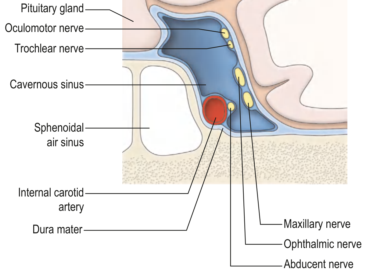
Coronal section of the cavernous sinus showing the ICA, pituitary gland, and cranial nerve relations (Gray's Anatomy for Students)
Memory Mnemonics
Incoming ("ESOME"):
- Superficial middle cerebral vein
- Emissary veins (pterygoid plexus)
- Sphenoparietal sinus
- Ophthalmic veins (superior + inferior)
- Meningeal veins (via sphenoparietal)
- Each other (via intercavernous sinuses)
Outgoing: The cavernous sinus drains posteriorly - superior petrosal sinus → transverse sinus → sigmoid sinus → internal jugular vein; and inferior petrosal sinus → directly to internal jugular vein.
Sources:
- Gray's Anatomy for Students, Table 8.3 and Cavernous Sinuses section (pp. 1025-1027)
- Gray's Anatomy for Students, Veins of the Orbit section (p. 1082)
- Scott-Brown's Otorhinolaryngology, Cavernous Sinus section (p. 1247)
- Neuroanatomy through Clinical Cases, 3rd ed., Venous Drainage of the Cerebral Hemispheres
- Goldman-Cecil Medicine, Anatomy and Pathophysiology section
This is a shared conversation. Sign in to Orris to start your own chat.