
Osteosarcoma
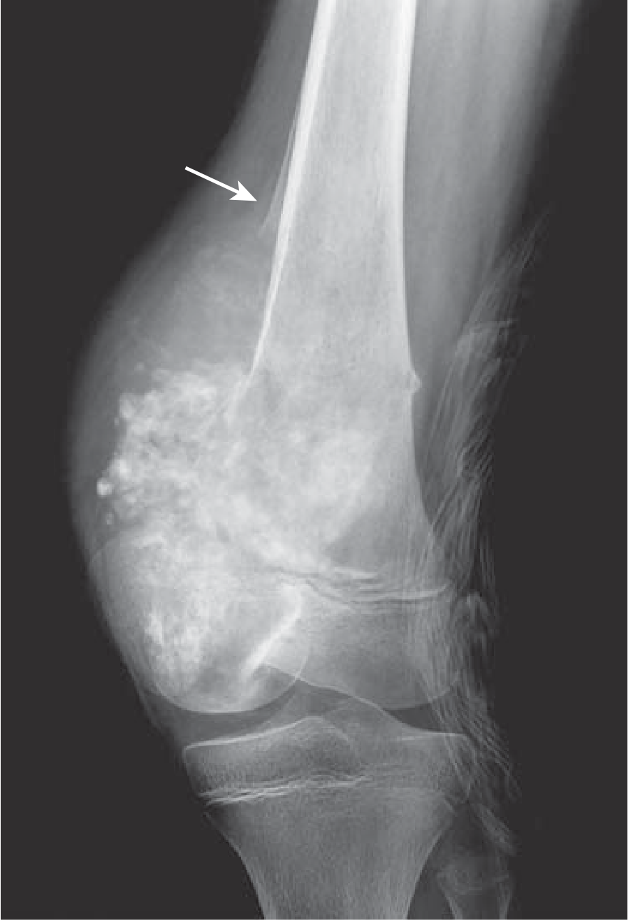
Osteosarcoma conventional high grade radiograph metaphysis knee sunburst periosteal reaction
osteosarcoma treatment
NCI osteosarcoma treatment PDQ diagnosis treatment
https://www.cancer.gov/types/bone/hp/osteosarcoma-treatment-…
Osteosarcoma
Typical epidemiology and site
- Most commonly affects adolescents, especially during the pubertal growth spurt.
- A second, smaller peak occurs in older adults, often associated with Paget disease, prior radiotherapy, bone infarction, or pre-existing bone disease.
- Usually arises in the metaphysis of a long bone, especially around the knee:
- Distal femur
- Proximal tibia
- Proximal humerus
Presentation
- Persistent, progressive localized bone pain
- Swelling or enlarging mass
- Reduced joint movement if near a joint
- Occasionally a pathological fracture
Imaging features
- Aggressive, destructive mixed lytic-sclerotic lesion
- Ill-defined/infiltrative margins
- Cortical destruction with an associated soft-tissue mass
- Periosteal reaction, including:
- Codman triangle: lifted periosteum producing triangular reactive bone
- “Sunburst” pattern: radiating new bone formation
Diagnosis and staging
- X-ray of the involved bone
- MRI of the entire involved bone and adjacent joint for local extent and surgical planning
- Chest CT, because the lungs are the most frequent metastatic site
- Bone scan or PET-based imaging where appropriate
- Carefully planned core-needle or incisional biopsy, ideally performed by the team that will undertake definitive surgery
Histology and types
-
Telangiectatic osteosarcoma
-
Low-grade central osteosarcoma
-
Small-cell osteosarcoma
-
Parosteal osteosarcoma
-
Periosteal osteosarcoma
-
High-grade surface osteosarcoma
-
Grainger & Allison’s Diagnostic Radiology, p. 1048
Genetics and associations
-
RB1 abnormalities, including hereditary retinoblastoma
-
TP53 abnormalities, including Li-Fraumeni syndrome
-
Less commonly, Rothmund-Thomson syndrome
-
Robbins & Kumar Basic Pathology, p. 782
Treatment
- Neoadjuvant multi-agent chemotherapy
- Wide surgical resection with tumor-free margins, often limb-salvage surgery where feasible
- Postoperative chemotherapy
Prognostic factors
- Localized disease at diagnosis
- Complete surgical resection with clear margins
- Good histologic response to preoperative chemotherapy
- Extremity site rather than pelvis or axial skeleton
Open fracture and wound debridment
open fracture debridement
BOAST 4 open fractures wound debridement guidelines antibiotics timing 2025
https://www.boa.ac.uk/resource/boast-4-pdf.html
Open fracture and wound debridement
Gustilo-Anderson classification
| Grade | Description |
|---|---|
| I | Clean wound <1 cm, low-energy injury |
| II | Wound 1-10 cm, moderate soft-tissue injury/contamination |
| IIIA | High-energy injury with extensive damage, but adequate soft-tissue coverage of bone |
| IIIB | Exposed bone or inadequate soft-tissue coverage, often requiring flap reconstruction |
| IIIC | Open fracture with arterial injury requiring repair |
Initial emergency management
-
ATLS resuscitation first
- Treat hemorrhage, shock, head/chest/abdominal injuries.
- Assess and document distal pulses, capillary refill, motor and sensory function before and after reduction/splinting.
-
Give IV antibiotics immediately
- Ideally within 1 hour of injury.
- Choice follows local protocol and allergy profile. Gram-positive cover is standard for lower-grade injuries; severe Grade III injuries commonly need broader coverage. Add specific anaerobic/clostridial cover for farm, soil, sewage, or fecal contamination as directed by local protocol. BOAST open-fracture standard and EAST antibiotic guidance
-
Tetanus prophylaxis
- Vaccinate or give immunoglobulin when indicated by immunization status and wound risk.
-
Handle the wound minimally
- Remove only gross surface contamination.
- Photograph when appropriate, cover with saline-soaked sterile gauze and an occlusive dressing.
- Do not probe the wound, repeatedly expose it, or perform “mini-washouts” in the emergency department. BOAST guidance
-
Realign and splint
- Gentle reduction and immobilization reduce pain, bleeding, ongoing soft-tissue damage, and neurovascular compromise.
- Obtain appropriate radiographs without delaying life- or limb-saving interventions.
-
Urgent specialist referral
- Major open fractures need coordinated orthopedic and plastic surgery care, termed an orthoplastic approach.
Wound debridement
Definition
-
Reduce bacterial and foreign-body burden
-
Remove devitalized tissue that permits bacterial adhesion and biofilm formation
-
Assess the full injury zone, including bone, muscle, fascia, skin, vessels, and nerves
-
Enable stable fracture fixation and early definitive soft-tissue closure
-
Rockwood and Green’s Fractures in Adults, 10th ed., pp. 631-632
Principles of surgical debridement
- Perform in an operating theatre by an experienced trauma/orthoplastic team.
- Extend the wound adequately, preferably along appropriate surgical or fasciotomy lines, to expose the whole zone of injury.
- Remove:
- Dirt, clothing, vegetation, and foreign material
- Dead skin and subcutaneous tissue
- Devitalized fascia and muscle
- Detached or minimally attached bone fragments that cannot contribute to reconstruction
- Preserve viable tissue, especially major nerves and vessels.
- Assess muscle viability clinically by its color, consistency, contractility, and capacity to bleed, while recognizing these assessments can be imperfect.
- Irrigate generously with appropriate fluid to remove residual debris.
- Eliminate dead space and hematoma where possible.
- After completion, treat fixation and reconstruction as clean surgery: re-prep and re-drape the limb, use new instruments and gloves. BOAST open-fracture standard
Timing of debridement
-
Immediately: vascular compromise/ischemia, compartment syndrome, or heavily contaminated wounds such as agricultural, aquatic, or sewage injuries
-
Within 12 hours: other high-energy open fractures
-
Within 24 hours: low-energy open fractures
-
-
Rockwood and Green’s Fractures in Adults, 10th ed., p. 632
After debridement
Fracture stabilization
- External fixation is often used for severe contamination, major soft-tissue injury, vascular injury, or damage-control situations.
- Internal fixation can be appropriate when adequate debridement and definitive soft-tissue coverage can be achieved safely.
Soft-tissue coverage
- Primary closure is considered only when the wound is clean, tension-free, and adequately debrided.
- Severe injuries may require local or free flap coverage.
- If not performed at initial surgery, definitive closure/coverage should generally occur within 72 hours. BOAST guidance
Exam-style summary
Fat embolism
fat embolism syndrome
fat embolism syndrome diagnosis management review guideline
PMID: 38760078
Fat embolism and fat embolism syndrome
Pathogenesis
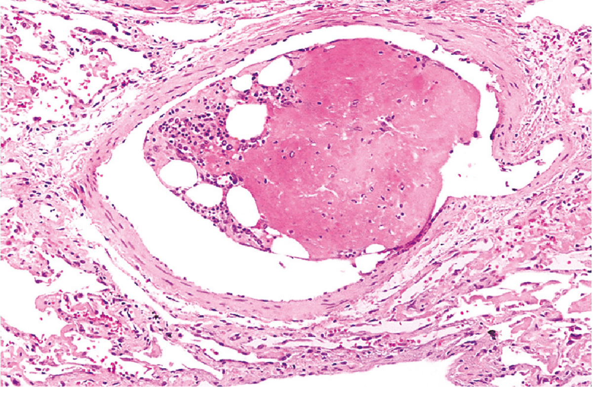
- Mechanical obstruction: fat droplets enter venous channels from disrupted marrow and obstruct pulmonary and sometimes cerebral microcirculation.
- Biochemical injury: free fatty acids cause endothelial damage, inflammation, platelet activation, capillary leak, and ARDS-like lung injury.
- Robbins & Kumar Basic Pathology, p. 94
- Murray & Nadel's Textbook of Respiratory Medicine, p. 1853
Classic clinical triad
- Respiratory distress: tachypnea, dyspnea, hypoxemia, cyanosis, and potentially ARDS
- Neurologic features: agitation, confusion, drowsiness, delirium, seizures, or coma
- Petechial rash: classically on the conjunctivae, oral mucosa, neck, upper chest, shoulders, and axillae
Other findings
- Tachycardia and fever
- Thrombocytopenia
- Falling hemoglobin/hematocrit
- Retinal changes
- Diffuse bilateral pulmonary infiltrates
- Acute kidney injury or oliguria in severe cases
Diagnosis
- Respiratory insufficiency
- Cerebral involvement not explained by head injury or another cause
- Petechial rash
- Tachycardia
- Fever
- Retinal changes
- Jaundice or renal changes
- Thrombocytopenia
- Anemia
- Raised ESR
- Fat macroglobulinemia
- Arterial blood gas: hypoxemia
- CBC: thrombocytopenia and anemia
- Chest radiograph or CT: nonspecific diffuse infiltrates
- CT pulmonary angiography: may help exclude thrombotic pulmonary embolism but can be normal in FES
- Brain MRI for prominent neurologic disease may show a diffusion-weighted “starfield” pattern
Management
- Administer oxygen and monitor continuously
- Escalate to high-flow oxygen, noninvasive support, or mechanical ventilation if needed
- Maintain adequate circulation with careful fluid resuscitation and vasopressors when indicated
- Treat associated trauma, hemorrhage, and complications
- Monitor platelet count, hemoglobin, renal function, blood gases, and neurologic status
- Admit moderate or severe cases to ICU
- Consider lung-protective ventilation if ARDS develops
Prevention
- Prompt immobilization of fractures
- Gentle handling and appropriate resuscitation
- Early stabilization/fixation of long-bone fractures when the patient is physiologically suitable
- Careful operative technique during intramedullary procedures
Important distinction
- Fat embolism: fat droplets in the circulation, commonly asymptomatic.
- Fat embolism syndrome: symptomatic systemic illness, usually developing after a delay and characterized by hypoxemia, neurologic changes, petechiae, thrombocytopenia, and anemia.
Distal radius fracture
distal radius fracture x ray Colles fracture dorsal angulation radiograph
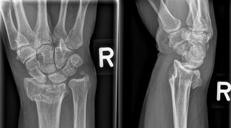
This diagnostic image consists of a two-view conventional radiograph (X-ray) of the right wrist, presenting both anteroposterior (AP) and lateral views. The image demonstrates a distal radius fracture with significant displacement. In the AP view (left), a transverse fracture line is visible across the distal metaphysis of the radius, accompanied by an associated fracture of the ulnar styloid process. The lateral view (right) clearly illustrates dorsal (posterior) displacement and dorsal angulation of the distal radial fragment, characteristic of a Colles-type fracture pattern. There is a disruption of the normal radiocarpal alignment, with the carpal bones following the dorsally displaced distal radius segment. Key anatomical structures visible include the distal radius and ulna, the proximal and distal carpal rows, and the bases of the metacarpals. This visual material is primarily used in orthopedic and emergency medicine education to teach fracture identification, terminology for displacement and angulation, and the assessment of wrist joint congruity.
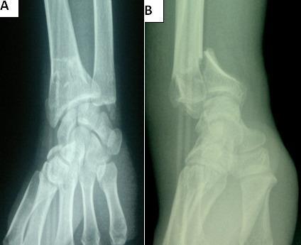
This diagnostic image consists of two preoperative X-ray views of the wrist, illustrating an unstable intra-articular distal radius fracture. View A is an anteroposterior (AP) radiograph showing comminution and a clear sagittal fracture line extending into the radiocarpal joint space. View B is a lateral radiograph demonstrating a Goyrand-Smith fracture, characterized by significant volar (palmar) displacement and angulation of the distal fracture fragment relative to the radial shaft. The alignment between the distal radius, carpal bones, and distal ulna is severely disrupted, with the carpus following the volar displacement of the distal radial fragment. This case highlights key orthopedic findings for surgical planning, specifically the involvement of the articular surface and the direction of fragment displacement, which distinguishes the Smith fracture from the more common dorsal displacement seen in Colles fractures.
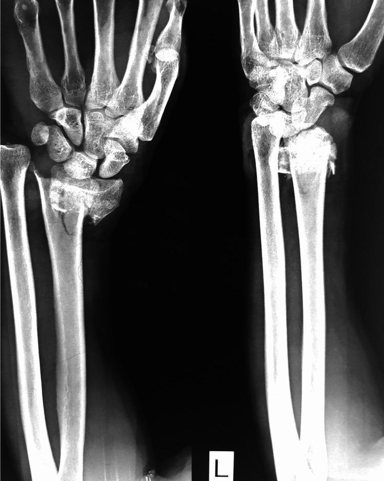
This diagnostic image is a plain radiograph (X-ray) of the left wrist in two views (oblique and lateral) demonstrating a complex distal radius fracture. The visual evidence shows a comminuted fracture of the distal radial metaphysis with intra-articular extension. Key pathological features include significant radial shortening, dorsal displacement, and dorsal angulation of the distal fracture fragments (characteristic of a Colles-type injury pattern). The alignment of the radiocarpal joint is disrupted, with the carpal bones shifted dorsally in relation to the main radial shaft. There is visible fragmentation and irregularity at the fracture site, indicating instability. The distal ulna appears intact, though the distal radioulnar joint (DRUJ) relationship is altered due to the radial shortening. This image illustrates a common orthopedic trauma scenario requiring surgical evaluation for anatomic reduction and stabilization, often via external fixation or internal plate osteosynthesis.
distal radius fracture management
AAOS distal radius fractures clinical practice guideline management radiographic indications
https://www.aaos.org/drfcpg
PMID: 41945667
Distal radius fracture
Clinical presentation
-
Wrist pain, swelling, bruising, and limited movement
-
Deformity in displaced fractures, classically “dinner-fork” deformity in a Colles fracture
-
Possible numbness or tingling in the thumb, index, middle, and radial half of ring finger due to median nerve compression
-
Check skin integrity, capillary refill, pulses, sensation, and finger movement
-
Firestein & Kelley’s Textbook of Rheumatology, section “Distal Radius Fractures”
Important patterns
| Pattern | Description |
|---|---|
| Colles fracture | Extra-articular distal radius fracture with dorsal displacement/angulation, usually after a fall on an extended wrist |
| Smith fracture | Volar displacement/angulation, often after a fall on a flexed wrist |
| Barton fracture | Intra-articular rim fracture with radiocarpal subluxation/dislocation |
| Chauffeur fracture | Radial styloid fracture |
| Intra-articular comminuted fracture | Higher instability risk and may require fixation |
Assessment and imaging
- AP and lateral wrist radiographs, often with an oblique view
- Assess:
- Extra-articular vs intra-articular involvement
- Radial height/shortening
- Radial inclination
- Volar or dorsal tilt
- Articular step-off or gap
- Comminution
- Distal radioulnar joint stability
- Associated ulnar styloid, carpal, TFCC, or ligament injury
- CT is useful for complex intra-articular fractures and operative planning.
Initial treatment
- Analgesia, elevation, ice, and immobilization in a splint.
- Urgent reduction if the fracture is displaced, skin is threatened, neurovascular status is impaired, or there is acute median nerve compression.
- Repeat clinical neurovascular examination and post-reduction radiographs.
- Arrange fracture clinic or hand/orthopedic follow-up.
Nonoperative management
- Closed reduction if needed
- Below-elbow cast or splint
- Serial radiographs early in treatment when loss of reduction is a concern
- Finger, elbow, and shoulder movement during immobilization
- Wrist range-of-motion and strengthening after immobilization
When surgery is considered
- Radial shortening >3 mm
- Dorsal tilt >10 degrees
- Intra-articular step-off/displacement >2 mm
- Volar locking plate fixation
- Percutaneous K-wire pinning
- External fixation
- Bridge plating in selected complex injuries
Complications
- Loss of reduction and malunion
- Wrist stiffness and reduced grip strength
- Median nerve compression/carpal tunnel syndrome
- Complex regional pain syndrome
- Extensor pollicis longus tendon rupture
- Post-traumatic osteoarthritis, especially after intra-articular incongruity
- Distal radioulnar joint dysfunction and ulnar-sided wrist pain
Practical points
- A low-energy distal radius fracture in an older adult should prompt assessment of bone health and falls risk.
- Avoid tight circumferential casts in a markedly swollen wrist.
- Seek urgent care for increasing pain, numbness, pale/cold fingers, inability to move fingers, or a cast that feels too tight.
Septic shock
septic shock management
Surviving Sepsis Campaign adult septic shock guidelines 2025 treatment norepinephrine fluid resuscitation
https://www.sccm.org/clinical-resources/guidelines/guideline…
PMID: 39516335
Septic shock
Pathophysiology
-
Vasodilation and reduced systemic vascular resistance
-
Capillary leak and relative/absolute intravascular hypovolemia
-
Myocardial depression
-
Microcirculatory and mitochondrial dysfunction
-
Inflammatory activation that can cause ARDS, acute kidney injury, liver dysfunction, encephalopathy, and DIC
-
Rosen’s Emergency Medicine, p. 59
Clinical features
- Hypotension, cool or sometimes warm peripheries
- Tachycardia, tachypnea, hypoxemia
- Fever or hypothermia
- Altered mental status
- Oliguria
- Raised lactate or metabolic acidosis
- Mottled skin, delayed capillary refill
- Signs of organ dysfunction, such as AKI, thrombocytopenia, jaundice, or ARDS
Immediate management: first-hour priorities
-
ABCDE assessment and ICU-level monitoring
- Oxygen as needed, secure the airway if necessary.
- Insert IV access, monitor ECG, blood pressure, urine output, oxygen saturation, and mental status.
- Send blood cultures and appropriate cultures, but do not delay treatment.
-
Give broad-spectrum IV antimicrobials urgently
- In septic shock, start empiric antibiotics immediately after cultures are obtained if this does not cause meaningful delay.
- Tailor therapy to the suspected source, local resistance patterns, prior antibiotics, immune status, and later culture results.
- De-escalate once microbiology and the clinical course permit.
-
Fluid resuscitation
- Start IV crystalloid resuscitation promptly.
- Balanced crystalloids are generally preferred over 0.9% saline when appropriate.
- Reassess frequently using blood pressure, capillary refill, urine output, lactate trend, bedside ultrasound/echocardiography, and dynamic fluid-responsiveness tests.
- Avoid indiscriminate fluid loading, particularly in heart failure, renal failure, or established pulmonary edema.
-
Vasopressor support
- Start norepinephrine promptly if hypotension persists during or after initial fluids. Do not delay vasopressors while giving excessive fluid.
- Initial target: MAP 65 mmHg, individualized where needed.
- If norepinephrine requirements escalate: add vasopressin.
- If MAP remains inadequate despite norepinephrine and vasopressin: consider epinephrine.
- If cardiac dysfunction with ongoing hypoperfusion persists despite adequate volume and pressure: consider dobutamine with norepinephrine, or epinephrine alone in selected cases.
- Source control
Identify and control the source as early as possible, for example:
- Drain an abscess or empyema
- Remove an infected vascular catheter
- Debride necrotizing soft-tissue infection
- Relieve obstructed infected urinary or biliary tract
- Operate for perforation or intra-abdominal sepsis
Investigations
- CBC with differential, renal/liver function, glucose, coagulation profile
- Blood gas and serial lactate
- Blood cultures before antibiotics if feasible
- Urinalysis and urine culture
- Chest radiograph
- ECG and cardiac enzymes when indicated
- Ultrasound, CT, or other imaging directed to the suspected source
- Bedside echocardiography to assess cardiac function and volume status when shock is unclear or refractory
Adjunctive care
- Mechanical ventilation using lung-protective strategy for ARDS/respiratory failure
- Renal replacement therapy when indicated
- Glycemic control avoiding severe hyperglycemia and hypoglycemia
- DVT and stress-ulcer prophylaxis when appropriate
- Consider IV corticosteroids, typically hydrocortisone, in vasopressor-dependent shock that remains refractory after fluids and vasopressors
- Frequent reassessment of perfusion: capillary refill, urine output, mentation, lactate trajectory, and skin perfusion
Key exam summary
Cultures → immediate broad-spectrum antibiotics → IV crystalloids → norepinephrine for MAP ≥65 → early source control → ICU organ support.