
Tympanic membrane perforation
tympanic membrane perforation otoscopy
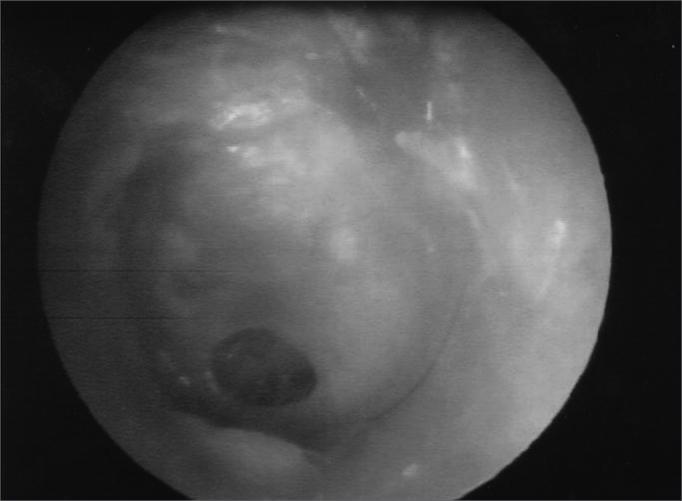
This clinical photograph, captured via otoscopy or endoscopy, displays a human tympanic membrane (eardrum) with a prominent central perforation. The tympanic membrane appears grayish-white and translucent in intact regions, with visible light reflections indicating a smooth surface texture. The central feature is a dark, well-demarcated, roughly circular or oval opening (perforation) located within the pars tensa. The edges of the perforation are sharp and appear non-inflamed, with no immediate evidence of active infection, drainage, or cholesteatoma in the visible field. This image serves as a classic educational representation of chronic otitis media or traumatic eardrum injury, illustrating the loss of membrane integrity that can lead to conductive hearing loss. The visualization provides a clear example for medical students and otolaryngologists to study eardrum morphology and the anatomical localization of tympanic defects prior to surgical intervention, such as tympanoplasty.
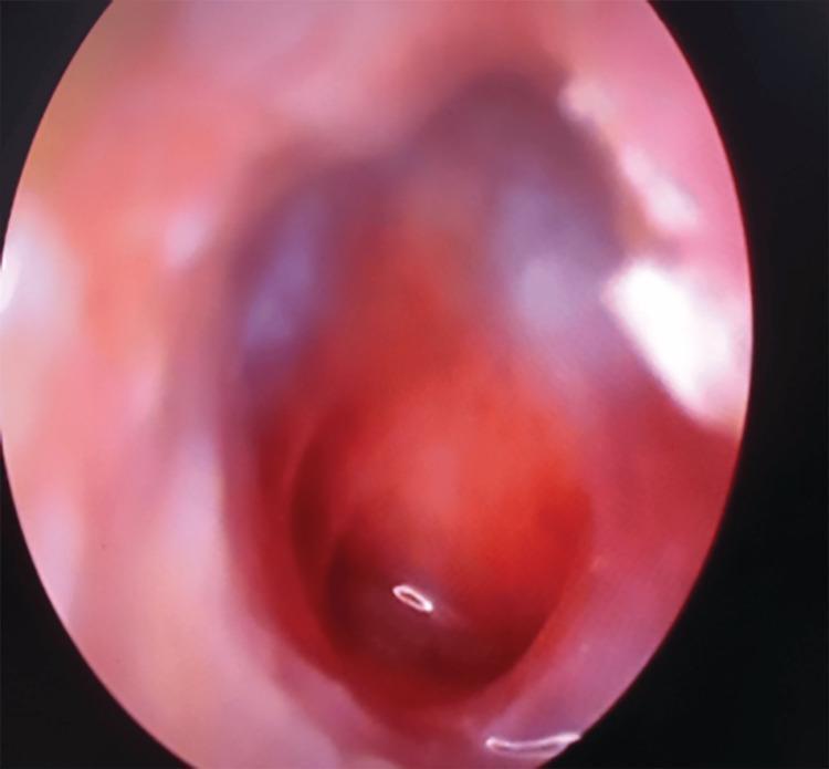
This clinical photograph, obtained via otoscopy/endoscopy, depicts a human tympanic membrane with a large central perforation. The perforation occupies a significant portion of the pars tensa, appearing as a dark, well-demarcated void that reveals the underlying middle ear space. The surrounding tympanic membrane remnant is erythematous and thickened, indicating active or chronic inflammation. The middle ear mucosa visible through the defect appears congested and reddish. Key anatomical landmarks, such as the handle of the malleus and the light reflex, are obscured or distorted due to the extensive tissue loss and inflammatory changes. This visual is characteristic of chronic suppurative otitis media (CSOM) or significant acoustic/mechanical trauma. The image serves as an educational example of middle ear pathology and the visual assessment of tympanic membrane integrity in an otolaryngology context.
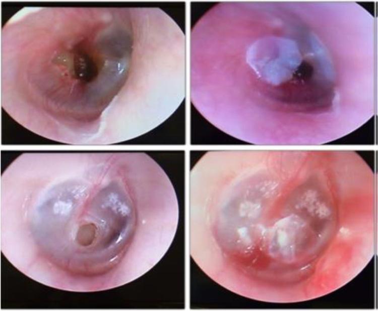
A comparison chart consisting of four video-otoscopy clinical photographs organized into two rows. The left column displays pre-treatment views of the tympanic membrane in two different patients, each exhibiting a clear central perforation. In the top left image, a medium-sized irregular perforation is visible with mildly inflamed margins. In the bottom left image, a smaller, circular, well-defined central perforation is seen in a membrane showing signs of myringosclerosis (white calcific plaques). The right column shows the same ears post-procedure, demonstrating the temporary closure of the defects using a translucent Bionext® cellulose film. The film is visible as an opaque-to-translucent patch covering the previous perforation sites, effectively sealing the middle ear space from the external auditory canal. This visual comparison illustrates a conservative clinical approach to managing symptomatic tympanic membrane perforations to assess immediate functional and symptomatic improvement before considering permanent surgical repair like myringoplasty.
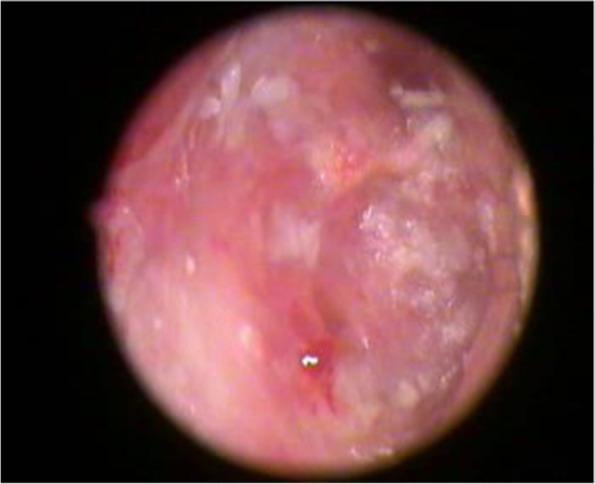
This clinical photograph, obtained via otoscopy or laryngoscopy, displays a postoperative view of a tympanic membrane graft (likely temporalis fascia) one month following myringoplasty. The image shows the graft positioned within the middle ear space, exhibiting a predominantly reddish-pink hue, which indicates early neovascularization and tissue integration. The membrane appears semi-translucent with an irregular, uneven surface texture and scattered whitish patches representing keratinization or residual surgical material. A small, persistent perforation or focal thinning is visible in the lower central quadrant. The surrounding ear canal walls appear healthy. This visual serves as an educational example of early-stage graft uptake and provides a clinical reference for assessing surgical success and potential residual defects in the immediate postoperative period for patients treated for chronic otitis media or tympanic membrane perforation.
"tympanic membrane perforation" AND management
Tympanic Membrane Perforation
Anatomy Recap
- Pars tensa - the large, taut inferior portion attached to the bony canal via the fibrous annulus. It is only a few cell layers thick and therefore easily torn.
- Pars flaccida (Shrapnell's membrane) - the small, loose superior portion above the notch of Rivinus; less rigid.
Classification
By Location
| Type | Description | Clinical Significance |
|---|---|---|
| Central | Does not reach the fibrous annulus; a rim of TM persists at the periphery | Lower risk of cholesteatoma; amenable to repair |
| Marginal | Reaches or involves the annulus | Higher risk of cholesteatoma; involves posterosuperior quadrant most dangerously |
| Pars flaccida | In the attic/superior segment | Strongly associated with acquired cholesteatoma |
| Subtotal / Total | Very large; may involve most or all of pars tensa | Greater conductive loss |
By Cause
- Infective - spontaneous rupture from acute otitis media (AOM) or chronic suppurative otitis media (CSOM)
- Traumatic - direct (cotton swabs, slap), barotrauma (blast, diving), penetrating trauma
- Blast / Acoustic - primary blast injury; TM perforation is a marker for concurrent concussive head injury (relative risk ~2.76)
- Caustic - button batteries (especially in children; consider non-accidental injury)
- Iatrogenic - during ear surgery (elevation from annular sulcus)
Etiology and Epidemiology
- In AOM, spontaneous rupture occurs in ~5% of cases; the most common pathogen in children is nontypeable H. influenzae. About 90% of these heal without additional intervention.
- Traumatic perforations are almost always in the pars tensa, occurring anteriorly or inferiorly.
- In blast injuries, only 16% of blast-exposed soldiers had TM perforations (50% bilateral); perforation alone is a poor predictor of primary blast injury (PBI) - only 7% of blast injuries had PBI, and only half of those had TM perforation.
Pathophysiology of Hearing Loss
- Loss of sound-pressure difference across the TM - this provides the primary drive to ossicular motion. A perforation equalizes pressure, reducing the trans-TM pressure gradient.
- Perforation size - larger perforations produce greater loss; losses are greatest at low frequencies and decrease at higher frequencies.
- Middle-ear air space volume - smaller volumes (e.g., sclerotic mastoid) result in larger air-bone gaps for the same perforation. A well-pneumatized mastoid buffers the pressure equalization effect.
- Perforation location - counterintuitively, location (anterior vs. posterior) has no significant effect on degree of hearing loss for perforations of the same size, as confirmed by theoretical calculations, temporal bone experiments, and clinical data.
- Wet vs. dry state - air-bone gap is often smaller when the perforation is dry; infection reduces middle-ear air volume, worsening the gap.
The same-sized perforation in two different ears can produce air-bone gaps differing by 20-30 dB solely based on differences in mastoid air space volume (normal range: 2-20 cm³). - Shambaugh Surgery of the Ear
Pathological Features (Chronic Inactive Perforations)
- TM perforated; no active middle-ear inflammation
- Edges may be surrounded by healthy TM, tympanosclerosis, dimeric membrane, or scar tissue
- The mucocutaneous junction is typically at the perforation edge - but squamous epithelial cells can migrate medially into the middle ear
- If the TM is surgically repaired without removing these migrated epithelial cells, an iatrogenic cholesteatoma forms
Clinical Features
| Symptom/Sign | Notes |
|---|---|
| Otalgia | Acute onset; prominent in traumatic perforation |
| Conductive hearing loss | Proportional to size of perforation |
| Otorrhoea | May be bloody (traumatic) or mucopurulent (infective) |
| Tinnitus | Usually transient |
| Vertigo | Transient unless inner ear injury, round/oval window rupture |
| Visible perforation on otoscopy | Dark opening; edges may be inflamed or clean |
Otoscopic Appearance
Management
Conservative / Emergency (Acute Traumatic)
- Most traumatic TM perforations heal spontaneously - reassure and refer
- Instruct patient to keep ear dry (no water entry)
- Topical or systemic antibiotics NOT routinely needed unless contaminated foreign material suspected in the canal or middle ear
- Formal audiogram as soon as possible after injury
- Posterosuperior quadrant perforations or penetrating trauma: refer to otolaryngologist within 24 hours (ossicular chain damage risk)
Acute Infective (AOM with Perforation)
- Topical antibiotics are the treatment of choice for the acute infection
- The TM typically heals without further intervention (~90%)
Surgical (Persistent / Chronic)
| Procedure | Description | Use |
|---|---|---|
| Paper patch / Gelfoam overlay | Office-based closure of small perforations | Small acute iatrogenic or traumatic perforations |
| Myringoplasty | Repair of TM only, using underlay graft (temporalis fascia, perichondrium, fat) | Isolated TM perforation without ossicular damage |
| Tympanoplasty | TM repair + middle ear reconstruction | Perforation with ossicular chain involvement |
Complications
| Complication | Notes |
|---|---|
| Persistent conductive hearing loss | If perforation does not heal |
| Recurrent otitis media | Water contamination; pathogens entering middle ear |
| Cholesteatoma | Especially with marginal or pars flaccida perforations; epithelial migration |
| Ossicular chain erosion | From chronic infection or pressure necrosis |
| Sensorineural hearing loss | If inner ear involved (blast, penetrating trauma, oval/round window rupture) |
| Meningitis | Rare, via direct spread or in cochlear implant patients |
Key Points to Remember
- Central perforations have a rim of TM at the periphery; marginal perforations reach the annulus and carry higher cholesteatoma risk.
- Hearing loss magnitude depends on perforation size and mastoid air volume - NOT on location (anterior vs. posterior).
- In AOM, 90% of spontaneous perforations heal without surgery.
- Posterosuperior quadrant perforation = suspect ossicular damage = urgent ENT referral.
- Keep ears dry; antibiotics only if contamination suspected.
- TM perforation from head trauma is a strong predictor of concussive injury (RR ~2.76).
Definition of perforation of tm
Definition of Tympanic Membrane Perforation
Structural Basis
- Outer layer - stratified squamous epithelium (continuous with the skin of the EAC)
- Middle layer - fibrous lamina propria (gives the TM its tensile strength)
- Inner layer - mucosa (continuous with the middle-ear lining)
Textbook Definitions
"The tympanic membrane has been ruptured... Perforations can be in the pars flaccida or pars tensa of the tympanic membrane, and can be marginal, central, subtotal, or total. Pathologically, the tympanic membrane is perforated but there is no inflammation of the middle-ear space or mucosa [in the inactive form]."
"Chronic suppurative otitis media, by definition, involves a perforated tympanic membrane (spontaneous perforation or the presence of a tympanostomy tube) and ear discharge (otorrhea)."
In Brief
| Element | Detail |
|---|---|
| What it is | Full-thickness hole/tear in the eardrum |
| Location | Pars tensa (most common) or pars flaccida |
| Extent | Central, marginal, subtotal, or total |
| Consequence | Loss of sound-pressure transmission across the drum; direct access of external environment to middle ear |
Definition of types of perforation
types of tympanic membrane perforation central marginal diagram
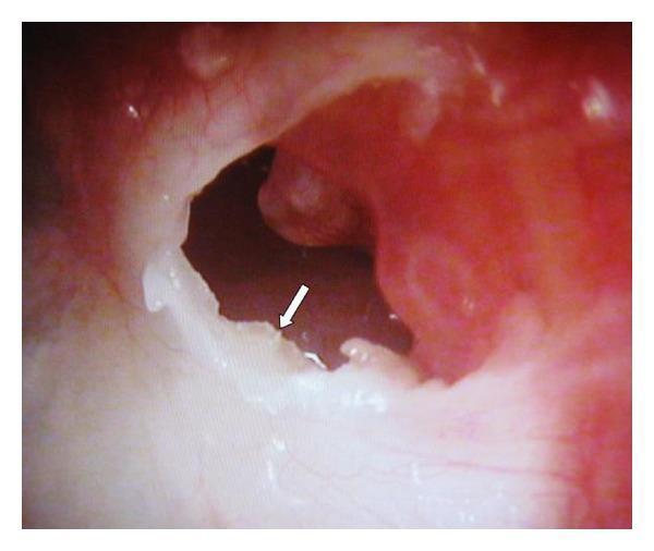
This clinical photograph shows an otoendoscopic view of a left tympanic membrane (TM) exhibiting a marginal perforation in the posterosuperior quadrant. The TM appears thickened, opaque, and creamy-white in color, suggesting chronic inflammatory changes or myringosclerosis. A prominent feature is the presence of white, flaky keratin debris (indicated by a white arrow) emerging from the medial surface of the TM through the perforation. The surrounding ear canal and margins of the perforation appear erythematous and inflamed. The image illustrates findings characteristic of chronic otitis media, potentially involving a cholesteatoma or keratoma, as evidenced by the accumulation of keratin in the middle ear space. This visual is relevant for otolaryngology education, specifically for identifying structural ear defects, distinguishing between central and marginal perforations, and recognizing clinical signs of keratin pearl or cholesteatoma formation.
This clinical photograph, captured via otoscopy or endoscopy, displays a human tympanic membrane (eardrum) with a prominent central perforation. The tympanic membrane appears grayish-white and translucent in intact regions, with visible light reflections indicating a smooth surface texture. The central feature is a dark, well-demarcated, roughly circular or oval opening (perforation) located within the pars tensa. The edges of the perforation are sharp and appear non-inflamed, with no immediate evidence of active infection, drainage, or cholesteatoma in the visible field. This image serves as a classic educational representation of chronic otitis media or traumatic eardrum injury, illustrating the loss of membrane integrity that can lead to conductive hearing loss. The visualization provides a clear example for medical students and otolaryngologists to study eardrum morphology and the anatomical localization of tympanic defects prior to surgical intervention, such as tympanoplasty.
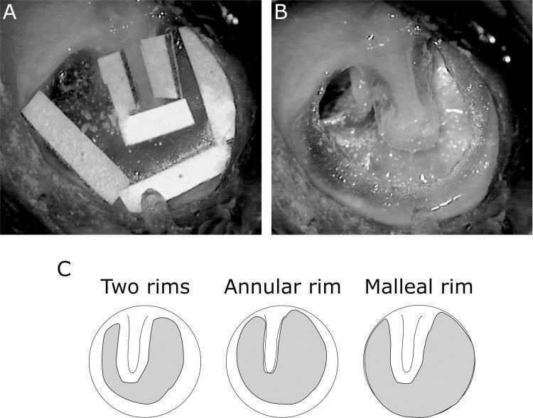
This composite educational image illustrates experimental tympanic membrane (TM) perforation models used in otolaryngology research. Panel A is a clinical endoscopic photograph showing a temporal bone preparation with 1mm rectangular white paper patches applied to the surface of the TM. These patches serve as protective markers to delineate and preserve specific TM remnants, including the annular rim and the malleal rim adjacent to the umbo. Panel B displays the resulting large subtotal central perforation after surgical excision. It demonstrates a 'two rims' condition, where two distinct segments of native TM tissue remain: a peripheral rim along the annulus and a central rim attached to the malleus and umbo. Panel C is a schematic diagram illustrating three experimental rim conditions: 'Two rims' (preserving both annular and malleal tissue), 'Annular rim' (preserving only the periphery), and 'Malleal rim' (preserving only the central attachment). Shaded grey areas indicate the regions of membrane removal. This visual material is used to study middle ear mechanics, sound conduction, and the efficacy of different grafting materials in myringoplasty.
Types of Tympanic Membrane Perforation - Definitions
1. By Location Relative to the Annulus
Central Perforation ("Safe")
- Definition: A perforation entirely within the pars tensa in which a rim of intact tympanic membrane persists between the edge of the perforation and the fibrous annulus all around.
- The perforation does not reach the annulus at any point.
- Associated with tubotympanic (mucosal) disease - "safe" chronic otitis media.
- Cholesteatoma is extremely rare.
- Discharge is profuse, mucoid, and odorless.
- Hearing loss is conductive and usually mild.
Marginal Perforation ("Unsafe")
- Definition: A perforation in which the edge reaches or involves the fibrous annulus at one or more points - there is no rim of TM between the perforation edge and the bony canal wall.
- Most commonly in the posterosuperior quadrant of pars tensa.
- Associated with atticoantral disease and a high risk of cholesteatoma - squamous epithelium can migrate medially through the defect.
- From Cummings: "Pockets and perforations that are marginal in location (involving the annulus) are more likely to result in retained debris and cholesteatoma."
2. By Part of the TM Involved
Pars Tensa Perforation
- Involves the large, taut, lower portion of the TM.
- Can be central or marginal.
- Most traumatic perforations occur here (anteriorly or inferiorly).
Pars Flaccida Perforation ("Attic Perforation") - Unsafe
- Definition: A perforation (or more accurately a deep retraction pocket) in the pars flaccida - the small, loose superior portion above the notch of Rivinus.
- In reality, this is usually a retraction pocket - likened to "the cut finger of a surgical glove" - whose blind distal end lies hidden deep in the middle-ear space.
- Invariably associated with cholesteatoma (atticoantral/unsafe disease).
- The cholesteatoma enlarges lateral to the head of malleus and body of incus, causing scutum erosion and medialization of the ossicular chain.
3. By Size
| Type | Definition |
|---|---|
| Small | Involves a minor portion of the pars tensa (conventionally <25% of TM area) |
| Medium | Involves ~25-50% of TM area |
| Large / Subtotal | Involves most of the pars tensa; a peripheral rim of membrane remains |
| Total | Complete absence of the TM; entire pars tensa destroyed; may involve the annulus |
4. Safe vs. Unsafe - The Key Clinical Classification
| Feature | Tubotympanic (Safe) | Atticoantral (Unsafe) |
|---|---|---|
| Perforation | Confined to pars tensa (central) | "Perforation" of pars flaccida OR posterosuperior pars tensa (retraction pocket) |
| Discharge | Profuse, mucoid, odorless; responds to treatment | Scanty, thick, foul-smelling; does NOT respond to treatment |
| Cholesteatoma | Extremely rare | Common |
| Complications | Unusual except in neglected cases | Frequent |
| Hearing loss | Conductive, usually mild | Larger conductive loss ± SNHL |
| Granulations | Uncommon | Common around mouth of retraction pocket |
| CT findings | Haziness of middle ear in active disease | Scutum erosion (pars flaccida); lateralization/medialization of ossicular chain |
| Bacteriology | P. aeruginosa, S. aureus, Proteus | P. aeruginosa, S. aureus, anaerobes (Bacteroides, Peptostreptococcus) |
Clinical Images
Summary
| Type | Rim Intact? | Part of TM | Cholesteatoma Risk | Safety |
|---|---|---|---|---|
| Central | Yes (all around) | Pars tensa | Very low | Safe |
| Marginal | No (reaches annulus) | Pars tensa - posterosuperior | High | Unsafe |
| Pars flaccida | N/A (retraction pocket) | Pars flaccida (attic) | Very high | Unsafe |
| Subtotal/Total | Partial/None | Pars tensa (large) | Depends on edges | Variable |