
Treatment of loose stool frequency is 4 times a day age is 3.5 yr
acute diarrhea children treatment ORS zinc
WHO UNICEF acute diarrhea treatment children ORS zinc guidelines 2024
Treatment of Acute Diarrhea in a 3.5-Year-Old Child (4 loose stools/day)
Step 1 - Assess Dehydration Status First
| Sign | No Dehydration | Some Dehydration | Severe Dehydration |
|---|---|---|---|
| Eyes | Normal | Sunken | Very sunken |
| Thirst | Normal | Thirsty, drinks eagerly | Unable to drink |
| Skin pinch | Returns quickly | Returns slowly | Returns very slowly |
| Urine | Normal | Decreased | Very little/none |
Step 2 - Oral Rehydration (FIRST-LINE Treatment)
- Sodium: 75 mmol/L
- Chloride: 65 mmol/L
- Glucose: 75 mmol/L
- Potassium: 20 mmol/L
- Citrate: 10 mmol/L
- Give 50-100 mL of ORS after each loose stool
- For mild dehydration: 50 mL/kg over 3-4 hours
- If ORS is not available, mix in 1 L of water: ½ tsp salt + ¼ tsp baking soda + 8 tsp sugar
Step 3 - Zinc Supplementation (MANDATORY per WHO)
- Zinc 20 mg/day for 10-14 days (dispersible tablets or syrup)
- For a 3.5-year-old: standard dose is 20 mg/day
Step 4 - Continue Feeding (Do NOT fast)
- Do not withhold food - fasting prolongs recovery
- Resume age-appropriate diet as tolerated: rice, dal, banana, potato, bread
- The BRAT diet (banana, rice, applesauce, toast) is traditionally recommended
- Avoid dairy products temporarily (transient lactase deficiency can occur)
- Breastfed children should continue breastfeeding throughout
Step 5 - Antibiotics (Targeted, Not Routine)
- Bloody diarrhea (suspected Shigella, EHEC, Campylobacter)
- High fever with systemic toxicity
- Confirmed Giardia, E. histolytica, or C. difficile
- Immunocompromised child
| Organism | Drug of Choice (Pediatric) |
|---|---|
| Shigella | Azithromycin or 3rd-gen cephalosporin |
| Giardia | Metronidazole or Tinidazole |
| Cholera | Azithromycin |
| C. difficile | Metronidazole (mild) or Vancomycin (severe) |
IMPORTANT: Do NOT use antidiarrheals (loperamide) in children - risk of toxic megacolon, ileus, and hemolytic uremic syndrome with STEC. - Symptom to Diagnosis, 4th ed.
Step 6 - Probiotics (Adjunct)
- Lactobacillus rhamnosus GG or Saccharomyces boulardii
- Can reduce duration of diarrhea by ~1 day
Red Flags - When to Refer/Hospitalize
- Sunken eyes, dry mouth, no tears, lethargy (signs of moderate-severe dehydration)
- Bloody diarrhea
- Persistent vomiting preventing oral intake
- Diarrhea >7 days without improvement
- High fever (>39°C)
- Suspected surgical cause (intussusception, appendicitis)
Summary Management at a Glance
| Intervention | Dose/Detail |
|---|---|
| ORS | 50-100 mL after each stool |
| Zinc | 20 mg/day x 10-14 days |
| Diet | Continue normal food; avoid dairy |
| Antibiotics | Only for specific pathogens |
| Antidiarrheals | Contraindicated in children |
| Probiotics | Optional adjunct |
Note: This guidance covers typical acute infectious diarrhea. If diarrhea is chronic (>2 weeks), recurrent, or associated with weight loss/blood/mucus, further evaluation for malabsorption, inflammatory bowel disease, or parasitic infection is needed.
Cause of umblical swelling in 2 month old child and what could be treatment modalities
umbilical hernia infant newborn
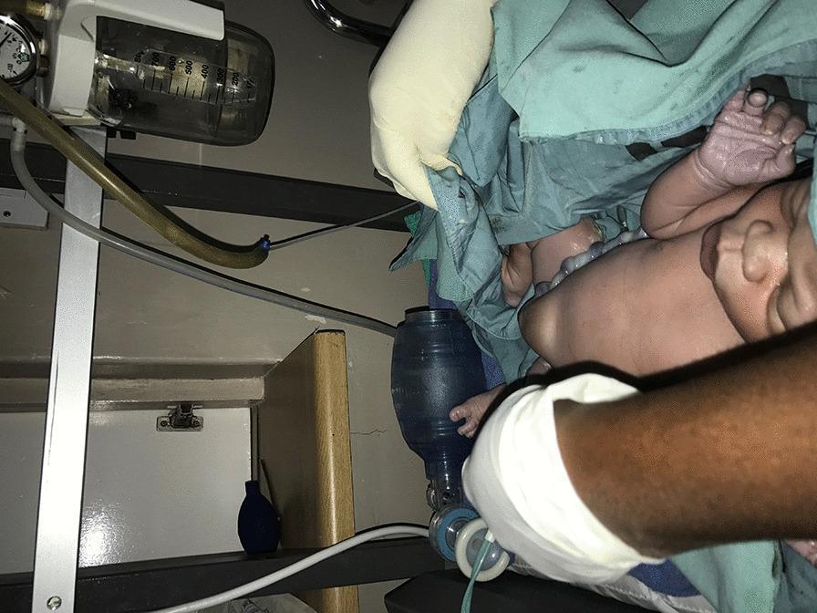
Clinical photograph of a full-term newborn in a delivery or neonatal resuscitation setting, positioned on a warming table. The infant displays a prominent soft tissue protrusion at the umbilicus, consistent with a congenital umbilical hernia. The newborn's skin tone appears light with visible peripheral cyanosis (acrocyanosis) of the hands and feet, which are slightly purplish in color. Essential neonatal resuscitation equipment is visible in the frame, including a blue manual resuscitation bag (Ambu bag) with a clear mask held by a healthcare provider's gloved hand, prepared for positive pressure ventilation. In the background, medical suction apparatus is visible, comprising a graduated collection canister and a pressure gauge mounted on a stand with tubing. This procedural image illustrates the immediate postnatal assessment and management of an infant born with an abdominal wall defect in a clinical environment.
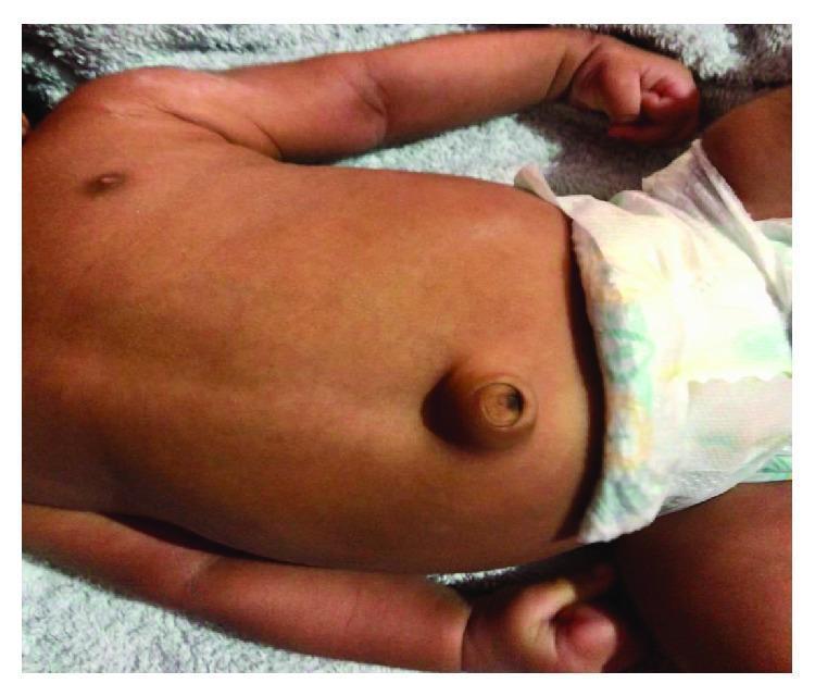
This clinical photograph displays the torso of an infant in a supine position, highlighting a classic infantile umbilical hernia. The primary finding is a prominent, rounded protrusion localized at the umbilical region. The hernia is covered by intact, smooth skin that is continuous with the abdominal wall, showing normal coloration without signs of inflammation, erythema, or strangulation. The umbilicus is visible at the apex of the swelling. The abdomen appears slightly distended, and the surrounding skin texture demonstrates healthy turgor. This image serves as a pedagogical example of a common pediatric congenital condition, illustrating the characteristic 'outie' appearance caused by a defect in the abdominal wall fascia through which intra-abdominal contents, such as small bowel loops or omentum, can bulge. It is used in medical education to teach visual diagnosis of reducible umbilical hernias and to distinguish them from other abdominal wall defects like omphalocele or gastroschisis.
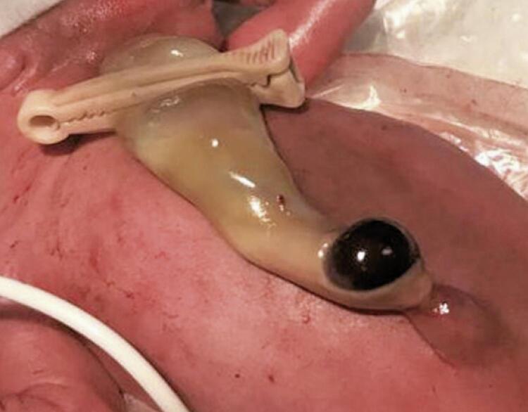
This clinical photograph shows a close-up view of a newborn's abdominal wall and umbilical cord, demonstrating a localized umbilical cord hematoma (UCH). The cord is secured distally with a white plastic umbilical clamp. The proximal portion of the cord near the insertion site at the umbilicus features a distinct, rounded, tense, brownish-purple swelling, characteristic of an acute hematoma. The surrounding cord tissue appears pale yellow and slightly edematous. A small amount of clear serous fluid is visible near the base of the lesion. The newborn's skin is erythematous with visible superficial vasculature, consistent with a preterm infant. A white medical tube or line is visible in the lower left periphery, indicating a neonatal intensive care setting. This image serves as an educational example of neonatal umbilical cord pathology, specifically differentiating a benign hematoma from other umbilical masses such as omphalocele or umbilical hernia.
umbilical granuloma omphalitis newborn infant
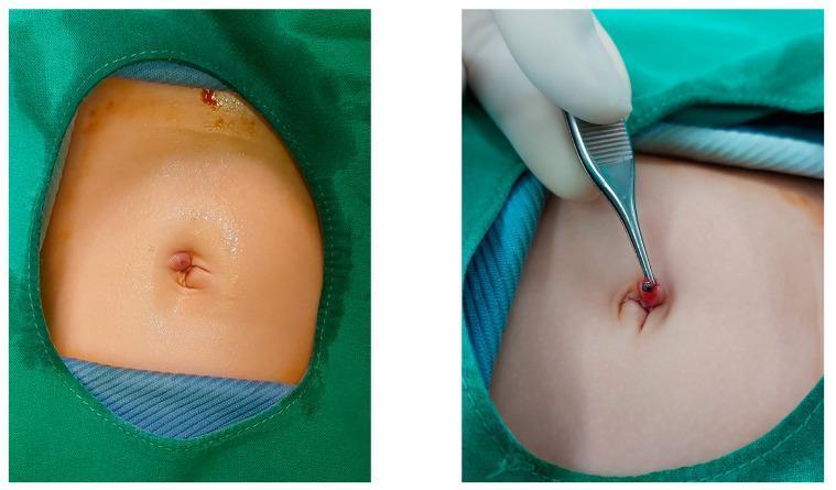
This clinical photograph consists of two panels illustrating postoperative omphalitis (umbilical infection) in a pediatric patient following a surgical procedure. The first panel shows an infant's abdomen prepped for examination, highlighting an erythematous and slightly edematous umbilicus. The second panel depicts a close-up of the umbilical region where a clinician, using metallic surgical forceps, is manipulating the inflamed tissue to reveal a suture reaction. The umbilical site shows localized intense redness and a small incision, consistent with a stitch-induced inflammatory response or granuloma. The images serve as an educational example of minor surgical complications following single-incision laparoscopic surgery, emphasizing the clinical appearance of omphalitis and foreign body reactions to non-absorbable multifilamentous sutures in neonatal or infant patients.
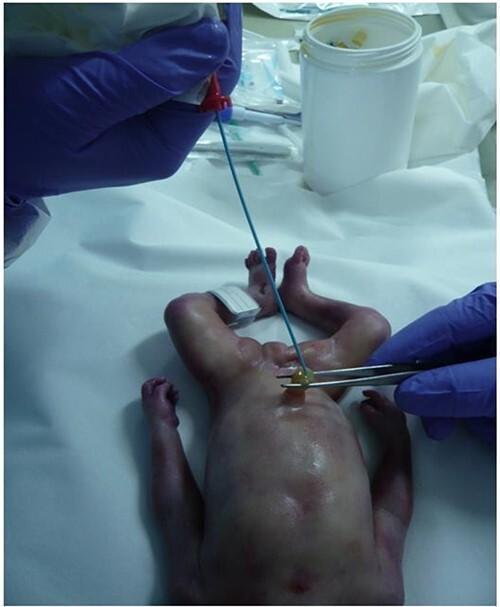
This clinical photograph demonstrates the procedure of umbilical vein catheterization in a newborn in a post-mortem clinical setting. The infant is positioned supine on a sterile white drape, exhibiting skin findings of pallor, generalized mottling, and peripheral cyanosis of the hands and feet. A gloved healthcare provider uses surgical forceps to stabilize the umbilical cord stump while inserting a 5 French radiological sheath (thin blue catheter) into the umbilical vein. The catheter is equipped with a red proximal adapter for the administration of contrast media, such as Angiofil, to facilitate post-mortem computed tomography angiography (PMCTA). This procedure is part of a forensic or diagnostic investigation to visualize the vascular system and evaluate for anomalies, such as those associated with hydranencephaly. The image captures key anatomical landmarks including the umbilical cord and the abdominal wall, highlighting the technique for establishing vascular access in neonatal pathology.
Causes of Umbilical Swelling in a 2-Month-Old Child & Treatment
Causes (Differential Diagnosis)
1. Umbilical Hernia (Most Common)
- Occurs due to failure of the umbilical ring to close after cord separation, causing a fascial defect in the linea alba through which abdominal contents (omentum, small bowel loops) can bulge out
- Seen in up to 10-50% of infants - incidence is up to 8x higher in Black infants compared to white infants
- Also more common in premature babies
- The hernia appears within a few weeks of birth and classically increases in size with crying, straining, or coughing - forming a conical protrusion
- The mass is soft, compressible, and easily reducible through the fibrous ring
- Usually asymptomatic at this age; incarceration is extremely rare under 3 years
2. Umbilical Granuloma
- A small, moist, pink/red fleshy nodule at the base of the umbilicus after cord separation
- Results from overgrowth of granulation tissue at the healing cord stump
- Child is afebrile, no systemic signs
- May have slight serous or mucoid discharge, but NOT foul-smelling or purulent (distinguishes from omphalitis)
3. Omphalitis
- Bacterial infection of the umbilical stump and surrounding skin
- Presents with erythema, warmth, swelling, and foul-smelling purulent discharge from the umbilicus
- The periumbilical redness/induration spreading onto the abdominal wall is a key sign
- Most common organisms: Staphylococcus aureus, Streptococcus, gram-negative enteric bacteria
- Can spread to cause necrotizing fasciitis or septicemia - a potentially life-threatening emergency if untreated
4. Patent Urachus
- Persistence of the urachal tract (embryologic connection between bladder and umbilicus) that normally obliterates before birth
- Presents as persistent moisture or clear urine-like fluid draining from the umbilicus
- May also present as a urachal cyst - an inflammatory mass inferior to the umbilicus
- Associated with recurrent UTIs
- Confirmed by umbilical exploration or imaging (ultrasound)
5. Patent Vitelline (Omphalomesenteric) Duct
- Persistence of the duct connecting the midgut to the yolk sac
- Presents with intestinal content or greenish discharge from the umbilicus, or a visible intestinal mucosa at the umbilicus (umbilical polyp/fistula)
- Often associated with a Meckel's diverticulum
- Can also present as an umbilical sinus (blind-ended tract) or cyst
6. Omphalocele (should have been noted at birth)
- A congenital defect where bowel and solid viscera are covered by peritoneum and amniotic membrane and protrude through the umbilical ring
- This is diagnosed at birth (not typically a new finding at 2 months), but may have been repaired and a residual defect could present later
- Distinguished from umbilical hernia: in omphalocele, the sac contains viscera and the cord inserts into the sac
Summary Comparison Table
| Cause | Key Feature | Age of Onset |
|---|---|---|
| Umbilical hernia | Reducible, soft, cries larger | Few weeks of life |
| Umbilical granuloma | Pink moist nodule, serous discharge | After cord separation |
| Omphalitis | Red, warm, purulent, foul smell | First weeks of life |
| Patent urachus | Urine draining from umbilicus | Newborn/infancy |
| Patent vitelline duct | Intestinal discharge/mucosa visible | Newborn/infancy |
Treatment Modalities
Umbilical Hernia
- Observation and parental reassurance: 95% close spontaneously
- No taping, binding, or coin application (these are ineffective and potentially harmful)
- Spontaneous closure: Most hernias close within 2-3 years of life
- Surgical repair is indicated if:
- Hernia persists beyond 3-5 years of age (most surgeons wait until age 5)
- Defect is very large (>2 cm) or cosmetically disfiguring
- Signs of incarceration (tender, hard, irreducible mass + bilious vomiting + abdominal pain) - requires immediate emergency surgery
- Recurrent episodes of incarceration
- Small curved infra-umbilical incision
- Hernia sac dissected free, contents reduced to peritoneal cavity
- Sac excised, fascial defect closed with interrupted absorbable sutures in a transverse plane
- Umbilical skin sutured to fascial closure for cosmesis
- Performed as day-case/outpatient procedure under general anesthesia
Umbilical Granuloma
- Silver nitrate application (75-95% AgNO3 stick) to the granuloma - first-line treatment
- Applied carefully to avoid burning surrounding normal skin
- May need 1-3 applications over 1-2 weeks
- Salt (table salt) application has also been shown effective (small studies)
- If large or not responding: surgical excision or ligation under local anesthesia
Omphalitis
- This is a medical emergency in a 2-month-old
- IV antibiotics are required (do not rely on topical treatment alone)
- Cover gram-positive and gram-negative organisms
- Regimen: Cloxacillin/Flucloxacillin + Gentamicin (or Ampicillin + Gentamicin)
- Add Metronidazole if anaerobic infection/necrotizing fasciitis is suspected
- Local wound care, keep umbilicus clean and dry
- Admit for IV therapy and close monitoring for spread
- Surgical debridement if necrotizing fasciitis develops
Patent Urachus
- Initial treatment: Drainage of any infected urachal cyst, then elective surgical excision of the urachal tract after inflammation resolves
- The urachal tract is excised and the bladder closed with absorbable suture
- Confirm diagnosis with ultrasound first
Patent Vitelline Duct
- Surgical treatment: Umbilical exploration with resection of the duct remnant
- If associated Meckel's diverticulum found, resect it simultaneously
Red Flags Requiring Urgent Referral
- Irreducible or tense swelling + vomiting = suspect incarceration - emergency surgery
- Erythema spreading onto abdominal wall from umbilicus = omphalitis/necrotizing fasciitis - admit immediately
- Urine or intestinal contents draining from umbilicus = patent urachus/vitelline duct - needs surgical evaluation
- High fever, lethargy, poor feeding with umbilical swelling = sepsis workup + IV antibiotics
At 2 months of age, the most likely diagnosis is umbilical hernia, which is benign and managed conservatively with reassurance. However, the character of the swelling (reducibility, discharge, overlying skin changes, associated symptoms) should guide the diagnosis and urgency of treatment.
How to confirm diagnosis of umblical hernia in this baby
umbilical hernia physical examination infant diagnosis
This clinical photograph displays the torso of an infant in a supine position, highlighting a classic infantile umbilical hernia. The primary finding is a prominent, rounded protrusion localized at the umbilical region. The hernia is covered by intact, smooth skin that is continuous with the abdominal wall, showing normal coloration without signs of inflammation, erythema, or strangulation. The umbilicus is visible at the apex of the swelling. The abdomen appears slightly distended, and the surrounding skin texture demonstrates healthy turgor. This image serves as a pedagogical example of a common pediatric congenital condition, illustrating the characteristic 'outie' appearance caused by a defect in the abdominal wall fascia through which intra-abdominal contents, such as small bowel loops or omentum, can bulge. It is used in medical education to teach visual diagnosis of reducible umbilical hernias and to distinguish them from other abdominal wall defects like omphalocele or gastroschisis.
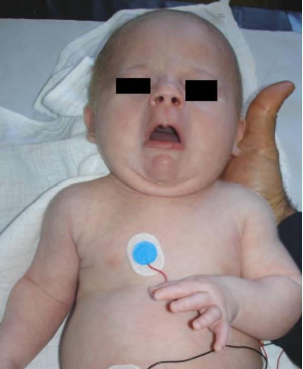
This clinical photograph displays an infant with a cluster of dysmorphic features and physical findings characteristic of a systemic syndrome, such as Stuve-Wiedemann syndrome. Key facial findings include an expressionless (mask-like) facies, blepharophimosis (narrowing of the palpebral fissures, here obscured by black privacy boxes), and a distinctive small, pursed mouth that remains partially open. Musculoskeletal examination reveals multiple joint contractures, most notably camptodactyly (permanent flexion) of the fingers on the left hand. The abdomen shows fullness, and the clinical history notes an umbilical hernia. The infant is placed in a clinical setting, evidenced by the presence of pediatric ECG monitoring electrodes on the chest and abdomen. The skin appears pale and smooth with subcutaneous fullness. This image serves as a teaching tool for recognizing neonatal dysmorphology, neuromuscular contractures, and specific facial phenotypes associated with rare genetic bone dysplasias or signaling pathway mutations (e.g., LIFR gene).
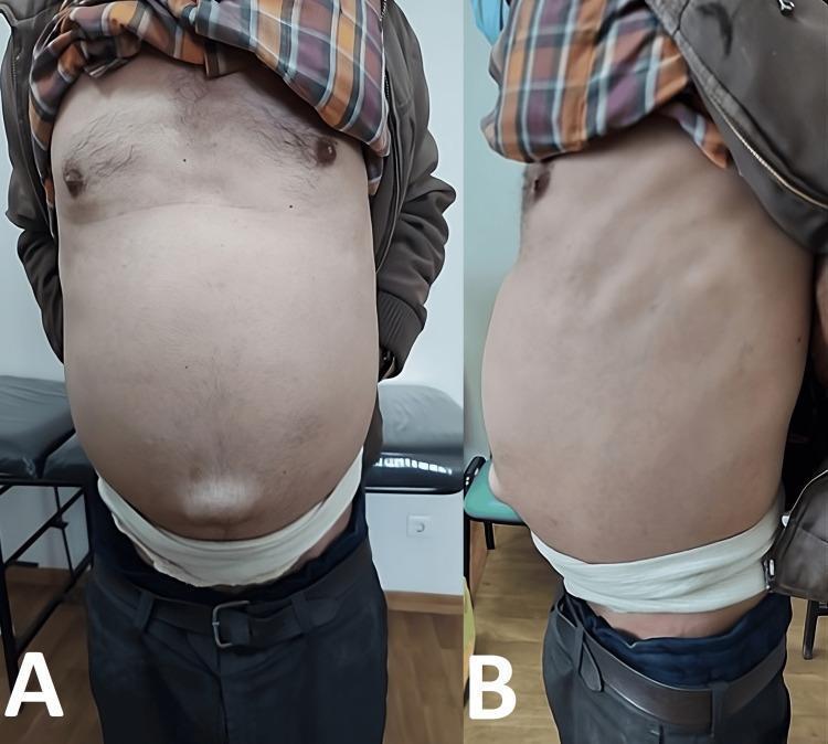
Clinical photograph showing the anterior (View A) and lateral (View B) profile of a patient's abdomen during physical examination. The image demonstrates massive abdominal distension, often associated with severe ascites or intra-abdominal masses. A prominent bulging umbilical hernia is clearly visible at the center of the abdominal wall. The skin appears taut and smooth due to the underlying pressure. A white support belt is positioned around the lower abdomen, used by the patient to provide structural stability and alleviate discomfort from the weight of the distended abdomen. This visual presentation is characteristic of pathology causing significant increase in intra-abdominal volume, such as Pseudomyxoma Peritonei (PMP), which leads to 'jelly belly' through the accumulation of mucinous fluid.
Confirming Diagnosis of Umbilical Hernia in a 2-Month-Old
Umbilical Hernia is a CLINICAL Diagnosis
Step 1 - History
| Feature | Umbilical Hernia |
|---|---|
| Onset | Appears within a few weeks of birth, after cord separation |
| Behavior | Swelling increases when baby cries, strains, or is held upright; reduces when calm and supine |
| Symptoms | Usually painless and symptomless at 2 months |
| Family history | Familial tendency present |
| Prematurity | More common in premature infants |
Step 2 - Physical Examination (The Diagnostic Cornerstone)
2a. Inspection
- A soft, rounded or conical protrusion at the umbilicus, covered by intact, normal-colored skin
- Visibly enlarges when the baby cries or strains - this is a classic, pathognomonic feature
- Skin over the swelling is normal - no erythema, no warmth, no discharge
- Typically ranges from a few mm to several cm in diameter
2b. Palpation - The Key Clinical Test
- Reducibility - The hernia should reduce easily back into the abdomen with gentle pressure. This is the single most important sign confirming a hernia.
- Feel the fascial defect (umbilical ring) - After reducing the contents, you can feel the edge of the fascial ring (defect in the linea alba) with your fingertip. Measure the ring diameter in cm - this guides prognosis and management.
- Contents - On reduction you may feel a soft gurgle (bowel) or a soft pad (omentum) slipping back in
- Impulse on crying - Place a finger over the reduced hernia and ask someone to make the baby cry - you will feel an expansile cough/cry impulse as the hernia re-protrudes
2c. Key Negative Findings That Confirm It Is Simple Hernia
- No tenderness over the swelling
- No erythema of overlying skin
- No discharge from umbilicus
- Not hard or fixed (hard/fixed = suspect incarceration)
- Fully reducible every time
Step 3 - Clinical Features That Differentiate from Other Causes
| Feature | Umbilical Hernia | Umbilical Granuloma | Omphalitis | Patent Urachus |
|---|---|---|---|---|
| Reducible | YES | No | No | No |
| Increases with crying | YES | No | No | No |
| Skin colour | Normal | Normal/pink nodule | Red, inflamed | Normal |
| Discharge | None | Serous/mucoid | Purulent, foul | Clear fluid/urine |
| Tenderness | No | No | YES | Usually no |
| Fever | No | No | Possibly | No |
| Fascial defect felt | YES | No | No | No |
Step 4 - Measure and Document
- Ring/defect diameter (in cm) - fingertip assessment
- Maximum size of protrusion
- Reducibility - complete or partial
- Defects <1 cm - high chance of spontaneous closure by 4-5 years
- Defects 1-2 cm - likely to close but monitor
- Defects >2 cm - less likely to close spontaneously; may need earlier surgery
Step 5 - When Is Imaging (Ultrasound) Needed?
- Diagnosis is uncertain (cannot confirm reducibility)
- Swelling is irreducible or tender (rule out incarceration, strangulation)
- Concern about patent urachus or vitelline duct remnant (fluid-filled tract on US)
- Associated abdominal distension or other anomalies
- Underlying condition suspected (e.g., hypothyroidism causing umbilical hernia - check TSH)
Step 6 - Associated Conditions to Screen For
| Condition | Clue |
|---|---|
| Hypothyroidism | Prolonged jaundice, hypotonia, poor feeding, large fontanelle - check TSH/T4 |
| Down syndrome (Trisomy 21) | Dysmorphic features, hypotonia |
| Beckwith-Wiedemann syndrome | Macroglossia, macrosomia, ear creases |
| Mucopolysaccharidoses | If presenting later in infancy |
| Prematurity | History of preterm birth |
Summary: Diagnostic Checklist at the Bedside
| Step | What to Do | Positive Finding |
|---|---|---|
| Inspection | Look at umbilicus while baby is crying | Conical protrusion appearing/enlarging |
| Palpation | Gently press swelling back in | Easily reduces - contents slip back |
| Finger over ring | Feel rim of defect | Palpable fascial ring |
| Cry impulse | Hold finger over area, baby cries | Protrusion re-emerges under finger |
| Skin check | Inspect overlying skin | Normal colour, no warmth/erythema |
| Discharge check | Check umbilicus | No discharge |
| Defect size | Measure ring diameter | Document in cm |
In a 2-month-old with a soft, painless, reducible umbilical swelling that enlarges with crying and has a palpable fascial defect - the diagnosis of umbilical hernia is confirmed clinically. No further investigations are needed unless associated features suggest a secondary cause. - Schwartz's Principles of Surgery 11th ed.; Bailey & Love's 28th ed.