HIV manifestations of ENT according to standard ENT textbook
Reading File
Finding Sources
Finding Sources
Finding Sources
Finding Sources
Finding Sources
Reading File
Reading File
Reading File
Reading File
Reading File
Reading File
Reading File
Reading File
Reading File
Loading Image
Loading Image
Loading Image
I now have all the content needed for a comprehensive, well-illustrated answer. Here is the full reference from Scott-Brown's Otorhinolaryngology, Head & Neck Surgery:
HIV Manifestations of the ENT System
Source: Scott-Brown's Otorhinolaryngology Head & Neck Surgery (Vol 1, Chapter 23)
Overview
ENT manifestations of HIV infection are extremely common and can affect every region of the head and neck. Otolaryngologists should test for HIV whenever it enters the differential diagnosis - specifically in patients with unexplained oral candidiasis, hairy leukoplakia, atypical head and neck cancers, parotid cysts, cervical lymphadenopathy, chronic parotitis, and pyrexia of unknown origin.
1. Cervical Lymphadenopathy
Persistent generalized lymphadenopathy is a hallmark of early symptomatic HIV infection, with cervical nodes most commonly involved. Nodes are typically small, symmetrical, and non-tender. Biopsy reveals follicular hyperplasia; no treatment is required unless the diagnosis is uncertain.
Causes of cervical lymphadenopathy in HIV:
- Non-Hodgkin's lymphoma (NHL)
- Hodgkin's lymphoma
- Tuberculosis
- Kaposi's sarcoma (KS)
- Syphilis
- Metastatic malignancies
- Mycobacterium avium complex (MAC)
- Dimorphic fungi, toxoplasmosis (less common)
Castleman disease - A rare lymphoproliferative disorder associated with HHV-8 (the same virus that causes KS). In HIV patients it presents as multicentric disease with fever, weight loss, and lymphadenopathy. Diagnosis is by excision biopsy.
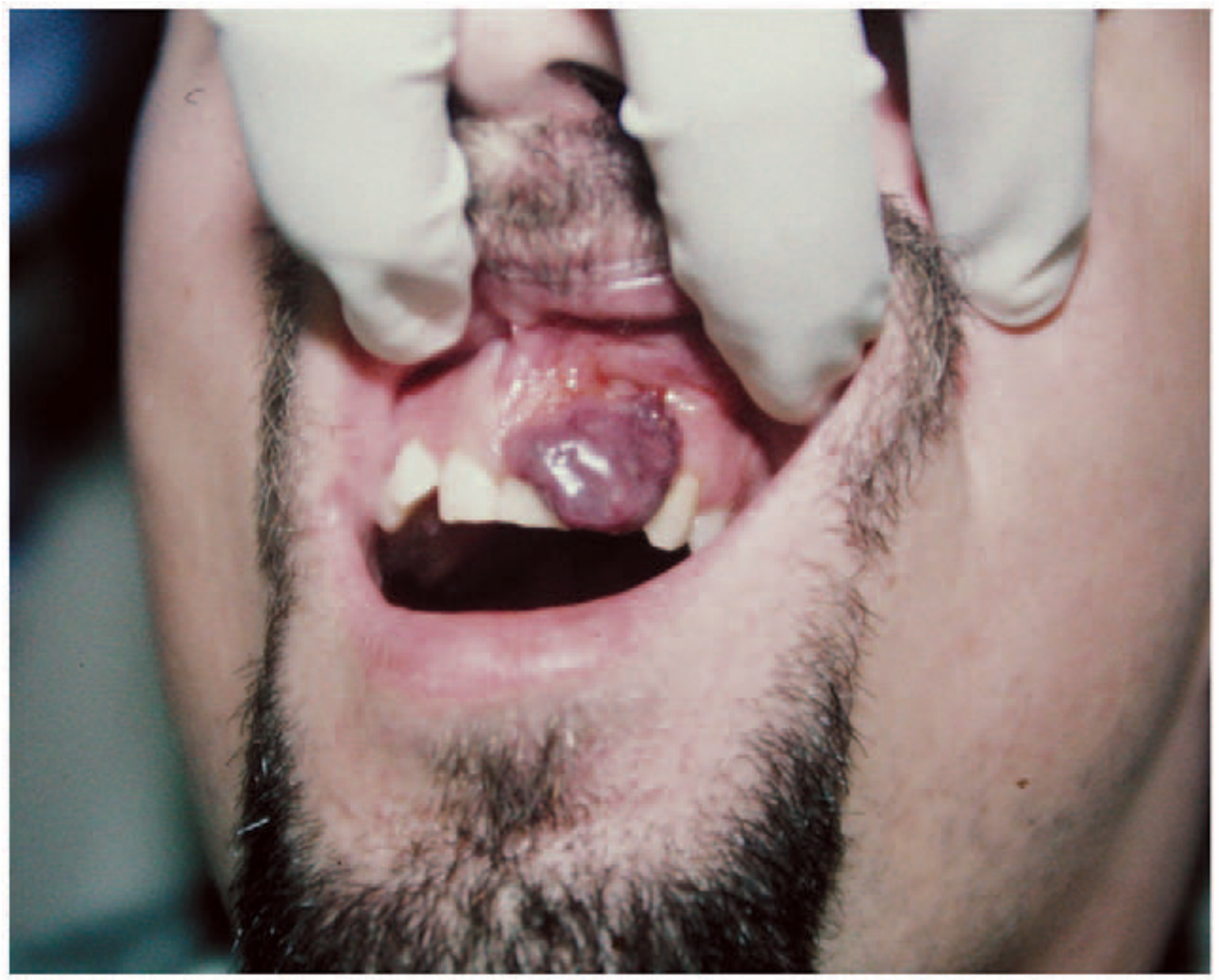
2. Kaposi's Sarcoma (Head & Neck)
KS is a malignant spindle cell tumour caused by co-infection of HIV and HHV-8. It is an AIDS-defining condition and the most common HIV-related tumour.
- Presents with cutaneous or mucosal lesions - most commonly in the head and neck
- Cutaneous lesions: color varies from purple to dark brown; may be flat, raised, or nodular
- Oral mucosa lesions are classically raised, red, and may ulcerate - typically on the hard and soft palate
- Visceral and lymph node involvement also occurs
- HAART is the mainstay of treatment - the condition regresses as immunity recovers
- Advanced cases require: localized radiotherapy, surgical excision, topical retinoids, intra-lesional vinblastine, or systemic chemotherapy
Bacillary angiomatosis mimics KS and must be differentiated. It is a Bartonella infection causing vascular proliferation with angiomatous lesions. Biopsy and microbiology will distinguish it. Treatment is with erythromycin.
3. Otologic Manifestations
External Ear
- Malignant otitis externa (MOE): Higher incidence in HIV patients. Osteomyelitis of the skull base causes facial nerve/cranial nerve dysfunction. CT of temporal bone or Tc-99m bone scan confirms diagnosis. Causative agent is usually Pseudomonas aeruginosa; with very low CD4, Aspergillus is common and highly invasive. Treatment: 6-8 weeks antipseudomonal antibiotics (3rd-gen cephalosporin + ciprofloxacin or aminoglycoside); Aspergillus requires IV amphotericin B +/- surgical debridement.
- Pneumocystis jiroveci (formerly P. carinii): Can infect the external auditory canal, middle ear, and mastoid bone. Presents as cyst or polyp; large cysts can occlude the EAC or spread to middle cranial fossa. Treated with TMP/SMX and/or dapsone.
Middle Ear
- Serous and recurrent otitis media are the most common otologic entities. Aetiologies include Eustachian tube dysfunction, nasopharyngeal neoplasms, adenoid hypertrophy, and sinusitis/allergies. Nasopharyngeal neoplasms must always be excluded in HIV patients, especially with unilateral otitis media.
- Tuberculous otitis media: Should be considered when OM is unresponsive to conventional therapy. Classical signs (multiple TM perforations, granulations, painless otorrhoea, bone necrosis) may not be evident in HIV. Minimum 6 months antituberculous chemotherapy.
- Mycobacterium avium complex (MAC): Also reported to cause otitis media.
Inner Ear
- Vestibular and cochlear symptoms are common.
- Sensorineural hearing loss (SNHL): Can be caused by: direct HIV neurotoxicity, CNS lymphoma, CMV labyrinthitis, cryptococcal meningitis, and drug toxicity (macrolides, aminoglycosides, isoniazid). HIV patients appear more susceptible to aminoglycoside ototoxicity.
- Otosyphilis: Rare but important. In HIV, syphilis can have an abbreviated course with early neurological involvement. Classic presentation: SNHL sparing mid-range frequencies + tinnitus + vertigo (mimics Meniere's disease). Treat with high-dose procaine penicillin + probenecid.
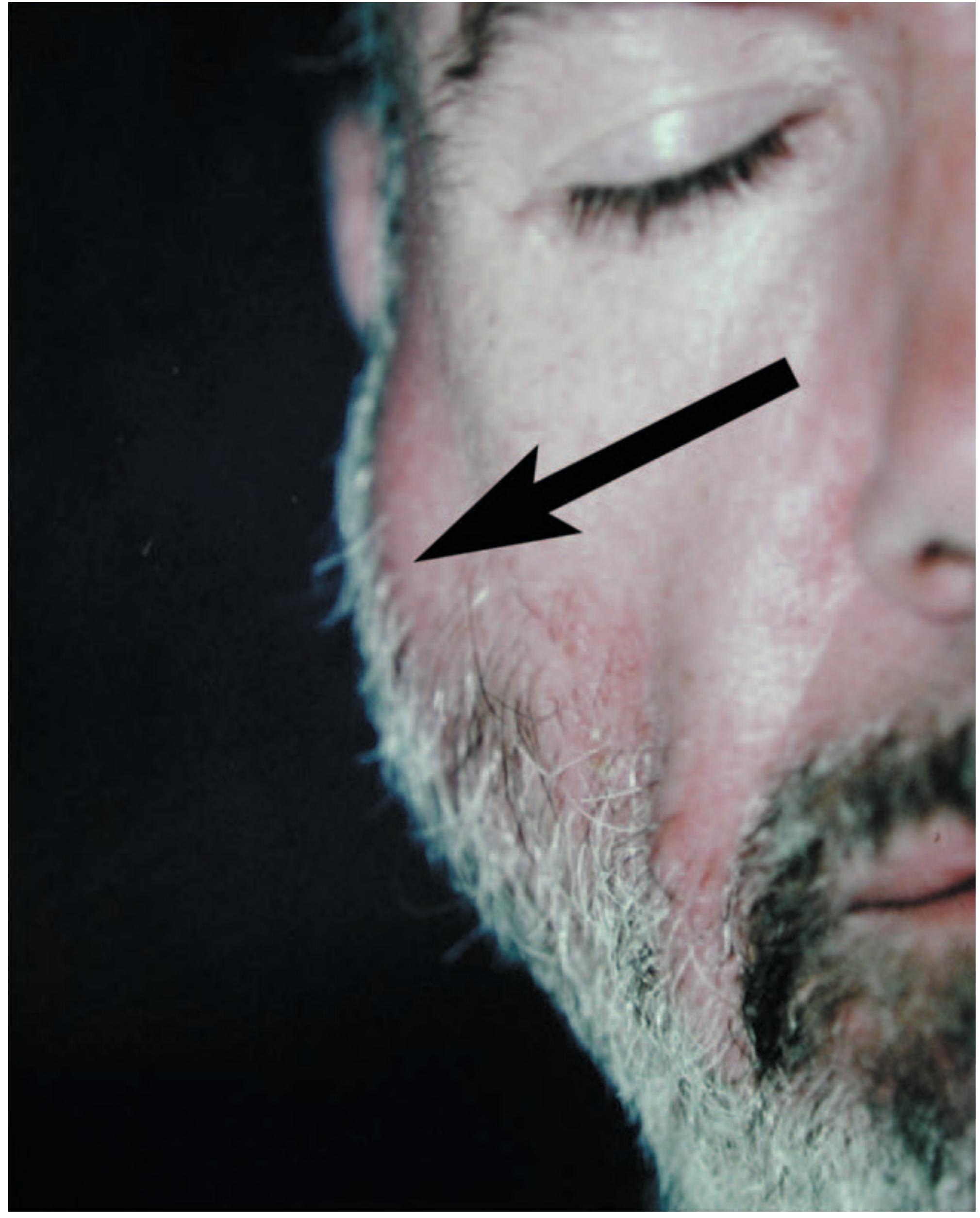
Facial Nerve
- Facial nerve palsy occurs in up to 7.2% of HIV patients. Causes: direct HIV infection, neoplasms, opportunistic infections, AIDS encephalopathy, toxoplasmosis.
- Bell's palsy: Most frequent diagnosis for CN VII paralysis in HIV (postulated due to HSV). Complete recovery usually within 3 weeks to 3 months.
- Ramsay Hunt syndrome (herpes zoster oticus): More common in AIDS patients. Triad: peripheral facial nerve palsy + herpetic vesicles along CN VII dermatome/conchal bowl + severe herpetic pain. Treat with high-dose acyclovir + high-dose steroids (when not contraindicated).
- Bilateral facial nerve palsies indicate systemic disease. Serology for syphilis, CMV, HSV, HZV, and EBV should be sent. MRI brain + CSF analysis needed.
4. Nose and Paranasal Sinuses
30-68% of HIV-positive patients will develop nasal and/or sinus symptoms.
Nasal Obstruction
- Adenoidal hypertrophy - Any non-paediatric patient with adenoid hypertrophy should be tested for HIV
- Hypertrophy of any tissue in Waldeyer's ring
- Neoplasms: KS (including nasal septum involvement) or Non-Hodgkin's lymphoma (arising from nasal cavity, nasopharynx, or sinuses)
Sinusitis
- Acute sinusitis pathogens are similar to non-infected persons: S. pneumoniae, M. catarrhalis, H. influenzae
- Chronic sinusitis (CD4 < 200/mL): S. aureus, P. aeruginosa, Aspergillus
- Other reported pathogens: CMV, Microsporidia, Acanthamoeba castellani, Legionella pneumophila
- In HIV patients with fever of unknown origin, the sinuses must always be investigated
- Management: antibiotics + decongestants; topical nasal steroids; guaifenesin as adjunct
- Surgical indication: persistent symptoms despite medical therapy, OR any extra-sinus signs (mental status changes, cranial nerve dysfunction, intracranial extension, abscess). Endoscopic sinus surgery (ESS) is safe and effective.
5. Oral Cavity
Oral manifestations are reported in up to 100% of AIDS patients and are often the first manifestation of HIV in otherwise asymptomatic individuals.
Recurrent Aphthous Ulcers
- Larger than in non-immunocompromised patients
- Solitary or multiple, with an erythematous halo; may have pseudomembrane
- Giant aphthous ulcers cause severe odynophagia and can worsen anorexia/wasting
- Treatment: topical anaesthetics/steroids; topical tetracycline or systemic clindamycin if superinfection; intralesional triamcinolone acetonide; oral thalidomide (heals completely in 55%, partially in 90% - but risk of increased viral load, teratogenic)
Oral Candidiasis
- Most prevalent form: pseudomembranous candidiasis (thrush) - white plaques on erythematous erosive mucosa
- Chronic hypertrophic form: heaped-up plaques not easily scraped
- Also presents as angular cheilitis and atrophic (erythematous) form
- Oesophageal involvement may coexist - must be asked about
- Treatment: nystatin mouthwash (mild); clotrimazole or amphotericin lozenges; fluconazole (systemic, for advanced disease); amphotericin B or echinocandins IV (severe cases)
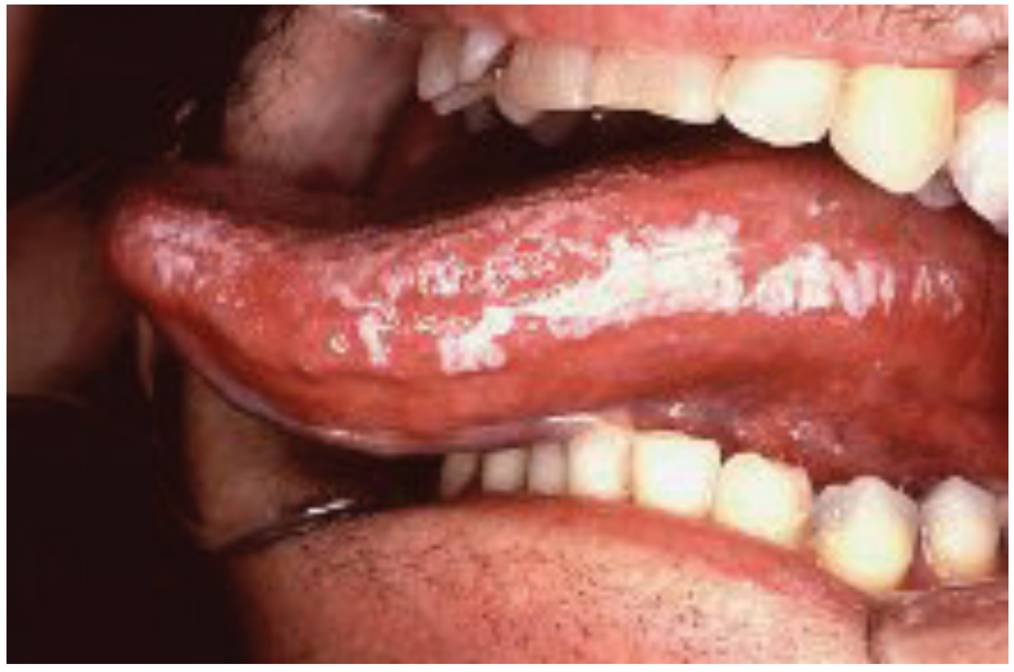
Hairy Leukoplakia
- Unique to HIV-positive individuals (EBV-related)
- White, raised, corrugated/filiform lesion on the lateral border of the tongue; can also affect dorsal tongue, buccal/labial mucosa, floor of mouth, soft palate
- Usually asymptomatic - no treatment required
- Must be differentiated from other serious oral disorders
Gingival Disease
- Gingival tissue loss, tooth loosening/loss, bone exposure
- Requires close dental evaluation and frequent management
- Advanced cases require aggressive antibiotics and/or surgery
Non-Hodgkin's Lymphoma (Oral)
- Ulcerative lesions or red exophytic masses
- Typically involve the gingiva, alveolar ridges, and Waldeyer's ring
- Usually a poor prognostic indicator; recurrences common despite chemo/radiation
Summary Table of Common Oral HIV Manifestations (from Scott-Brown's Table 23.1):
| Disease | Features | Management |
|---|---|---|
| Gingival disease | Tissue loss, tooth loosening, bone exposure | Frequent dental cleanings; aggressive antibiotics/surgery when advanced |
| Oral thrush | Pseudomembranous plaques, atrophic erythema, angular cheilitis | Topical clotrimazole; fluconazole for refractory/advanced disease |
| Aphthous ulcers | Large, painful ulcers; biopsy confirms non-neoplastic aetiology | Topical steroids; intralesional/systemic steroids; thalidomide; ARV therapy |
| Kaposi's sarcoma | Hard/soft palate, occasionally gingiva/buccal mucosa | Radiotherapy, intralesional vinblastine, or systemic chemotherapy |
| Hairy leukoplakia | Lateral tongue, EBV-related | No treatment if asymptomatic |
| Radiation mucositis | Side effect of treating other oral lesions | Topical steroids/antifungals; supportive care |
6. Pharynx, Larynx and Oesophagus
Many of the same processes that affect the oral cavity extend to the pharynx, larynx, and oesophagus, presenting with odynophagia, dysphagia, and chest pain.
- Candidiasis of the oesophagus/pharynx: presents with odynophagia; oesophageal involvement requires systemic antifungals
- CMV oesophagitis: Large solitary ulcerations with raised indurated margins, usually at severe immunocompromise (CD4 <100). Treatment: IV ganciclovir/foscarnet
- HSV oesophagitis: Discrete vesicles with mucosal ulceration. Usually occurs when CD4 is well below 100/mm³. Treatment: IV acyclovir followed by oral agents
- Idiopathic oesophageal ulcer: Same frequency as CMV oesophagitis; treatment with systemic steroids or thalidomide
- Kaposi's sarcoma of the larynx/pharynx: presents with dysphagia, odynophagia, hoarseness, shortness of breath, or severe airway compromise
7. Salivary Glands
Both major and minor salivary glands can be involved. Most common complaint is xerostomia.
Lymphoepithelial Cysts
- Almost diagnostic of HIV infection
- Present as progressive, multiple, bilateral, non-tender parotid swellings
- FNAB reveals fluid with lymphocytes and squamous epithelial cells
- CT/ultrasound shows multiple thin-walled cysts
- Facial nerve dysfunction should raise suspicion of malignancy
- Cysts usually resolve with HAART; surgery only for enlarged lesions that are cosmetically troublesome or diagnostically equivocal
- Needle aspiration is not curative (recurrence almost always occurs)
- Without HAART: sclerotherapy with alcohol or sodium morrhuate can be effective
When to Test for HIV (ENT Triggers)
An otolaryngologist should test for HIV in any patient with:
- Unexplained oral candidiasis
- Hairy leukoplakia of the tongue
- Atypical head and neck cancers
- Parotid cysts
- Cervical lymphadenopathy (unexplained)
- Chronic parotitis
- Pyrexia of unknown origin
- Non-paediatric adenoid hypertrophy
Source: Scott-Brown's Otorhinolaryngology Head & Neck Surgery, Vol 1, Chapter 23 (HIV & ENT)
This is a shared conversation. Sign in to Orris to start your own chat.