Shoulder mri

Shoulder MRI covers a lot of ground. Let me narrow this down so I can give you the most useful answer.
Asking for Preferences

What would you like to know about shoulder MRI?

Shoulder MRI covers a lot of ground. Let me narrow this down so I can give you the most useful answer.

Yes

Reading File
Finding Sources
Searching Images

shoulder MRI rotator cuff tear

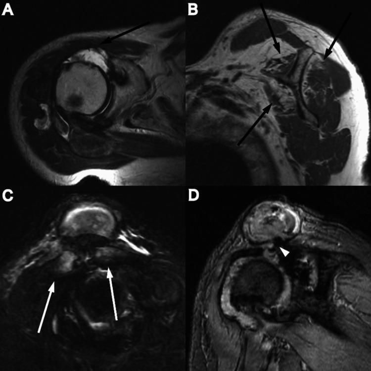
This composite diagnostic image features four MRI views (A-D) of a human shoulder, illustrating advanced degenerative joint disease and rotator cuff pathology. Panels A and B are axial and coronal sections showing a massive rotator cuff tear accompanied by severe muscle atrophy. Black arrows indicate fatty infiltration and volume loss in the supraspinatus, infraspinatus, and subscapularis muscles. Panel C highlights the acromioclavicular (AC) joint with white arrows pointing to osteoarthritic changes, including joint space narrowing and subchondral irregularities. Panel D is a T2-weighted MRI demonstrating a 'Geyser sign' (white arrowhead), where a large synovial cyst communicates directly with a markedly degenerated glenohumeral joint through a full-thickness rotator cuff defect. The images collectively demonstrate the progression of chronic rotator cuff insufficiency leading to secondary osteoarthritis and superior joint fluid leakage. This material is suitable for orthopedic and radiology education regarding shoulder instability and end-stage cuff tear arthropathy.
This composite diagnostic image features four MRI views (A-D) of a human shoulder, illustrating advanced degenerative joint disease and rotator cuff pathology. Panels A and B are axial and coronal sections showing a massive rotator cuff tear accompanied by severe muscle atrophy. Black arrows indicate fatty infiltration and volume loss in the supraspinatus, infraspinatus, and subscapularis muscles. Panel C highlights the acromioclavicular (AC) joint with white arrows pointing to osteoarthritic changes, including joint space narrowing and subchondral irregularities. Panel D is a T2-weighted MRI demonstrating a 'Geyser sign' (white arrowhead), where a large synovial cyst communicates directly with a markedly degenerated glenohumeral joint through a full-thickness rotator cuff defect. The images collectively demonstrate the progression of chronic rotator cuff insufficiency leading to secondary osteoarthritis and superior joint fluid leakage. This material is suitable for orthopedic and radiology education regarding shoulder instability and end-stage cuff tear arthropathy.

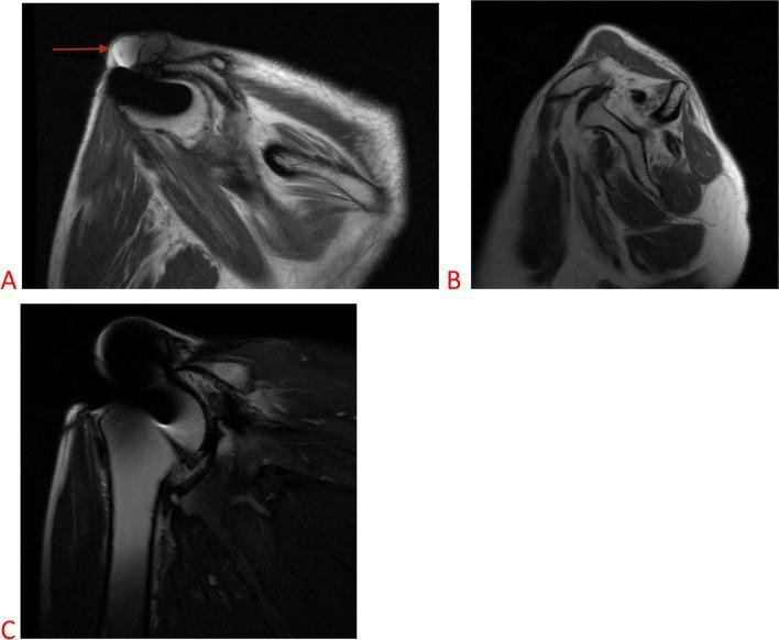
This composite figure presents three MRI views of a right shoulder, illustrating end-stage rotator cuff arthropathy and its secondary manifestations.
This composite figure presents three MRI views of a right shoulder, illustrating end-stage rotator cuff arthropathy and its secondary manifestations.

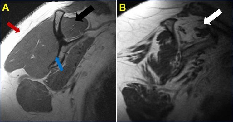
This diagnostic image consists of two side-by-side sagittal T1-weighted MRI scans of the shoulder, illustrating the differences between healthy rotator cuff musculature and a chronic massive rotator cuff tear. Panel A shows a healthy shoulder: a black arrow identifies the supraspinatus muscle belly with characteristic low signal intensity (dark appearance); a red arrow indicates hyperintense (bright) subcutaneous fat; and a blue arrow marks the body of the scapula. Panel B demonstrates pathological changes associated with a chronic, massive rotator cuff tear. A white arrow highlights significant fatty infiltration and muscle atrophy within the supraspinatus. In this pathological state, the muscle tissue exhibits a high signal intensity similar to the surrounding subcutaneous fat, indicating replacement of muscle fibers with adipose tissue. This comparison is clinically significant for diagnosing Goutallier stages of fatty degeneration in musculoskeletal radiology and orthopedics, helping to assess the prognosis and surgical reparability of chronic rotator cuff injuries.
This diagnostic image consists of two side-by-side sagittal T1-weighted MRI scans of the shoulder, illustrating the differences between healthy rotator cuff musculature and a chronic massive rotator cuff tear. Panel A shows a healthy shoulder: a black arrow identifies the supraspinatus muscle belly with characteristic low signal intensity (dark appearance); a red arrow indicates hyperintense (bright) subcutaneous fat; and a blue arrow marks the body of the scapula. Panel B demonstrates pathological changes associated with a chronic, massive rotator cuff tear. A white arrow highlights significant fatty infiltration and muscle atrophy within the supraspinatus. In this pathological state, the muscle tissue exhibits a high signal intensity similar to the surrounding subcutaneous fat, indicating replacement of muscle fibers with adipose tissue. This comparison is clinically significant for diagnosing Goutallier stages of fatty degeneration in musculoskeletal radiology and orthopedics, helping to assess the prognosis and surgical reparability of chronic rotator cuff injuries.

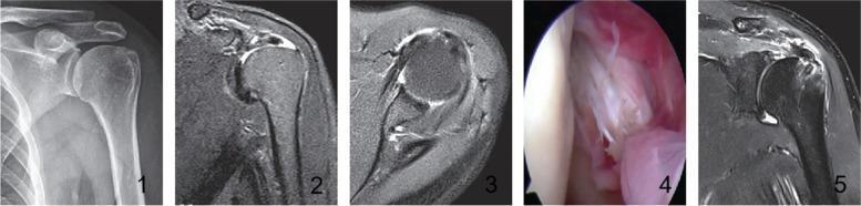
This composite figure illustrates the preoperative, intraoperative, and postoperative findings of a complex rotator cuff tear in a left shoulder. Panel 1 is an anteroposterior radiograph demonstrating an elevated acromion, often indicative of chronic rotator cuff pathology. Panels 2 and 3 provide preoperative T2-weighted MRI scans in oblique and transverse planes, respectively; Panel 2 shows a supraspinatus tendon tear with Patte stage 2 retraction, while Panel 3 exhibits mixed hyperintensities within the joint space. Panel 4 presents an intraoperative arthroscopic view confirming a Lafosse I injury of the superior third of the subscapularis tendon. Panel 5 is a follow-up T2-weighted MRI showing successful surgical intervention, characterized by a suture anchor visible in the greater tuberosity of the humerus and evidence of continuous supraspinatus tendon healing. This sequence highlights the diagnostic pathway from radiography and MRI to arthroscopic confirmation and subsequent postoperative evaluation of rotator cuff repair using suture anchors.
This composite figure illustrates the preoperative, intraoperative, and postoperative findings of a complex rotator cuff tear in a left shoulder. Panel 1 is an anteroposterior radiograph demonstrating an elevated acromion, often indicative of chronic rotator cuff pathology. Panels 2 and 3 provide preoperative T2-weighted MRI scans in oblique and transverse planes, respectively; Panel 2 shows a supraspinatus tendon tear with Patte stage 2 retraction, while Panel 3 exhibits mixed hyperintensities within the joint space. Panel 4 presents an intraoperative arthroscopic view confirming a Lafosse I injury of the superior third of the subscapularis tendon. Panel 5 is a follow-up T2-weighted MRI showing successful surgical intervention, characterized by a suture anchor visible in the greater tuberosity of the humerus and evidence of continuous supraspinatus tendon healing. This sequence highlights the diagnostic pathway from radiography and MRI to arthroscopic confirmation and subsequent postoperative evaluation of rotator cuff repair using suture anchors.

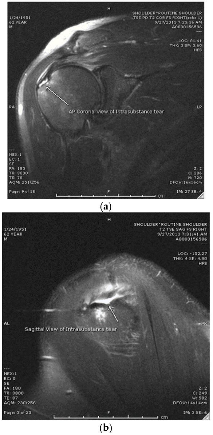
This diagnostic imaging composite displays two T2-weighted magnetic resonance imaging (MRI) views of a right shoulder joint, specifically demonstrating an intrasubstance rotator cuff tear. Figure (a) is a coronal T2 Turbo Spin Echo (TSE) Fat-Saturated (FS) image, showing the glenohumeral joint and the supraspinatus tendon. A white arrow indicates a localized area of high signal intensity within the tendon substance, which does not communicate with the bursal or articular surfaces. Figure (b) is a sagittal T2 TSE FS image of the same shoulder, providing a cross-sectional view of the rotator cuff muscles and tendons. The arrow in this view confirms the high-intensity fluid signal within the tendon mid-substance, characteristic of a high-grade partial-thickness intrasubstance tear. Key anatomical structures visible include the humeral head, glenoid, and the rotator cuff complex. These images are used in orthopedics and radiology to differentiate intrasubstance tears from articular-sided or bursal-sided partial tears, which is critical for determining surgical versus non-operative management strategies like PRP injections.
This diagnostic imaging composite displays two T2-weighted magnetic resonance imaging (MRI) views of a right shoulder joint, specifically demonstrating an intrasubstance rotator cuff tear. Figure (a) is a coronal T2 Turbo Spin Echo (TSE) Fat-Saturated (FS) image, showing the glenohumeral joint and the supraspinatus tendon. A white arrow indicates a localized area of high signal intensity within the tendon substance, which does not communicate with the bursal or articular surfaces. Figure (b) is a sagittal T2 TSE FS image of the same shoulder, providing a cross-sectional view of the rotator cuff muscles and tendons. The arrow in this view confirms the high-intensity fluid signal within the tendon mid-substance, characteristic of a high-grade partial-thickness intrasubstance tear. Key anatomical structures visible include the humeral head, glenoid, and the rotator cuff complex. These images are used in orthopedics and radiology to differentiate intrasubstance tears from articular-sided or bursal-sided partial tears, which is critical for determining surgical versus non-operative management strategies like PRP injections.

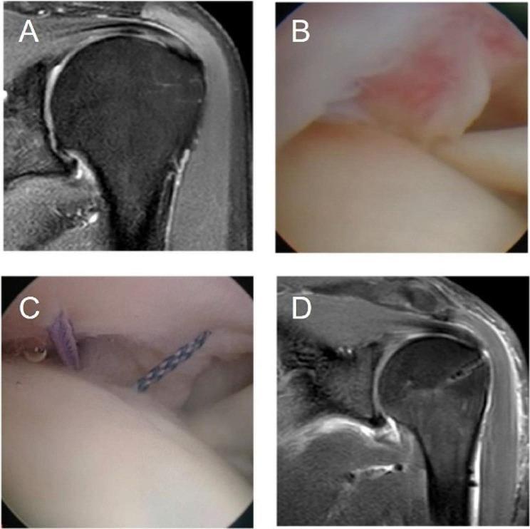
A multi-panel medical image sequence illustrating the diagnosis and surgical repair of a rotator cuff injury. (A) Coronal T2-weighted MRI of the shoulder showing an Ellman III partial articular-sided supraspinatus tendon tear, characterized by a focal high-signal defect at the tendon's footprint. (B) Intraoperative arthroscopic view from the glenohumeral joint confirming the partial articular-sided supraspinatus tendon tear with visible tissue fraying and hyperemia. (C) Intraoperative arthroscopic view demonstrating the surgical intervention, where purple and braided sutures are introduced through the tendon into the joint space as part of a trans-tendon repair technique. (D) Postoperative coronal T2-weighted MRI showing a restored tendon footprint with resolved signal intensity defects, indicating a successful rotator cuff repair. A hypointense linear artifact in the humeral head suggests the presence of a suture anchor. This sequence is intended for orthopedic education, specifically focusing on the management of partial-thickness rotator cuff tears through arthroscopic techniques.

A multi-panel medical image sequence illustrating the diagnosis and surgical repair of a rotator cuff injury. (A) Coronal T2-weighted MRI of the shoulder showing an Ellman III partial articular-sided supraspinatus tendon tear, characterized by a focal high-signal defect at the tendon's footprint. (B) Intraoperative arthroscopic view from the glenohumeral joint confirming the partial articular-sided supraspinatus tendon tear with visible tissue fraying and hyperemia. (C) Intraoperative arthroscopic view demonstrating the surgical intervention, where purple and braided sutures are introduced through the tendon into the joint space as part of a trans-tendon repair technique. (D) Postoperative coronal T2-weighted MRI showing a restored tendon footprint with resolved signal intensity defects, indicating a successful rotator cuff repair. A hypointense linear artifact in the humeral head suggests the presence of a suture anchor. This sequence is intended for orthopedic education, specifically focusing on the management of partial-thickness rotator cuff tears through arthroscopic techniques.

Searching PubMed

"shoulder"[MeSH Terms] AND "magnetic resonance imaging"[MeSH Terms]

Searching the Web

shoulder MRI indications protocol rotator cuff labrum

Reading File
Reading File

Shoulder MRI: Overview

What it's for

MRI is the primary advanced imaging test for the shoulder because it directly visualizes soft tissues that X-rays can't show - the rotator cuff tendons, labrum, articular cartilage, and biceps tendon. Main indications:
  • Rotator cuff disease - tendinopathy, partial- or full-thickness tears, impingement
  • Labral/instability workup - Bankart lesions, SLAP tears, Hill-Sachs lesions (often with intra-articular contrast, i.e. MR arthrography)
  • Biceps tendon (long head) pathology - subluxation, tears
  • Unexplained shoulder pain not clarified by X-ray
  • Adhesive capsulitis (frozen shoulder) evaluation
  • Osteonecrosis, infection, masses, and post-surgical evaluation
Plain radiographs are still done first to check bony alignment and exclude glenohumeral arthrosis; MRI is added when tendon, labral, or cartilage detail is needed - Grainger & Allison's Diagnostic Radiology, p. 3434 (Rotator Cuff Disease chapter).

Technique

The patient lies supine, arm at the side in neutral or slight external rotation, with a dedicated surface coil over the shoulder. Standard sequences use T1 and T2/STIR with fat suppression across three planes:
  • Axial - the labrum, biceps tendon, subscapularis
  • Oblique coronal (parallel to supraspinatus, perpendicular to the glenoid) - the supraspinatus/infraspinatus footprint and AC joint
  • Oblique sagittal (parallel to the glenoid) - the "hamburger" view of the whole cuff and rotator interval
T2/STIR fat-suppressed sequences are most sensitive for cuff pathology, showing fluid signal within or through the tendon - THIEME Atlas of Anatomy, General Anatomy and Musculoskeletal System, p. 47 (Standard planes in MRI shoulder examination). When labral/instability is the question, gadolinium is often injected into the joint first (MR arthrogram), sometimes with the arm abducted and externally rotated (ABER position) to stress the anteroinferior labrum.

Key findings

Rotator cuff tears
  • Partial-thickness: articular-surface (most common) or bursal-surface signal defect that doesn't cross the whole tendon
  • Full-thickness: fluid signal extends from the joint side to the bursal side, creating communication between the glenohumeral joint and subacromial bursa
  • Massive tear: ≥5 cm or involving 2+ tendons, often with retraction and muscle atrophy/fatty infiltration (graded by Goutallier stages) - both MRI and ultrasound approach nearly 100% accuracy for full-thickness tears (Grainger & Allison's Diagnostic Radiology, p. 3434-3448).
Chronic massive rotator cuff tear with fatty infiltration on T1 MRI
Coronal T2 MRI showing full-thickness rotator cuff tear with muscle atrophy and a "Geyser sign"
Labral tears / instability - best seen on MR arthrography: Bankart lesion (anteroinferior labral tear from anterior dislocation), SLAP tear (superior labrum anterior-posterior), Hill-Sachs lesion (posterolateral humeral head impaction fracture).
Impingement/tendinopathy - tendon thickening, intrasubstance increased signal, subacromial-subdeltoid bursitis, and bony changes (acromial spur, AC joint osteoarthritis) that narrow the subacromial space.

Recent evidence (last 2 years)

A few systematic reviews/meta-analyses worth knowing about, since they update or quantify what's above:
  • MR arthrography shows high sensitivity/specificity for labral tears, rotator cuff tears, Hill-Sachs, and Bankart lesions (PMID: 39914604, 2025)
  • MRI features for diagnosing adhesive capsulitis were systematically reviewed (PMID: 40241075, 2025)
  • MRI vs CT for glenoid bone loss in shoulder instability was compared in a 2026 meta-analysis (PMID: 40952518)
These generally reinforce standard teaching rather than contradict it, but if you're working up instability or adhesive capsulitis specifically, they're worth a closer look.
If you want, I can go deeper into a specific piece of this - like how to read a shoulder MRI report, the Bankart/SLAP classification, or how findings map to surgical decisions.
This is a shared conversation. Sign in to Orris to start your own chat.