
CASE 4 A 55-year-old man presented to the general practitioner with swelling and severe pain in his left big toe for the last 1 week. He gives history of similar episodes in the past 6 months. Picture A: Clinical picture of patient’s foot Picture B: Microscopic picture of biopsy from the patient’s toe. 4.1 Describe the abnormalities seen in the Picture A. 4.2 Describe the microscopic features seen in the Picture B. 4.3 State the most probable diagnosis 4.4 State the predisposing factors for this disease
gout tophus big toe clinical photo
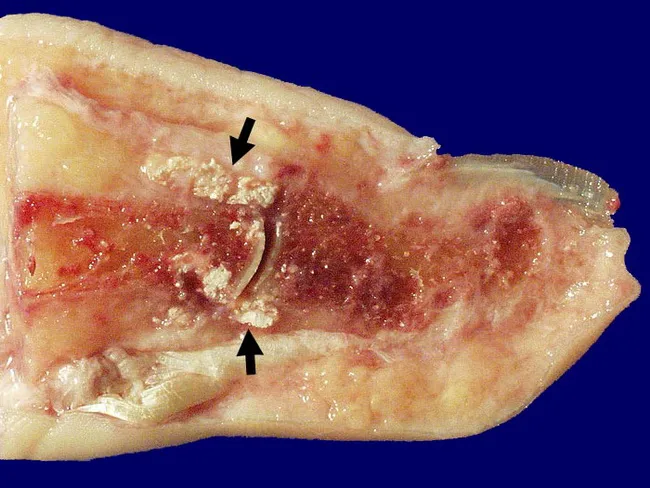
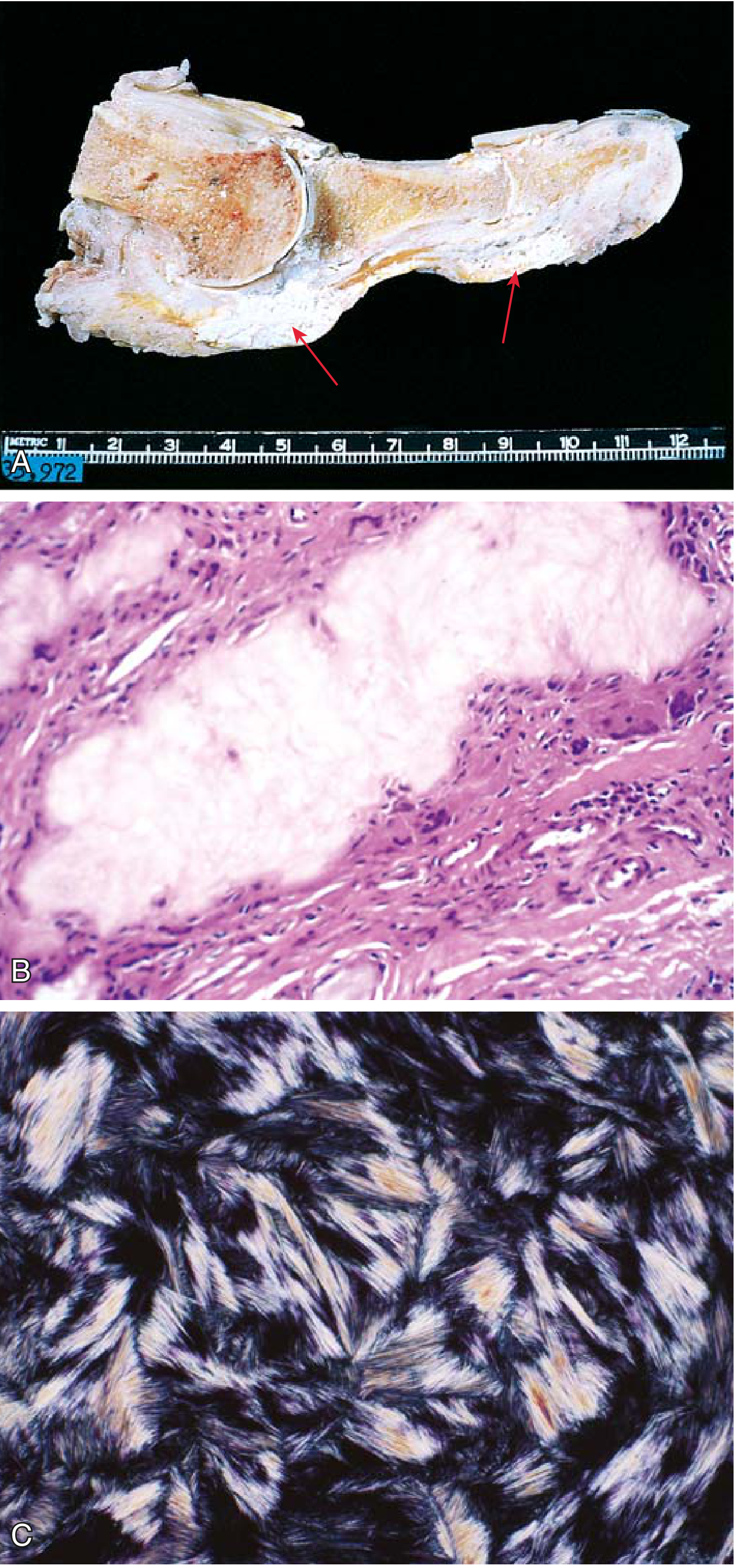
Gross pathology photograph of a toe joint showing chronic tophaceous gout with monosodium urate crystal deposition. Modality/technique: gross specimen imaging; cross‑sectional cut through the proximal interphalangeal joint (big toe) displaying soft tissue and juxta-articular bone. Anatomic site: digital toe, foot, musculoskeletal system; tissue components include tophus-contaminated soft tissues, articular cartilage and adjacent bone. Visual features: chalky, opaque white to pale yellow crystalline material adherent to articular surfaces and embedded within periarticular soft tissues; irregular joint surface erosion with subchondral bone destruction; thickened synovium with chronic inflammatory changes and fibrosis; superficial white crystalline deposits extend into surrounding ligaments and tendons; occasionally ulceration of overlying skin may be seen in advanced disease. Pathology: tophi composed of monosodium urate crystals with granulomatous inflammatory response, fibrocollagenous scar, and secondary degenerative changes; articular cartilage loss and joint-space narrowing. Clinical significance: hallmark of chronic gout and tophaceous arthritis; indicates long‑standing hyperuricemia and failed urate control; explains reduced range of motion due to fibrosis and ankylosis; aids differential diagnosis versus pseudogout, rheumatoid arthritis, osteoarthritis, and infection. Clinical/educational use: gross-pathology reference for medical education, radiologic-pathologic correlation, and therapeutic planning, including urate‑lowering treatment and tophus management. Correlates with serum urate levels, imaging findings, and clinical staging. important for prognosis.
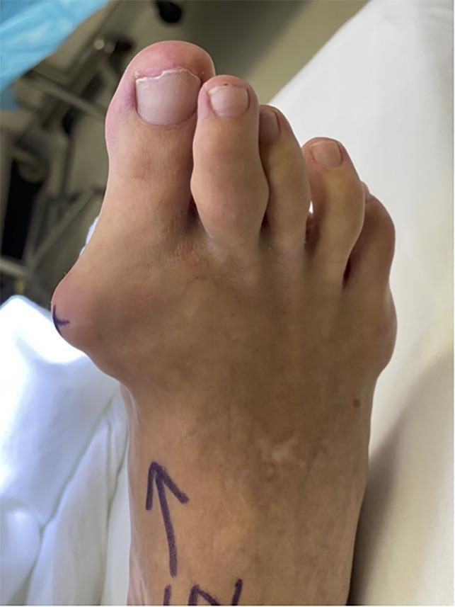
This clinical photograph displays a human right foot from a dorsal-medial perspective, illustrating advanced manifestations of chronic tophaceous gout and associated forefoot deformities. A large, prominent gouty tophus is visible as a firm, bulging subcutaneous mass on the medial aspect of the first metatarsophalangeal (MTP) joint. This mass creates a significant clinical hallux valgus deformity, characterized by the lateral deviation of the great toe toward the second digit. Additionally, the second toe exhibits soft tissue swelling and a smaller tophaceous deposit at the level of the proximal interphalangeal joint, resulting in a visible deformity. The skin over the primary tophus appears stretched and thin, though no ulceration is present. Surgical markings (an arrow) are visible on the dorsal surface of the foot, indicating preoperative preparation. The image serves as a clinical example of how monosodium urate crystal deposition can lead to significant structural joint distortion and secondary mechanical deformities in the foot.
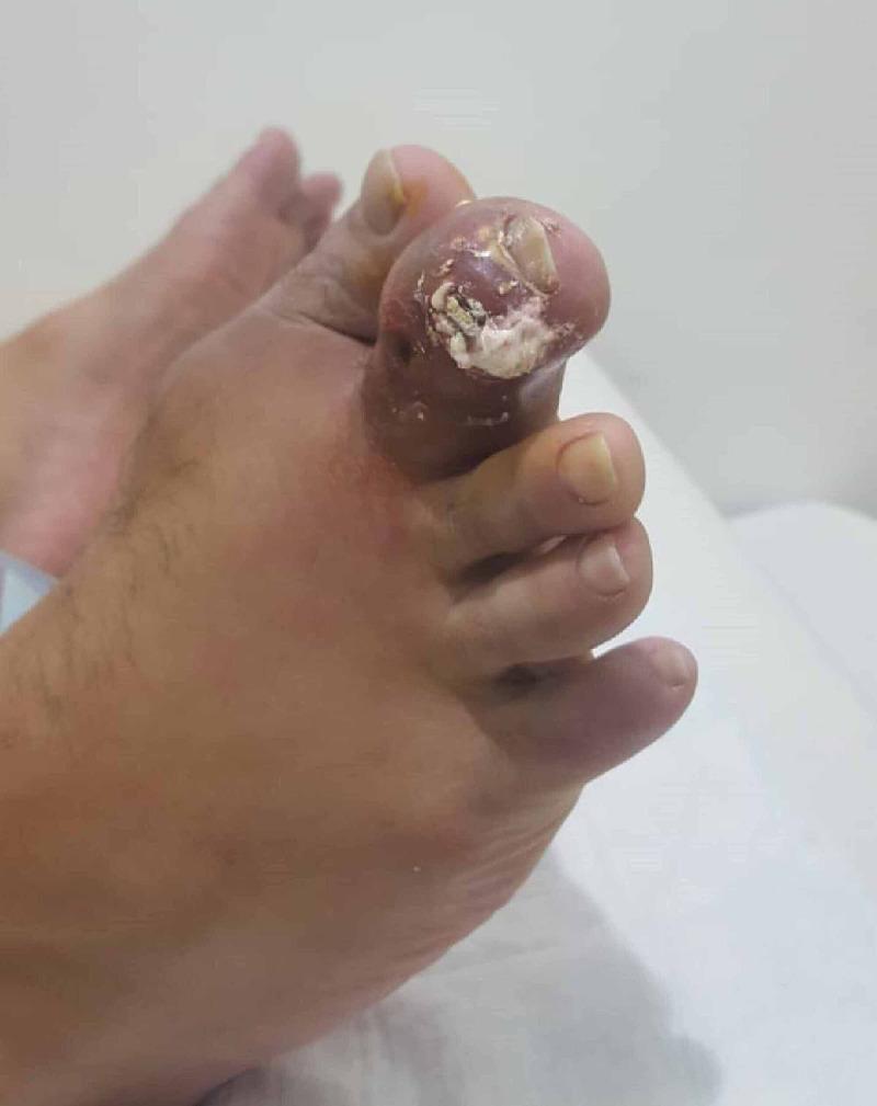
Clinical photograph of a human foot, focusing on the second toe, which exhibits severe gouty tophus formation. The distal portion of the toe is significantly enlarged with tense, erythematous skin, indicating acute inflammation. At the distal interphalangeal joint level, there is a visible rupture of the skin with the extrusion of chalky, white, toothpaste-like material characteristic of monosodium urate crystals. The surrounding soft tissue is edematous, and the nail appears dystrophic and partially elevated due to the underlying pressure of the tophaceous deposit. This image serves as a classic clinical representation of chronic tophaceous gout with an acute flare and ulceration. The condition demonstrates the typical presentation of urate crystal deposition in peripheral small joints, resulting in significant joint deformity and soft tissue involvement.
gout urate crystals microscopy biopsy needle shaped birefringent
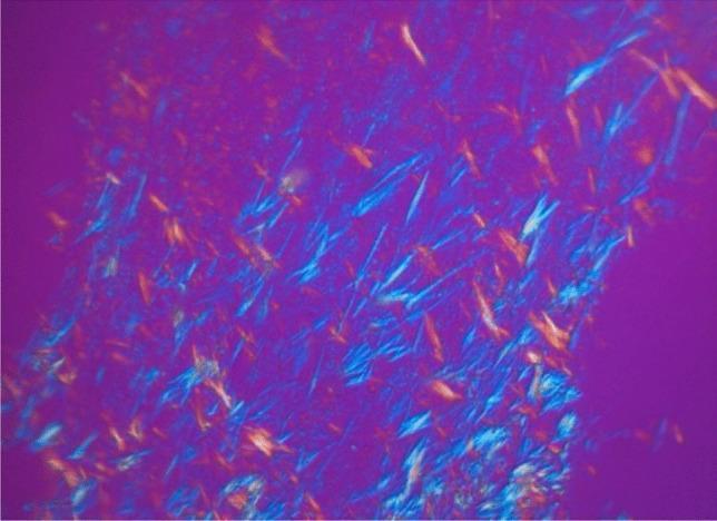
This diagnostic image demonstrates compensated polarized light microscopy (CPLM) of a tissue sample, identified as an aortic plaque biopsy. The image displays the characteristic optical properties of monosodium urate (MSU) crystals. Numerous elongated, needle-shaped crystals are distributed throughout the field of view, exhibiting strong birefringence. Against a magenta or pinkish-purple background, the crystals appear in contrasting colors, primarily bright blue and yellow-orange, depending on their orientation relative to the axis of the compensator. This finding is classically associated with gout but here illustrates cardiovascular MSU deposition within vascular plaques. The image serves as an educational example of how polarized light microscopy is used to confirm the presence of urate crystals at a microscopic level, highlighting their distinctive morphology and birefringent signature for clinical pathology and rheumatology education.
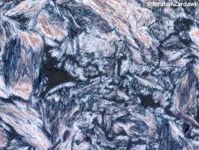
Polarized light microscopy image showing monosodium urate crystals in synovial fluid. Direct observation under polarized light reveals needle-shaped crystals that are colorless in ordinary light, but display birefringence when viewed with cross-polarized filters. The crystals are overlapping, forming bright sheaves and resulting in rainbow interference colors at crystal junctions. The sample is a synovial fluid aspirate from a joint; crystals appear strongly negatively birefringent (yellow when aligned parallel to the slow axis and blue when perpendicular). These features are characteristic of gout; presence of MSU crystals confirms crystal-induced arthritis and differentiates from calcium pyrophosphate crystal deposition disease, which yields rhomboid, positively birefringent crystals. The high-contrast appearance helps identify morphology (long, slender, needle-like; sharp apex) and distribution (clusters, elongated bundles). Clinically, PLM identification supports acute gout diagnosis, guides anti-inflammatory therapy, urate-lowering treatment decisions, and helps monitor crystal clearance over time. This image, courtesy of Dr. Ibrahim Zardawi, illustrates classic birefringent MSU crystals in synovial fluid with overlapping crystals creating iridescent colors due to light interference. The technique requires polarized light filters and proper illumination; no staining is required. Reproducible recognition of needle-shaped, colorless crystals with yellow/blue birefringence is crucial for accurate rheumatologic diagnosis and educational illustration. This image is widely used.
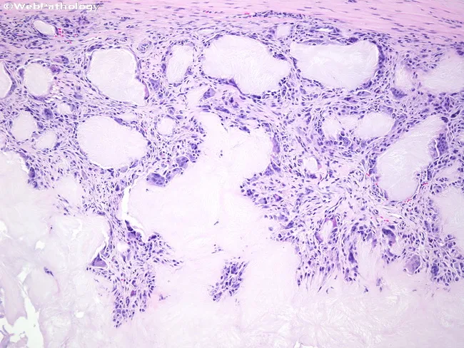
Light microscopy of a soft-tissue biopsy surrounding a gouty tophus shows extensive chalky, eosinophilic to basophilic amorphous deposits with adjacent granulomatous inflammation. Monosodium urate crystals are deposited within the periarticular soft tissue and synovium, appearing as refractile needle-shaped material that often dissolves during routine processing, leaving voids. The tophus is surrounded by a dense inflammatory capsule composed of macrophages, histiocytes, and numerous foreign body giant cells actively attempting to phagocytose crystalline material. Fibroblasts and scant lymphocytes form a fibrous stroma, with variable vascular proliferation. The gross histology may mimic a tumor or infection, but localization near joints and the characteristic granulomatous reaction supports chronic tophaceous gout. Polarized light examination would demonstrate negative birefringent crystals consistent with monosodium urate. Clinically, this pattern confirms chronic hyperuricemia with urate overproduction or reduced excretion and correlates with long-standing gout. The diagnostic significance lies in distinguishing inflammatory tophaceous gout from rheumatoid nodules, infectious arthritis, or soft-tissue neoplasms. Potential clinical use cases include confirming gout in patients with hyperuricemia, evaluating painful soft-tissue swellings around joints, and guiding urate-lowering therapy or surgical planning. This image emphasizes the pathognomonic granulomatous response to urate crystals and its impact on joint function. Correlation with serum uric acid improves diagnostic accuracy.
Case 4 - Analysis: Chronic Tophaceous Gout
4.1 Abnormalities in Picture A (Clinical photograph of the foot)
- Swelling and erythema of the first metatarsophalangeal (MTP) joint - the joint appears red, hot, and inflamed
- Tophus formation - firm, nodular, white-to-yellow subcutaneous deposits (chalky material representing monosodium urate crystal aggregates) visible over and around the joint and soft tissues
- Skin changes overlying the tophus: stretched, thinned skin that may appear white-yellow to skin-colored; in advanced cases, ulceration with discharge of chalky, toothpaste-like material
- Joint deformity at the first MTP joint due to repeated acute attacks and tophaceous deposits
- Soft tissue swelling involving periarticular structures
4.2 Microscopic Features in Picture B (Biopsy)
- Large aggregates of needle-shaped (acicular) monosodium urate crystals arranged radially
- In routine H&E-stained sections, the crystals are often dissolved out during aqueous processing, leaving behind empty needle-shaped clefts or voids (ghost spaces) within amorphous eosinophilic to pale basophilic material
- Under polarized light, if crystals are preserved (e.g., in alcohol-fixed tissue), they appear negatively birefringent - yellow when aligned parallel to the slow axis of the compensator, blue when perpendicular
- The crystal aggregates are encircled by a foreign body giant cell reaction - multinucleated giant cells attempting to phagocytose the crystals
- Macrophages and histiocytes surrounding the deposit
- Lymphocytes and plasma cells in the stroma
- Reactive fibroblast proliferation forming a fibrous capsule around the tophus
- The overall picture is that of a tophus: a central zone of amorphous/crystalline material surrounded by granulomatous inflammation and fibrous stroma
4.3 Most Probable Diagnosis
- 55-year-old male (classic demographic - middle-aged man)
- Severe pain and swelling of the left big toe (first MTP joint) = podagra - pathognomonic presentation; 50-75% of first attacks involve this joint
- Recurrent episodes over 6 months - indicates progression from acute gout to chronic/tophaceous stage with intercritical periods
- Clinical presence of tophi (chalky white nodules)
- Biopsy showing urate crystal aggregates with foreign body giant cell reaction - confirmatory
- Robbins & Kumar Basic Pathology, p. 797-798
4.4 Predisposing Factors for Gout
Patient-Related Factors
| Category | Specific Factors |
|---|---|
| Demographic | Male sex; older age; family history (genetic predisposition) |
| Metabolic/Dietary | Obesity; high-purine diet (red meat, organ meats, seafood); high-fructose foods; sugar-sweetened beverages |
| Lifestyle | Excessive alcohol intake (particularly beer and hard liquor - ethanol metabolism blocks renal uric acid excretion); sedentary lifestyle |
| Comorbid diseases | Hypertension; chronic kidney disease/renal insufficiency; diabetes mellitus; hyperlipidemia/hypercholesterolemia; heart disease; metabolic syndrome |
| Drugs/Medications | Diuretics (especially thiazides and loop diuretics - reduce renal urate excretion); cyclosporine; low-dose aspirin; pyrazinamide; ethambutol |
Enzyme/Metabolic Causes (Secondary Gout)
- Lesch-Nyhan syndrome (complete HGPRT deficiency) - overproduction of purines
- Partial HGPRT deficiency - overproduction
- Tumor lysis syndrome (rapid cell lysis during chemotherapy for leukemia/lymphoma) - massive release of purines
- Glucose-6-phosphatase deficiency (von Gierke disease)
Renal Causes
- Chronic renal disease - reduced excretion
- Lead nephropathy ("saturnine gout")
- Dermatology 2-Volume Set 5e - "Risk factors include obesity, excessive alcohol intake, renal insufficiency, and certain medications, most commonly diuretics."
- Firestein & Kelley's Textbook of Rheumatology - "Risk factors include older age; being male; family history; high consumption of alcohol and sugar-sweetened beverages; consumption of high-fructose foods, red meat, and seafood high in purines; chronic conditions (obesity, heart disease, diabetes, hypertension, hyperuricemia, elevated cholesterol, renal disease); and use of diuretics."
- Robbins & Kumar Basic Pathology, p. 796-798