
Prepare a neumonic for type of hallucinations from ks Narayan Reddy textbook of forensic medicine
Reading File
Finding Sources
The library has the KS Narayan Reddy textbook under ISBN 9789366165257 ("The Essentials of Forensic Medicine and Toxicology 36th edition"). Now let me search for hallucinations content in it.
Finding Sources
Found the section. Let me read the full hallucination passage from block 5.
Reading File
I now have all 9 types of hallucinations from KS Narayan Reddy. Let me craft the mnemonic.
The 9 types are:
- Visual
- Auditory
- Olfactory
- Gustatory
- Tactile (haptic)
- Psychomotor
- Command
- Microptic/Macroptic
- Sexual
Types of Hallucinations - KS Narayan Reddy (36th Ed.)
From the Textbook
Hallucination is a false sense perception without any external object or stimulus to produce it. Types listed:
| # | Type | Description (as per book) |
|---|---|---|
| 1 | Visual | Person imagines being attacked by a lion when none exists |
| 2 | Auditory | Person hears voices when no one is present |
| 3 | Olfactory | Person smells odor when none is present |
| 4 | Gustatory | Person tastes sweet/sour/bitter when no food is present |
| 5 | Tactile (Haptic) | Man imagines rats/mice crawling in bed; common in alcohol withdrawal and chronic cocaine poisoning |
| 6 | Psychomotor | Feeling of movement of a body part in its absence |
| 7 | Command | Ordered by hallucinatory voices to do things; can lead to suicide or homicide |
| 8 | Microptic/Macroptic | Objects appear smaller or bigger than they are |
| 9 | Sexual | Person feels sexually satisfied from self-imagined objects |
Mnemonic: "VAG TOP CMS"
Very Anxious Ghosts Trip On Pills, Causing Massive Shocks
| Letter | Type |
|---|---|
| V | Visual |
| A | Auditory |
| G | Gustatory |
| T | Tactile (Haptic) |
| O | Olfactory |
| P | Psychomotor |
| C | Command |
| M | Microptic/Macroptic |
| S | Sexual |
Alternative Mnemonic: "VAGO TAC-MS"
Very Anxious Ghosts Often Torment All Crazed Men Severely
Visual - Auditory - Gustatory - Olfactory - Tactile - A (drop) - Command - Microptic - Sexual
Easiest Recall Story
"VATO GPS-C-M" "A VATO uses GPS, Commands a Micro-Sexual fantasy"
Or the simplest clinical mnemonic:
"VAGOTP CMS"
"Virgins Are Generally Often Told: 'Please Control My Senses'"
| V | A | G | O | T | P | C | M | S |
|---|---|---|---|---|---|---|---|---|
| Visual | Auditory | Gustatory | Olfactory | Tactile | Psychomotor | Command | Microptic/Macroptic | Sexual |
Key clinical facts to pair with your mnemonic (from the book):
- Visual hallucinations = commonest in organic disorders (delirium tremens, CNS lesions, drug withdrawal)
- Auditory hallucinations = commonest in functional disorders (schizophrenia)
- Tactile = classic in alcohol withdrawal syndrome and chronic cocaine poisoning
- Command hallucinations = dangerous; can incite suicide or homicide
(Source: The Essentials of Forensic Medicine and Toxicology, KS Narayan Reddy, 36th Edition)
Head injury - definition, type, open and close head injury from ks Narayan Reddy textbook of forensic medicine
Searching sources...
Finding Sources
Finding Sources
The main content appears to be in block3. Let me search specifically for the head injury definition/types/open/closed sections.
Finding Sources
Reading File
I need to find the section with head injury definition, types, and open/closed classification. Let me search earlier in block3 for the introductory head injury chapter.
Finding Sources
Finding Sources
Finding Sources
Finding Sources
Let me look at the TOC/headings of block3 to find the chapter structure on head injuries, and also read the beginning of the head injury chapter.
Finding Sources
Found it - "HEAD INJURIES" starts at line 3498 in block3. Let me read that full section now.
Reading File
Excellent! I'm getting a lot of content. Let me continue reading to get the types of head injuries (open/closed classification) and further details.
Reading File
I need to find the open/closed head injury section. Let me read the section between line 3534 and 3654 which should have the classification.
Reading File
I see the chapter covers scalp, face, skull etc. The open/closed classification is likely within the skull fracture or overview section. Let me also fetch the flowchart image and read more from the skull fracture section area.
Reading File
Loading Image
Now I have almost everything I need. The book covers head injury under: definition, scalp, face, skull fractures (with types - fissured, depressed, comminuted, etc.), and brain injuries. The open/closed classification appears under the skull fracture discussion (simple vs compound). Let me also read the brain injury/classification section further to get the full open/closed head injury content.
Reading File
I now have comprehensive content. Let me read a bit further to get the brain injury classification (open/closed) and the rest of the chapter.
Reading File
I now have comprehensive content from the textbook. I have all the key information needed. Let me now compile a complete, well-organized answer from the textbook data I've collected.
Head Injuries - KS Narayan Reddy (Essentials of Forensic Medicine & Toxicology, 36th Ed.)
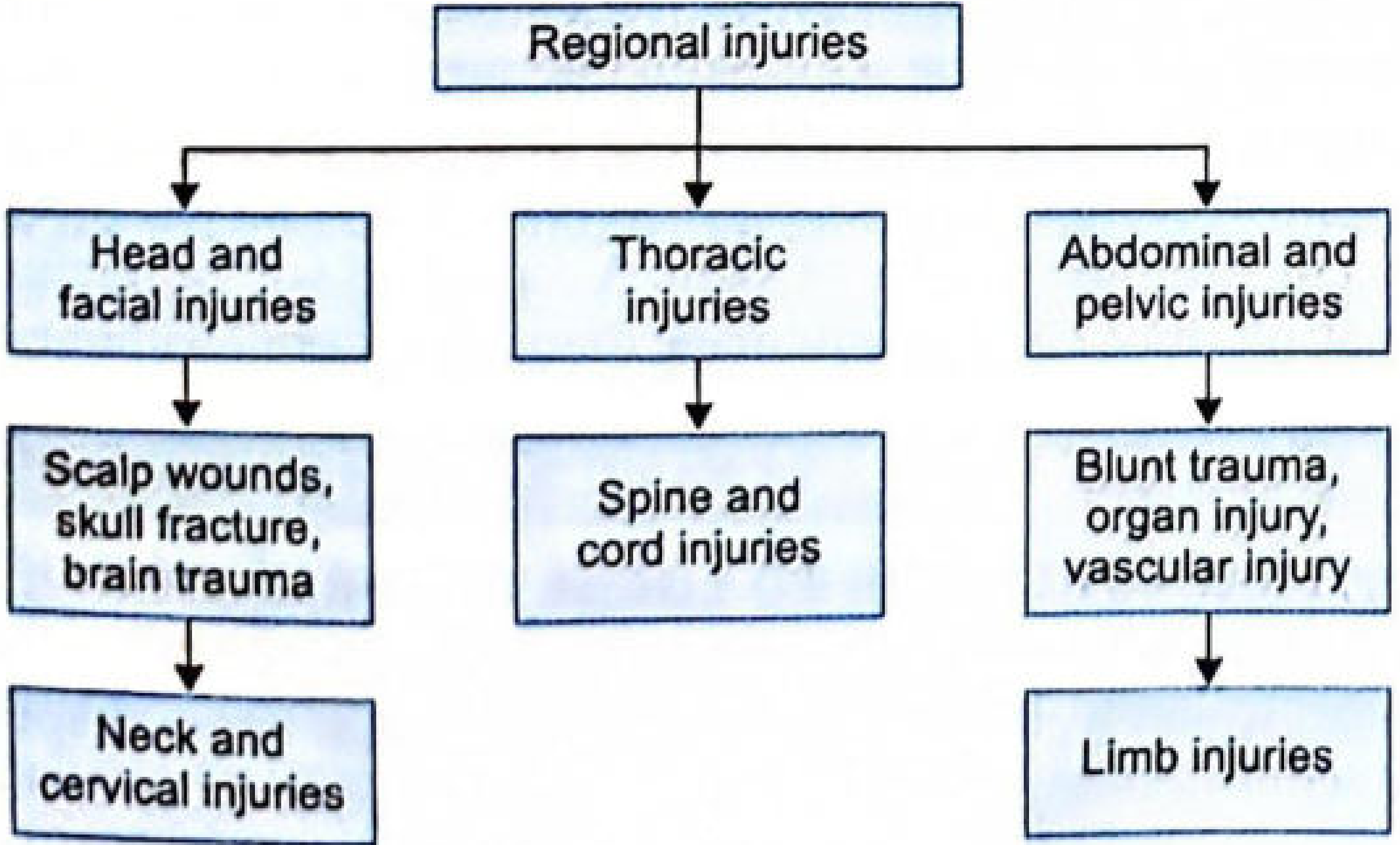
Definition
Head Injury is a morbid state, resulting from gross or subtle structural changes in the scalp, skull, and/or the contents of the skull, produced by mechanical forces.
Key points from the textbook:
- The application of blunt force to the head may result in injury to the skull contents, either alone or with fracture of the skull.
- The extent and degree of injury to the skull and its contents is not necessarily proportional to the amount of force applied.
- If a person is found unconscious with only a small abrasion externally, it indicates greater internal damage (lesser external violence = greater internal damage and vice versa).
Types of Head Injuries (Regional Classification)
Head and facial injuries include three main components:
1. Scalp Injuries
- Contusions - occur in the superficial fascia, temporalis muscles, or loose areolar tissue between the galea aponeurotica and pericranium.
- Subgaleal hematoma does not show any color change
- Temporal bruise may later appear behind the ear, suggesting primary neck injury
- Bruises of the scalp are better felt than seen
- Multiple contusions may join together and become confluent - making interpretation of exact number of blows difficult
- Lacerations - resemble incised wounds (called "split lacerated wounds") because thick hairy skin supported by bone splits instead of stretching. Hence, a guarded opinion is necessary about the causative weapon.
- Temporal arteries spurt freely as they are firmly bound and unable to contract - fatal blood loss can occur from extensive scalp laceration
- Avulsion of a large area of scalp can occur in traffic accidents or when hair becomes entangled in machinery
2. Skull Fractures
Mechanism of Skull Fracture
Fractures may be caused by direct or indirect violence.
Direct violence:
- Compression (e.g., midwifery forceps, crushing under a vehicle wheel)
- An object in motion striking the head (bullets, bricks, machinery, dagger)
- Head in motion striking an object (falls, traffic injuries)
Indirect violence: Fall on feet or buttocks with transmitted force.
Types of Skull Fractures
| # | Type | Description |
|---|---|---|
| 1 | Fissured (Linear) | Linear fracture involving whole thickness or inner/outer table only. ~70% of skull fractures are linear. Caused by blows with broad striking surface or falls |
| 2 | Depressed | A portion of the skull is driven inward below the normal level of skull surface. Caused by a small heavy object (stick, axe, hammer, chopper). Depth varies with velocity of impact |
| 3 | Elevated | One end of the fractured fragment is elevated over the surface of the skull, the other end is depressed into the cranial cavity. Caused by a heavy sharp weapon |
| 4 | Comminuted | Two or more intersecting fracture lines dividing the bone into 3 or more fragments. Caused by crushing injuries, vehicle accidents, falls from height, or repeated blows with a large surface weapon (iron bar, axe). Resembles a spider's web or mosaic |
| 5 | Pond (Indentation) | Saucer-like depression without a line of fracture; seen mainly in infants due to the pliable nature of the skull |
| 6 | Contre-coup fracture | Fracture at a site opposite to the point of impact |
| 7 | Ring fracture | Fracture around the foramen magnum; caused by forces transmitted into the base through the mandibular joints, or by a sudden violent turn of the head on the spine |
| 8 | Perforating fracture | Caused by firearms and pointed sharp weapons (daggers, knives, axe). The weapon passes through both tables of the skull, leaving a clean-cut opening corresponding to the cross-section of the weapon |
| 9 | Blowout fracture | Blunt trauma to the eye transmitting forces via the globe to the bony orbit causing fractures of medial wall and floor of orbit |
| 10 | Diastatic (Sutural) fracture | Separation of sutures; occurs only in young persons; usually seen in the sagittal suture; common in traffic accidents |
| 11 | Displaced fracture | Massive fragmentation/shattering of the skull with some pieces found outside the head; due to massive trauma (contact/close range firearm injury or bomb blasts) |
Fracture Base of Skull
Produced by:
- Force applied directly at the base
- General deformation of the skull
- Extension from the vault
- Force applied through the spinal column or face
3. Brain Injuries
Open vs. Closed Head Injury
The textbook classifies head injuries based on whether the dura is breached:
| Feature | Open (Compound) Head Injury | Closed Head Injury |
|---|---|---|
| Definition | The fracture is associated with injury to the overlying scalp or nasal sinuses, allowing communication with the external environment | No breach of the skull or overlying scalp; dura remains intact |
| Dura | May be torn/breached | Intact |
| Risk of infection | High (meningitis, brain abscess) | Low |
| Cause | Penetrating wounds - firearms, knives, sharp weapons; compound skull fractures | Blunt force injuries, falls, traffic accidents |
| Examples | Gunshot wounds to head, stab wounds, depressed fractures with scalp lacerations | Concussion, contusion, diffuse axonal injury, extradural/subdural hematoma (without scalp breach) |
| Forensic significance | Entry/exit wound analysis; weapon identification; trajectory determination | Analysis of coup/contrecoup pattern; intracranial hemorrhage |
Note from text: "The fracture may be simple or compound, i.e., associated with the injury to the scalp or nasal sinuses." - This is the textbook's definition of open (compound) vs. closed (simple) skull fracture.
Brain Injuries (Classification)
Primary Brain Injuries (at moment of impact)
-
Concussion - Temporary unconsciousness due to partial or complete paralysis of cerebral function; comes on immediately after injury; always followed by amnesia; tends to spontaneous recovery. Believed to be a transient electrophysiologic dysfunction of the reticular activation system in the upper midbrain caused by rotation of the cerebral hemispheres on the relatively fixed brainstem.
-
Contusion (Bruising of Brain) - More severe than concussion; structural damage to brain tissue.
- Coup injury - at the site of impact
- Contrecoup injury - opposite to site of impact; produced by formation of a vacuum/cavity in the cranial cavity on the opposite side of impact as the brain lags behind the moving skull
-
Laceration - Actual tearing of brain tissue
Secondary Brain Injuries (develop after impact)
- Cerebral edema/swelling
- Intracranial hemorrhages (extradural, subdural, subarachnoid, intracerebral)
Medico-Legal Importance (Key Points)
- A blow to the head produces coup contusions; contrecoup contusions are small or absent
- A fall on the head produces contrecoup contusions; coup contusions are small or absent
- Contrecoup injury is rare before age 3 years
- Skull fractures can occur without significant brain injury or impairment of consciousness
- Conversely, severe or fatal brain injury may occur without fracture of skull
- Admission to hospital after head injury: person should be under observation for at least 24-36 hours
- Head injury and acute alcoholic intoxication may coexist and may be confused
(Source: The Essentials of Forensic Medicine and Toxicology, KS Narayan Reddy, 36th Edition, Chapter 9 - Head Injuries)
Mechanism of brain injury from ks Narayan Reddy textbook of forensic medicine
Searching sources...
Finding Sources
Finding Sources
"MECHANISM OF CEREBRAL INJURY" starts at line 4097. Let me read it fully along with the concussion, contusion, and intracranial hemorrhage sections.
Reading File
Reading File
Reading File
Reading File
Now let me read the intracranial hemorrhage section and also fetch key diagrams.
Reading File
Reading File
I now have all the essential content. Let me also read the section just before intracranial hemorrhage to pick up the section on concussion features, and the earlier section on mechanism (lines 4144-4246) which was truncated.
Reading File
I now have all the content needed. Let me fetch the key mechanism diagram image.
Loading Image
I now have everything needed for a complete, detailed response. Here is the full compiled answer:
Mechanism of Brain Injury
KS Narayan Reddy - Essentials of Forensic Medicine & Toxicology (36th Ed.)
Fig. 9.11 - Mechanism of Cerebral Injury
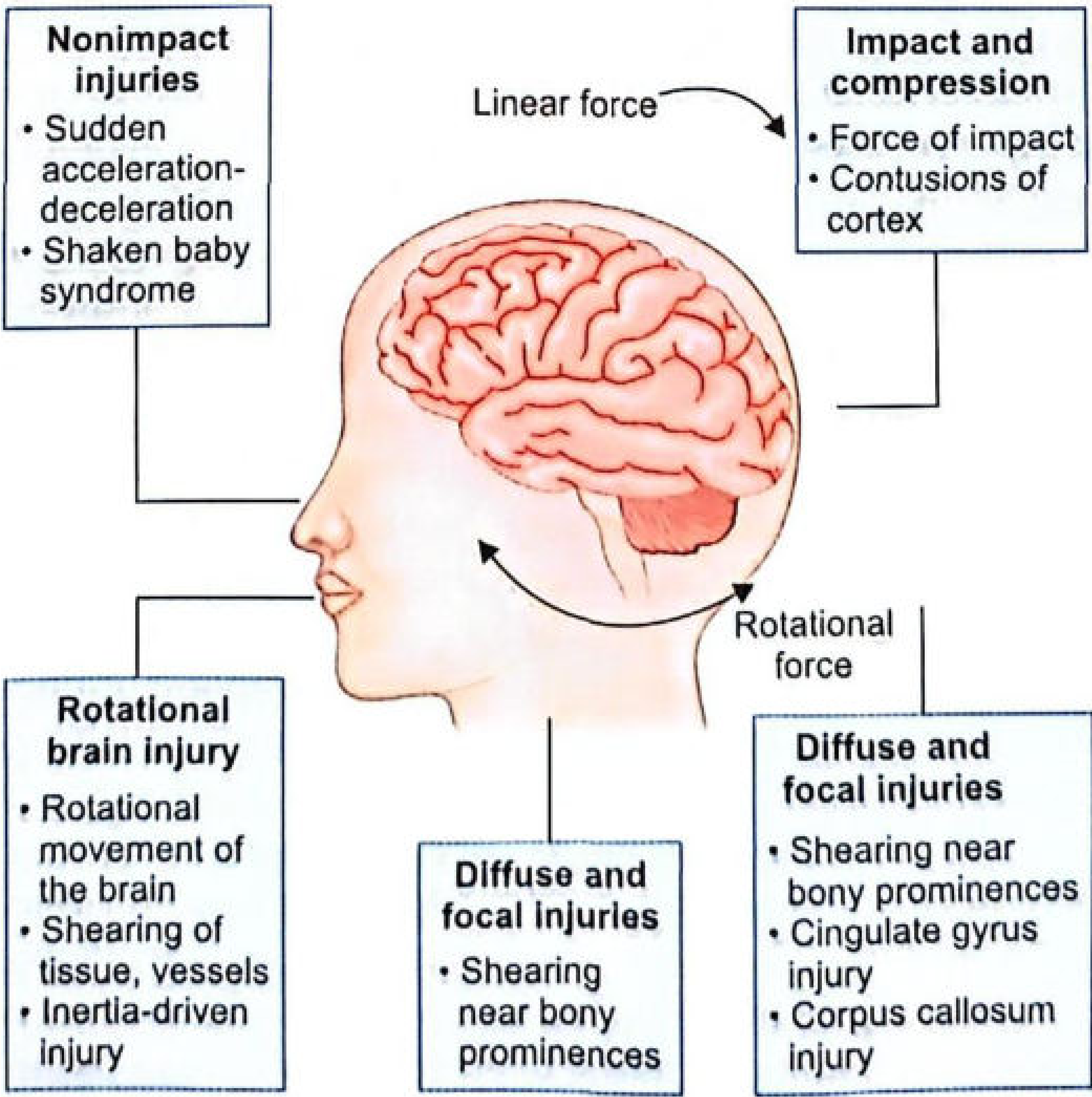
MECHANISM OF CEREBRAL INJURY (Point-by-Point, as in Textbook)
(1) Brain injury without a direct blow
Severe brain damage may be caused without an actual blow or fall on the head - e.g., by shaking an infant (child abuse) may cause subdural hemorrhage. A blow to the head causes either:
- Linear change in velocity - linear acceleration/deceleration forces that produce compressional or rarefactional (sound wave) forces which travel back and forth through the brain but do not cause damage on their own.
- Rotational change in velocity - the dangerous component.
(2) The brain is easily distorted but incompressible
Change in velocity (either acceleration or deceleration) with a rotational element causes brain damage. The initial sudden change in velocity sets the head in rotation, which is transmitted to the brain, causing the brain to glide within the dura.
(3) Momentary skull depression at impact
Even without fracture, the area of the skull beneath an impact becomes momentarily depressed and may strike the underlying brain, causing compression. This produces typical cone-shaped contusions on the cortex (base at the surface). Other areas of the skull bulge outward simultaneously to accommodate the deformation.
(4) Gliding/shear strains from angular rotation
The sudden arrest of the moving skull causes skull deceleration first, but the brain's momentum causes it to continue in motion. The skull and brain cannot change their velocities simultaneously. The brain slows down or speeds up only due to restraint by the falx and tentorium, causing damage to:
- Base of the cerebrum
- Corpus callosum
- Brainstem
(5) Diffuse cortical contusion from wide-area impact
Impact against a wide area of the skull may cause diffuse contusion of the cortex. Shearing is most severe where gliding is prevented by bony prominences, especially those of the anterior and middle fossae. The cerebellum (being smaller and lighter) is less liable to damage from rotatory movements.
(6) Side-of-head blow - cingulate gyrus and corpus callosum damage
A blow to the side of the head may damage the medial surface of the cerebral hemisphere on that same side, especially in the region of the cingulate gyrus, because the soft brain is pushed violently against the free edge of the falx. Shearing strains caused by unequal rotational gliding and twisting of the hemisphere may produce:
- Multiple hemorrhages
- Direct damage to fibers of the corpus callosum
(7) Holbourn's postulate (Shear Strain Theory)
Holbourn postulates that brain tissue is injured when its constituent particles are pulled so far apart that they do not join up again properly when the blow is over. In the brain, the amount of this pulling apart is proportional to the shear strains.
Types of Brain Injuries (Based on Mechanism)
A. CONTUSIONS OF THE BRAIN
Definition: Circumscribed areas of brain tissue destruction accompanied by extravasation of blood into affected tissues, without actual tearing of tissues (pia-arachnoid remains intact over surface contusions).
Mechanism:
- When a localized skull segment undergoes deformation at impact, shear strains develop in underlying brain tissue, producing a zone of contusion in the surface layers.
- When the head rotates, the layers of brain tissue slide over each other at different depths in the cortex, damaging blood vessels.
Locations most commonly affected:
- Frontal and temporal lobes (most common)
- Lateral and ventral surfaces of cerebral hemispheres
- Basal ganglia, midbrain, and brainstem (especially from impacts to forehead and vertex)
- Most hemorrhages occur at the crest of convolutions facing the dura of falx and tentorium
Types of Contusions (Lindenberg and Freytag Classification):
| Type | Description |
|---|---|
| Coup contusion | At the site of impact |
| Contrecoup contusion | On the opposite side of impact; formed by vacuum/suction effect as brain lags behind moving skull |
| Intermediary coup contusion | Deeper structures (white matter, basal ganglia, corpus callosum, brainstem) along the line between coup and contrecoup points |
| Fracture contusion | Contusions caused by fracture of the skull |
| Gliding contusion | Cortex and white matter of frontal and central convolutions near the upper margin of hemispheres; no relationship to area/direction of impact; caused by stretching and shearing forces during to-and-fro gliding of the brain; seen in falls and MVAs |
| Herniation contusion | In cerebellar tonsils and medulla oblongata; caused by momentary shifting of the brain toward the foramen magnum |
Coup vs. Contrecoup - Key ML Distinction:
| Blow to Head | Fall on Head | |
|---|---|---|
| Coup | Present (prominent) | Small or absent |
| Contrecoup | Small or absent | Present (prominent) |
Contrecoup injuries are rare before age 3 years. Contrecoup injury is seen in: skull, brain, liver, heart, and lungs.
Age of Contusions (Forensic Timing):
| Time | Finding |
|---|---|
| 1 hour | Ischemic changes in neurons; BAPP identifiable by immunohistology |
| 10-12 hours | Gross lesion visible as swollen gelatinous parenchyma (if purely ischemic) |
| 5 days | Capillary proliferation begins |
| 10-12 days | Capillary proliferation maximum |
| First 2 weeks | Fat-containing macrophages present in small numbers |
| Few weeks | Astrocytic proliferation |
| ~2 months | Scar formation (pale or golden yellow, depressed) |
B. CONCUSSION OF THE BRAIN
Definition: A state of temporary unconsciousness due to partial or complete paralysis of cerebral function, due to head injury, coming on immediately after injury, always followed by amnesia, and tending to spontaneous recovery.
Mechanism: A transient electrophysiologic dysfunction of the reticular activation system in the upper midbrain, caused by rotation of the cerebral hemispheres on the relatively fixed brainstem.
Features:
- True concussion may last for seconds or minutes
- Loss of consciousness is immediate
- Always followed by amnesia
Diffuse Axonal Injury (DAI) - the microscopic correlate of concussion:
- Axonal injuries NOT visible on microscopy for the first 12 hours
- After 12 hours: axons appear dilated → club-shaped → round balls called "retraction balls" (transected axons)
- After 2-3 weeks: retraction balls decrease; clusters of microglial cells appear, followed by astrocytosis and demyelination
C. LACERATIONS OF THE BRAIN
Definition: Traumatic lesions with actual loss of continuity (tearing) of the substance of brain. Pia-arachnoid is torn (unlike contusions where it remains intact).
They form small clefts, irregularly shaped holes, or trenches with sharply outlined walls (usually brown in color).
Features:
- Communicate with subarachnoid space; do not contain blood vessels
- On section they are triangular or wedge-shaped (wedge pointing into white matter)
- Area of necrosis is usually delineated by hemorrhage
- "Burst lobe" = combination of extensive contusion + associated subdural hematoma; usually seen in frontal or temporal lobe
D. INTRACRANIAL HEMORRHAGE
| Type | Vessel/Source | Mechanism | Classic Feature |
|---|---|---|---|
| Extradural (Epidural) | Middle meningeal artery (posterior branch); squamous temporal bone fracture | Skull moves relative to dura, stripping it from bone; creates empty extradural space; vessel bleeds into this space | Lucid interval (awake → unconscious → unconscious); biconvex/lens-shaped on CT |
| Subdural | Bridging cortical veins at their junction with dural sinuses | Shear stresses in upper cerebrum from rotational acceleration/deceleration; veins rupture at the cortical vein-dural sinus junction | Common in alcoholics, elderly (brain atrophy), infants; 70% due to falls/assaults |
| Subarachnoid | Cortical vessels in subarachnoid space | Trauma or spontaneous rupture of aneurysm | Blood in CSF; severe headache |
| Intracerebral | Deep brain vessels | Severe blunt force; hypertension | Often in basal ganglia, internal capsule |
Key ML point: "The application of a moderate force to the head may cause a severe intracranial hemorrhage, while a greater force may not produce hemorrhage."
E. CEREBRAL SWELLING (Edema)
May occur following significant head injury; may be:
- Focal (adjacent to area of brain injury)
- Diffuse (involving both cerebral hemispheres)
- Unilateral (involving one hemisphere)
Massive cerebral swelling can occur within 20 minutes following injury.
Summary: Forces Involved in Brain Injury
| Force Type | Effect |
|---|---|
| Linear acceleration/deceleration | Compressional/rarefactional waves - travel through brain but do not cause damage alone |
| Rotational acceleration/deceleration | THE MAIN CAUSE of brain damage - causes gliding of brain within dura, shear strains, DAI |
| Impact/compression | Direct contusions of cortex (coup); cone-shaped contusions |
| Shearing (Holbourn's theory) | Particles pulled apart beyond recovery; proportional to shear strains |
(Source: The Essentials of Forensic Medicine and Toxicology, KS Narayan Reddy, 36th Edition, Chapter 9)
Coup and contrecoup injuries from ks Narayan Reddy textbook of forensic medicine
Searching sources...
Finding Sources
Finding Sources
Your credits are exhausted. You can purchase a top-up or subscribe to a plan to continue using Orris. If you're already subscribed, your daily credits will reset tomorrow.
This is a shared conversation. Sign in to Orris to start your own chat.