I now have rich content. Let me compile the full, well-structured answer.
ANATOMY OF THE PANCREAS
(15 Marks Answer - Easy Scoring Format with Mnemonics)
1. LOCATION
Mnemonic: "RUPA lies behind the stomach"
Retroperitoneal, Upper abdomen, Posterior wall, Abdominal (L1-L2 level)
- The pancreas is a retroperitoneal organ lying on the posterior abdominal wall
- Obliquely oriented, from right (L2) to left (L1)
- Lies posterior to the stomach and anterior to the great vessels
- Length: 12-20 cm | Weight: 85-95 g in adults
- Soft, flattened, elongated, lobular gland with no true fibrous capsule
(Yamada's Textbook of Gastroenterology, p. 116)
2. EXTERNAL FEATURES (Parts)
Mnemonic: "HUNT" - Head, Uncinate, Neck, Tail (& Body)
| Part | Level | Key Feature |
|---|
| Head | L2 | Within C-shaped curve of duodenum |
| Uncinate Process | L2 | Posterior extension of head, hooks around SMA |
| Neck | L1-L2 | Lies anterior to SMA and SMV |
| Body | L1 | Passes left behind stomach |
| Tail | L1 | Reaches hilum of spleen |
Key facts about each part:
- Head: Largest part; CBD runs in a groove/tunnel on its posterior aspect
- Uncinate process: Latin "uncus" = hook; wraps around the superior mesenteric vessels
- Neck: Only 2 cm wide; the portal vein is formed behind it (SMV + splenic vein)
- Tail: Only intraperitoneal part; lies in lienorenal ligament
(Yamada's Textbook of Gastroenterology, p. 117)
3. INTERNAL FEATURES (Ducts)
Mnemonic: "WADS" = Wirsung (main), Accessory (Santorini), Duodenum, Sphincters
Main Pancreatic Duct (Duct of Wirsung)
- Runs the entire length of the gland from tail to head
- Joins the Common Bile Duct (CBD) to form the Hepatopancreatic ampulla (Ampulla of Vater)
- Opens into the 2nd part of duodenum at the major duodenal papilla
- Guarded by the Sphincter of Oddi
- Diameter: 2-3 mm (body), up to 3-4 mm (head)
Accessory Pancreatic Duct (Duct of Santorini)
- Drains the upper part of the head + uncinate process
- Opens at the minor duodenal papilla (2 cm above major papilla)
- Usually communicates with the main duct
Histological/Functional Units
- Exocrine (90-95%): Acinar cells (enzymes) + Ductal cells (bicarbonate)
- Endocrine (1-2%): Islets of Langerhans
Islet cells mnemonic - "ABGD":
| Cell | Hormone | Action |
|---|
| α (alpha) | Glucagon | Raises blood glucose |
| β (beta) | Insulin | Lowers blood glucose |
| δ (delta) | Somatostatin | Inhibits both α & β |
| PP cells | Pancreatic polypeptide | Inhibits exocrine secretion |
(Mulholland & Greenfield's Surgery, p. 2586)
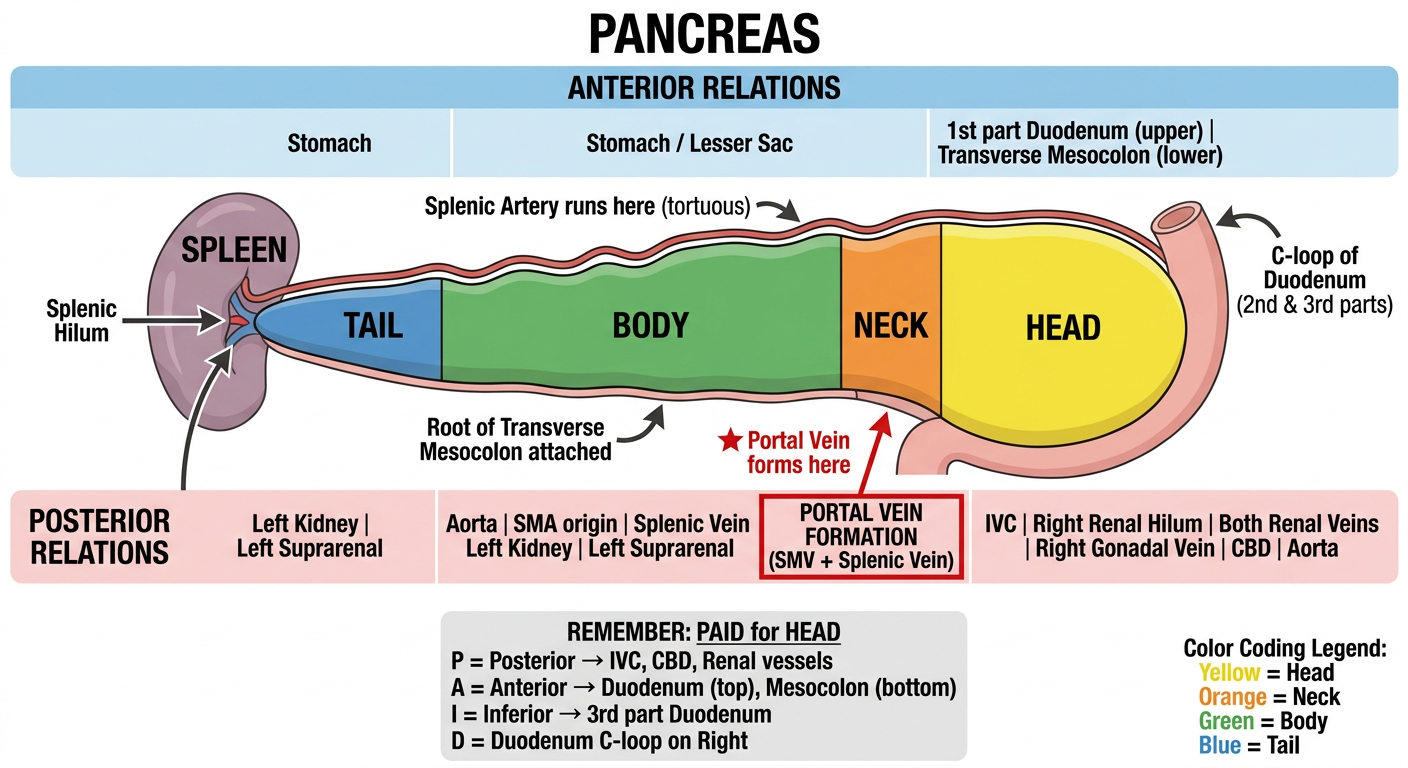
4. RELATIONS
Mnemonic: "PAID" = Posterior, Anterior, Inferior, Duodenal (for Head)
HEAD Relations:
| Surface | Structures |
|---|
| Anterior | 1st part duodenum (upper), transverse mesocolon (lower) |
| Posterior | IVC, right renal hilum, both renal veins, right gonadal vein, aorta, CBD |
| Right | 2nd & 3rd part duodenum (C-loop) |
| Superior | Epiploic foramen, portal vein, hepatic artery, CBD (in lesser omentum) |
NECK Relations:
- Anterior: Lesser sac, stomach
- Posterior: Formation of Portal Vein (SMV + splenic vein join here)
- The most surgically important landmark of the pancreas
BODY Relations:
| Surface | Structures |
|---|
| Anterior | Lesser sac, stomach |
| Posterior | Aorta, origin of SMA, splenic vein, left suprarenal, left kidney |
| Superior border | Splenic artery runs along it |
| Inferior border | Root of transverse mesocolon attached |
TAIL Relations:
- Lies in the lienorenal ligament with splenic vessels
- In contact with spleen (risk of splenic injury in pancreatectomy)
(Yamada's Textbook of Gastroenterology, pp. 119-120)
5. VASCULAR SUPPLY
Mnemonic: "Celiac Supplies the Head & Body, Splenic for Body & Tail"
"GDA makes PDA arcade for head; Splenic supplies body and tail"
ARTERIAL SUPPLY:
For HEAD:
- Anterior Superior Pancreaticoduodenal Artery (from gastroduodenal artery - GDA)
- Anterior Inferior Pancreaticoduodenal Artery (from SMA)
- These form the Anterior Pancreaticoduodenal Arcade
- Posterior Superior Pancreaticoduodenal Artery (from GDA)
- Posterior Inferior Pancreaticoduodenal Artery (from SMA)
- These form the Posterior Pancreaticoduodenal Arcade
For BODY & TAIL:
- Splenic artery (largest branch of celiac axis, 5-11 mm) - runs tortuous course along superior border
- Gives multiple side branches to neck, body, and tail
VENOUS DRAINAGE:
Mnemonic: "PASS into Portal" = Pancreaticoduodenal, All into SMV, Splenic, then Superior to Portal
- Head: Pancreaticoduodenal veins drain into SMV
- Body/Tail: Splenic tributaries into Splenic vein
- Final drainage: Portal vein (SMV + Splenic vein unite behind the neck of pancreas)
(Yamada's Textbook of Gastroenterology, pp. 198-200)
6. NERVOUS SUPPLY
Mnemonic: "CAVES" = Celiac, Autonomic, Vagus, Exocrine-Endocrine, Splanchnic
| Division | Source | Effect |
|---|
| Sympathetic | Greater & Lesser splanchnic nerves (T5-T10) via Celiac plexus | Inhibits secretion; vasoconstriction; mediates pain |
| Parasympathetic | Vagus nerve (CN X) via Celiac plexus | Stimulates exocrine & endocrine secretion |
Key points:
- The pancreatic plexus originates from the celiac plexus and runs posteriorly to innervate the gland
- Pain from the pancreas is referred to the epigastrium and back (T5-T10 dermatomes)
- Parasympathetics stimulate insulin, glucagon, and enzyme secretion
(Yamada's Textbook of Gastroenterology, p. 78)
7. LYMPHATIC DRAINAGE
Mnemonic: "SPLASH" = Splenic, Para-aortic, Lumbar, Aortocaval, Superior mesenteric, Hepatic
The pancreas has rich lymphatics that drain along its arterial supply:
| Region | Drains to |
|---|
| Head | Pancreaticoduodenal nodes → Hepatic nodes + Superior Mesenteric nodes |
| Body | Along splenic artery → Splenic hilar nodes |
| Tail | Along splenic artery → Splenic nodes + Celiac nodes |
| All regions | Ultimately drain to para-aortic (aortocaval) nodes → Cisterna chyli → Thoracic duct |
Surgical importance: Multiple lymphatic routes explain why pancreatic cancer spreads early and widely, making complete resection difficult.
8. APPLIED ANATOMY
Mnemonic: "PANC DOCS" for 8 important clinical points:
| Letter | Condition |
|---|
| P | Pancreatitis - Acute inflammation causes epigastric pain radiating to back (T5-T10) |
| A | Annular Pancreas - Congenital ring of pancreatic tissue encircling duodenum (causes duodenal obstruction in neonates) |
| N | Neck anatomy - Portal vein forms behind neck; critical in Whipple's operation |
| C | Carcinoma - Head cancer obstructs CBD → painless obstructive jaundice (Courvoisier's sign) |
| D | Divisum - Failed fusion of ventral & dorsal buds → most common congenital anomaly (4-10%) |
| O | Obesity/Spleen - Tail close to spleen; splenic injury during pancreatectomy/splenectomy |
| C | CBD - Runs in a groove/tunnel in posterior head; vulnerable in head pathology |
| S | SMA/SMV - Run behind neck; involvement = unresectable cancer |
Additional Applied Points:
- Whipple's Procedure (Pancreaticoduodenectomy): Uses the surgical plane anterior to SMV/SMA through the neck
- Retroperitoneal position: Pancreatic injuries (blunt trauma) are easily missed; serum amylase/lipase elevated
- Frey's/Puestow's procedure for chronic pancreatitis exploits the main pancreatic duct anatomy
- Pseudocyst of pancreas most commonly bulges into the lesser sac (anterior relation of body)
- Grey Turner's sign (flank bruising) and Cullen's sign (periumbilical bruising) in acute hemorrhagic pancreatitis - due to retroperitoneal hemorrhage tracking along fascial planes
QUICK REVISION SUMMARY TABLE
| Heading | Key Mnemonic | Must-Remember Fact |
|---|
| Location | "RUPA" | Retroperitoneal, L1-L2, 12-20 cm |
| External | "HUNT" | Head-Uncinate-Neck-Tail (& Body) |
| Internal | "WADS" | Wirsung + Santorini ducts; Islets "ABGD" |
| Relations | "PAID" | Portal vein forms behind neck |
| Arterial | GDA + SMA arcade | Splenic for body/tail |
| Venous | "PASS" | All into SMV/Splenic → Portal |
| Nerves | "CAVES" | Celiac plexus; T5-T10 pain |
| Lymphatics | "SPLASH" | Para-aortic final destination |
| Applied | "PANC DOCS" | Painless jaundice, divisum, Whipple |
Sources: Yamada's Textbook of Gastroenterology (7e), Mulholland & Greenfield's Surgery (7e)