
External jugular vein
Reading File
The external jugular vein (EJV) is a superficial vein of the neck that drains blood from the scalp and face. It runs obliquely across the sternocleidomastoid muscle and is commonly used for venous access and for estimating jugular venous pressure (JVP).
Searching sources...
Finding Sources
Finding Sources
Reading File
Reading File
Reading File
Loading Image
Reading File
Reading File
Here is a comprehensive overview of the external jugular vein based on Gray's Anatomy for Students, the Color Atlas of Human Anatomy, and Roberts & Hedges' Clinical Procedures in Emergency Medicine.
External Jugular Vein
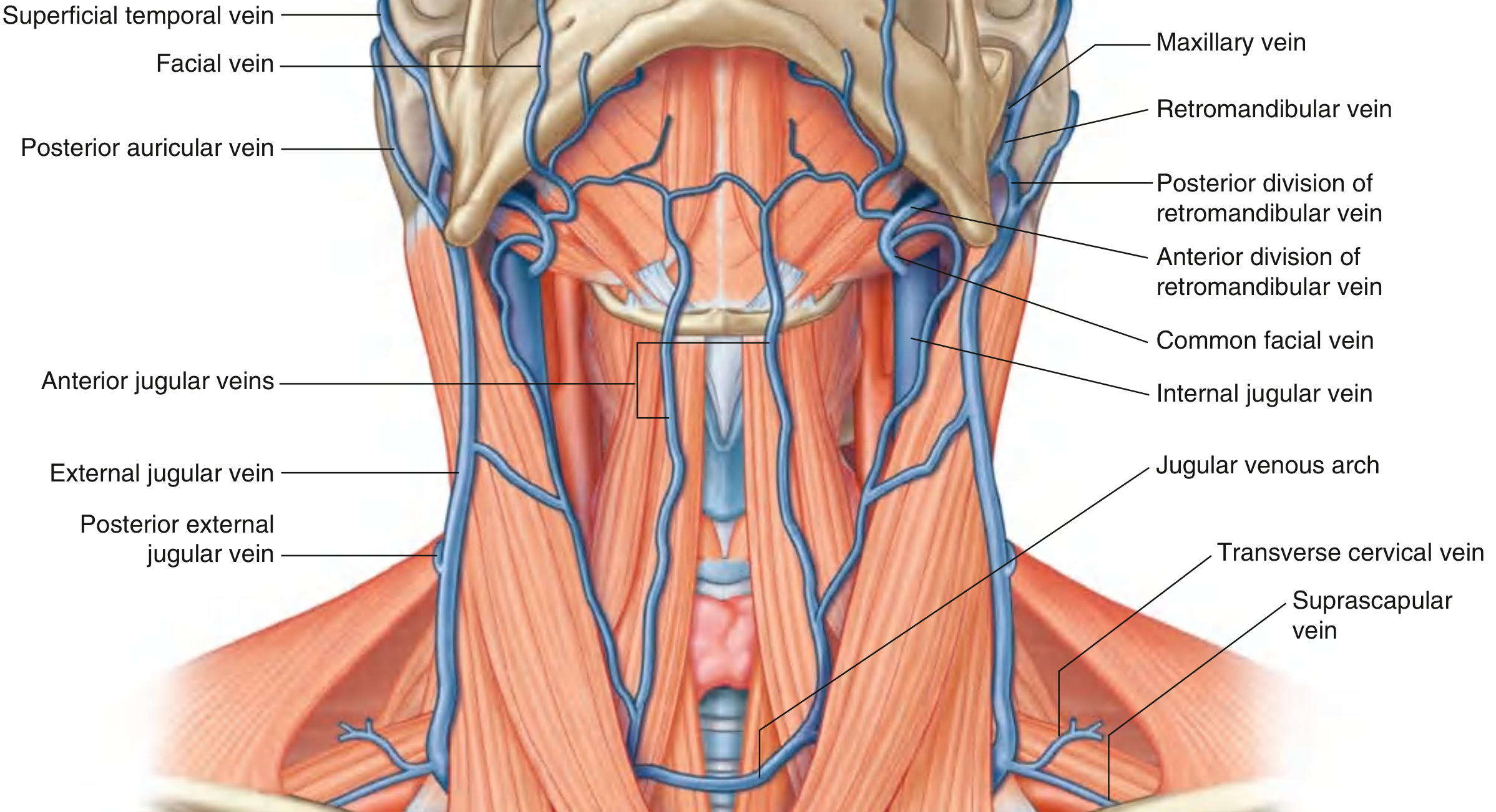
Formation
The external jugular vein (EJV) is formed posterior to the angle of the mandible by the union of two veins:
- Posterior auricular vein - drains the scalp behind and above the ear
- Posterior division of the retromandibular vein - the retromandibular vein itself is formed in the substance of the parotid gland when the superficial temporal and maxillary veins join; its posterior division combines with the posterior auricular vein to form the EJV
(The anterior division of the retromandibular vein, by contrast, joins the facial vein to form the common facial vein, which drains into the internal jugular vein.)
- Gray's Anatomy for Students, p. 1143
Course
-
Once formed, the EJV descends straight down the neck in the superficial fascia, lying entirely superficial to the sternocleidomastoid (SCM) muscle, crossing it diagonally as it descends.
-
It enters the posterior triangle of the neck after crossing the SCM.
-
In the lower neck, just superior to the clavicle and immediately posterior to the SCM, it pierces the investing layer of cervical fascia.
-
It passes deep to the clavicle and terminates by entering the subclavian vein (occasionally into the internal jugular vein near the venous angle).
-
Gray's Anatomy for Students, p. 1143; Color Atlas of Human Anatomy, p. 132
Tributaries
Along its descent, the EJV receives:
| Tributary | Drainage area |
|---|---|
| Posterior external jugular vein | Superficial areas of the back of the neck |
| Transverse cervical vein | Posterior scapular region |
| Suprascapular vein | Posterior scapular region |
| Anterior jugular vein (sometimes) | Anterior neck; occasionally drains into EJV just before it enters the subclavian |
- Gray's Anatomy for Students, p. 1144
Relations
- Lies in the superficial fascia, superficial to the SCM throughout
- Covered by platysma
- Pierces the investing (deep cervical) fascia just above the clavicle, which can cause air embolism risk during neck procedures or catheterization
- The anterior jugular veins may communicate with the EJV via the jugular venous arch in the suprasternal notch region
Valves
The EJV contains valves near its termination at the subclavian vein. These prevent backflow from the subclavian into the superficial system - Scott-Brown's Otorhinolaryngology Head and Neck Surgery.
Clinical Relevance
1. Jugular Venous Pressure (JVP)
The EJV is visible superficially on the neck and is used to estimate venous pressure. Distension at rest or elevation of the venous column suggests raised central venous pressure (e.g., right heart failure, cardiac tamponade, superior vena cava obstruction).
2. Peripheral IV Cannulation
In patients with limited peripheral venous access, the EJV can be cannulated as a peripheral vein:
- Place patient in Trendelenburg position to fill the vein
- Rotate head to the opposite side
- Align cannula toward the ipsilateral shoulder
- Puncture midway between the angle of the jaw and the midclavicular line
- Lightly compress above the clavicle with a free finger to act as a tourniquet
- Roberts and Hedges' Clinical Procedures in Emergency Medicine
3. Central Venous Access via EJV
The EJV can also be used for central venous catheterization, but this is technically difficult:
- Requires passage of a guidewire into the thorax
- Advancement of the wire is the most challenging step due to the angle at which the EJV meets the subclavian
- A small-radius J-tipped wire, distended lumen, exaggerated head tilt, and skin traction improve success
- Time-consuming; less preferred in emergencies compared to internal jugular or subclavian routes
4. As a Surgical Landmark
In neck surgery, the EJV serves as a landmark; for example, the subclavian vein puncture site is near where the EJV crosses the posterolateral border of the SCM. Surgeons take care not to injure it during dissection of the anterior triangle and subplatysmal plane.
Summary Table
| Feature | Detail |
|---|---|
| Formed by | Posterior auricular v. + posterior division of retromandibular v. |
| Location | Superficial fascia, crosses SCM obliquely |
| Terminates in | Subclavian vein (or occasionally internal jugular) |
| Covered by | Platysma |
| Pierces | Investing cervical fascia above the clavicle |
| Key tributaries | Posterior external jugular, transverse cervical, suprascapular, anterior jugular |
| Clinical uses | JVP assessment, peripheral IV access, central venous access (difficult) |
This is a shared conversation. Sign in to Orris to start your own chat.