
What is headache?
headache types classification causes overview
headache types classification diagram
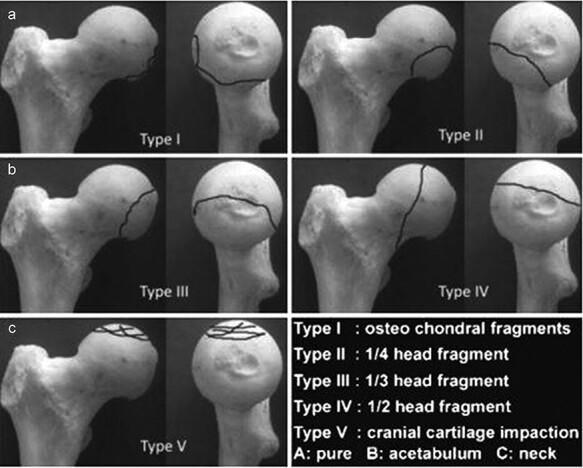
This orthopaedic anatomical diagram illustrates the Chiron classification (2004) for femoral head fractures, shown across five categorized types (Type I to Type V). Each type is presented using a dry bone model of the proximal femur from two perspectives: a lateral/oblique view and a frontal view of the femoral head. Fracture lines are demarcated by solid black lines to indicate the morphology and size of the fracture fragments. Type I displays small osteochondral fragments at the superior-lateral aspect. Type II shows a 1/4 head fragment on the anterior-superior surface. Type III demonstrates a 1/3 head fragment extending from superior to posterior. Type IV depicts a vertical fracture dividing approximately 1/2 of the femoral head. Type V illustrates cranial cartilage impaction with multiple intersecting lines across the superior articular surface. The classification is clinically relevant for orthopedic surgeons and residents for determining the severity of femoral head trauma and informing surgical decisions, such as fragment fixation versus excision based on size and location.
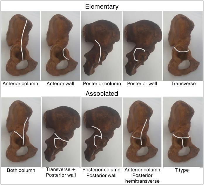
Educational diagram illustrating the Judet-Letournel classification of acetabular fractures using anatomical specimens of the hemipelvis. The visual is divided into two rows: 'Elementary' and 'Associated' fracture types. White lines superimposed on the specimens delineate specific fracture patterns. The 'Elementary' row features five simple patterns: Anterior column (vertical line through the pelvic brim), Anterior wall (localized articular segment), Posterior column (ischiadic notch through obturator foramen), Posterior wall (localized posterior rim), and Transverse (horizontal line bisecting the acetabulum). The 'Associated' row demonstrates complex configurations: Both column, Transverse plus posterior wall, Posterior column plus posterior wall, Anterior column with posterior hemitransverse (a vertical anterior column line joined by a horizontal posterior component), and T-type (a transverse fracture with an additional vertical stem extending through the obturator foramen). This orthopedic diagram serves as a clinical reference for orthopedic surgeons and medical students to understand pelvic trauma morphology and surgical planning.
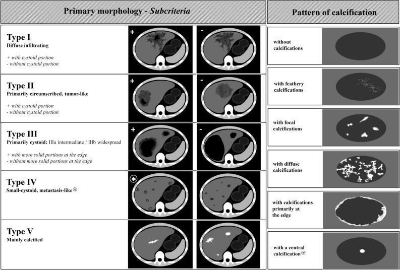
This educational infographic details the Echinococcus multilocularis Ulm Classification for Computed Tomography (EMUC-CT), a standardized system for evaluating hepatic Alveolar Echinococcosis (AE). The diagram is divided into two sections: Primary Morphology and Patterns of Calcification. The left section illustrates five morphological types (I-V) using axial CT cross-sections of the liver. Type I (diffuse infiltrating) and Type II (primarily circumscribed, tumor-like) are sub-categorized by the presence or absence of a cystoid portion. Type III (primarily cystoid) distinguishes between intermediate and widespread involvement, further categorized by solid peripheral portions. Type IV displays small-cystoid, metastasis-like lesions, while Type V represents mainly calcified, inactive lesions. The right panel illustrates six distinct calcification patterns within hepatic lesions: feathery (branching), focal (isolated spots), diffuse (widespread), edge-weighted (peripheral), and central. The classification system is designed to improve diagnostic accuracy and clinical assessment of disease activity in patients with Alveolar Echinococcosis.
Headache
Definition
- Harrison's Principles of Internal Medicine (22E), p. 1631
Anatomy and Physiology - How Headaches Arise
- The scalp, meningeal arteries, dural sinuses, falx cerebri, and proximal segments of large pial arteries
- Large intracranial vessels and dura mater - with peripheral terminals of the trigeminal nerve that innervate these structures
- The trigeminocervical complex - caudal trigeminal nucleus extending into the dorsal horns of the upper cervical spinal cord (receives input from C1-C2 nerve roots)
- Rostral pain-processing regions - ventroposteromedial thalamus and cortex
- Pain-modulatory systems - hypothalamus and brainstem structures (periaqueductal grey matter, rostral ventromedial medulla, locus ceruleus, dorsal raphe nuclei)
- Harrison's Principles of Internal Medicine (22E), p. 1634-1636
- Bradley and Daroff's Neurology in Clinical Practice, p. 4026
Classification (ICHD-3, 2018)
Part 1: Primary Headaches
| Type | Prevalence (%) |
|---|---|
| Tension-type headache | ~69% - most common overall |
| Migraine | second most common |
| Trigeminal autonomic cephalalgias (TACs) | includes cluster headache (least common) |
| Other primary headaches | thunderclap, stabbing, hypnic, cough, exercise-related, etc. |
Part 2: Secondary Headaches
| Category | Common Examples |
|---|---|
| Trauma/injury | Post-concussion headache, whiplash |
| Vascular disorders | Subarachnoid hemorrhage, stroke, giant cell arteritis |
| Non-vascular intracranial | Increased/decreased intracranial pressure, tumors |
| Substances/withdrawal | Medication overuse headache, caffeine withdrawal |
| Infection | Meningitis, encephalitis, sinusitis |
| Homeostasis disorders | Hypertension, hypoxia, dialysis |
| Cranial/cervical structures | Cervicogenic headache, glaucoma |
| Psychiatric disorders | Somatization, depression-associated |
Part 3: Painful Cranial Neuropathies and Other Facial Pain
- Trigeminal neuralgia
- Other cranial nerve pain syndromes
Red Flags (Require Urgent Investigation)
-
Thunderclap headache - sudden-onset severe headache (rule out aneurysm rupture)
-
"Worst headache of life"
-
New headache in a cancer patient
-
Progressive worsening in frequency or severity
-
Headache always localized to one area
-
Not responding to treatment
-
Associated with fever, altered mental status, or focal neurological deficit
-
Bradley and Daroff's Neurology in Clinical Practice, p. 2082
Epidemiology
- Primary headache disorders (migraine, tension, cluster) account for nearly 98% of all headaches
- Chronic daily headache (headache on 15+ days/month) affects about 4% of the population
- Secondary headaches, while less common, are important to recognize as they can be life-threatening
Key Takeaway
- Harrison's Principles of Internal Medicine (22E) | Bradley and Daroff's Neurology in Clinical Practice | ICHD-3 Classification (IHS)