
V-y junction suturing
Reading File
Finding Sources
Searching PubMed
"V-Y" AND suture
Searching the Web
V-Y junction suturing technique
V-Y junction suturing usually refers to closure of a V-Y advancement flap.
- A V-shaped incision or flap is designed adjacent to a tissue defect.
- The triangular tissue is advanced into the defect.
- The two limbs of the original V are then sutured together, converting the incision into a Y.
- It gains length and allows closure with less tension, rather than pulling the wound edges directly together.
Typical uses: fingertip defects, small facial/skin defects, and selected mucosal or reconstructive procedures. The flap should be advanced without excessive tension and with preservation of its blood supply. The apex is brought forward first, then the limbs are closed to form the Y. StatPearls description explains that the tissue is generally pushed or allowed to recoil into the defect rather than stretched.
In fingertip reconstruction, the flap base may be sutured to the nail bed and the remaining defect closed in the Y configuration. Campbell's Operative Orthopaedics, 15th ed., Fig. 21.8.
If you mean a V-Y closure at a specific site, such as lip after Le Fort I surgery, cleft palate, finger, Achilles tendon, or skin, tell me the site because the incision design and suture sequence differ.
Show the step-by-step suture sequence for a V-Y flap
Reading File
Finding Sources
Finding Sources
Searching Images
labeled V-Y advancement flap diagram showing suture closure sequence
Searching PubMed
"V-Y advancement flap"
Searching the Web
V-Y advancement flap closure suturing sequence surgical technique
V-Y advancement flap: generic suture sequence
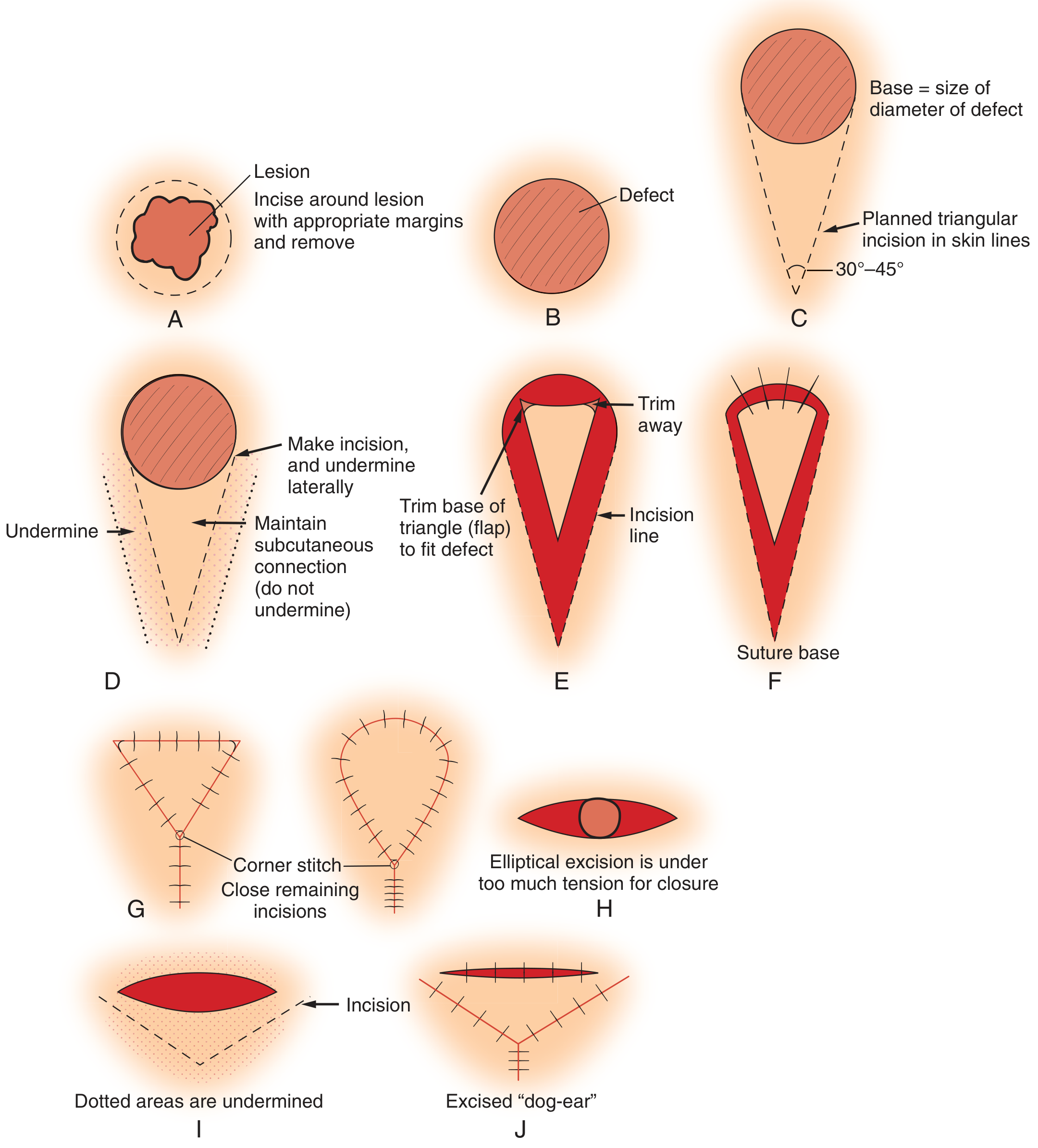
This is a generic skin V-Y advancement flap sequence. Exact needle, suture material, tissue plane, and order can differ for fingertip, face, lip, oral mucosa, or other sites.
-
Mobilize and check reach
After the V flap has been incised and released, gently advance it into the primary defect using skin hooks or fine forceps. It should move without blanching or significant tension. Do not undermine the central flap pedicle if it is an island flap, because its subcutaneous blood supply must be preserved. -
Place the leading-edge key suture
Advance the flap until its leading edge lies at the far edge of the defect. Place a key simple interrupted or deep dermal suture to fix the flap in its new position. This is the principal inset suture. -
Secure the two corners of the inset
Place interrupted sutures at each side of the advanced flap where it meets the recipient wound. These sutures align the corners and distribute tension rather than concentrating it at the flap tip. -
Complete closure of the recipient defect
Add interrupted sutures across the remaining leading edge as needed. The advanced triangular flap should now fill the original defect, with no excessive tension on the flap margin. -
Convert the V donor incision into the Y
Approximate the two limbs of the residual V-shaped donor wound. In some techniques, this is initiated by suturing from the apex of the V toward the flap base, which advances the flap forward. Continue closure until the donor incision becomes the vertical, common limb of the Y. -
Remove any standing cone only if necessary
If a dog-ear remains, excise it conservatively and close that short segment. Avoid over-trimming, which can compromise contour or perfusion. -
Final perfusion and tension check
Confirm capillary refill, color, absence of distal congestion, accurate edge eversion, and no strangulating sutures. The final scar is a Y: the two upper arms represent the flap/defect margins and the lower limb is the closed donor site.
Important principle: the flap is advanced by being pushed or allowed to recoil, not forcefully pulled into the defect. The closure should be tension-minimized and should not compromise the flap vascular pedicle. This sequence is consistent with Pfenninger and Fowler's Procedures for Primary Care, 3rd ed., Fig. 17-7 and the description of V-Y advancement in StatPearls.
For a flap where the V apex is remote from the defect, some surgeons close the apex first, progressing toward the base, until adequate advancement occurs, then inset the leading edge. Both descriptions follow the same principle: secure the flap in the defect and close the residual donor V to create the Y.
This is a shared conversation. Sign in to Orris to start your own chat.