
Describe the uses of drugs tolvaptan what is its mode of action and why it losses only water
tolvaptan clinical uses vasopressin antagonist
tolvaptan mechanism of action aquaresis V2 receptor clinical uses 2024
vasopressin V2 receptor aquaporin water channel mechanism collecting duct diagram
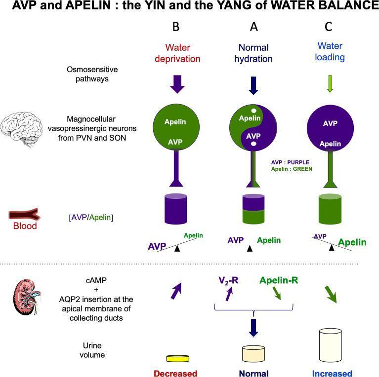
This pathophysiology diagram titled 'AVP and APELIN : the YIN and the YANG of WATER BALANCE' illustrates the neuroendocrine regulation of renal water excretion across three hydration states: Normal hydration (A), Water deprivation (B), and Water loading (C). The diagram tracks the relationship between Arginine Vasopressin (AVP, purple) and Apelin (green) within magnocellular neurons of the PVN and SON, their relative concentrations in the blood, and their physiological effects on the kidney. In Panel A, balanced levels of AVP and Apelin result in normal urine volume. In Panel B (Water deprivation), increased systemic AVP and decreased Apelin stimulate V2 receptors (V2-R), promoting cAMP-mediated Aquaporin-2 (AQP2) insertion at the apical membrane of collecting ducts, leading to decreased urine volume. In Panel C (Water loading), high systemic Apelin levels and low AVP levels antagonize this pathway via Apelin receptors (Apelin-R), resulting in increased aqueous diuresis. The 'yin-yang' motif represents the reciprocal storage and release mechanisms of these two peptides in response to osmosensitive pathways, visually summarized by balance scales showing the dominant circulating peptide for each condition.
This medical illustration presents a sagittal section of the human brain, detailing the regional distribution and cellular localization of water channel proteins Aquaporin-1 (AQP1) and Aquaporin-4 (AQP4). A central anatomical diagram uses blue markers for AQP1 and orange markers for AQP4, with four high-magnification insets (a-d) demonstrating specific cellular niches. Inset (a) shows AQP4 localized to the basolateral membrane of ependymal cells lining the ventricles. Inset (b) depicts AQP1 expressed at the apical membrane of choroid plexus epithelial cells, facilitating cerebrospinal fluid (CSF) secretion. Inset (c) illustrates AQP4 concentrated in astrocytic end-feet at the perivascular membrane surrounding brain capillaries and the glia limitans externa. Inset (d) shows AQP4 expression in the glial lamellae of the supraoptic nucleus and other osmosensitive regions. The diagram also illustrates regional parenchymal gradients, such as those in the neocortex and cerebellum. This schematic is designed for neuroanatomy and neurophysiology education, emphasizing the molecular mechanisms of water transport and CSF dynamics within the central nervous system.
This pathophysiology diagram illustrates the dysregulation of Aquaporin 5 (AQP5) in lacrimal gland acinar cells during Sjögren’s Syndrome Dry Eye (SSDE) compared to normal status. On the left, 'Normal Status' shows AQP5 channels primarily localized at the apical membrane, facilitating robust water transport (blue droplets) and tear secretion. On the right, 'Sjögren’s Syndrome Dry Eye' depicts chronic inflammation driven by cytokines (TNF-α, IFN-γ, and IL-17). These inflammatory mediators cause a distinct mislocalization of AQP5 from the apical membrane to basolateral and cytoplasmic regions, indicated by red arrows. This structural shift leads to reduced AQP5 expression at the apical surface and inhibited fluid transport, visually represented by blocked secretion pathways and sparse tear droplets. The comparison highlights how autoimmune-driven inflammation disrupts cellular polarity and water channel trafficking, providing a mechanistic explanation for the ocular dryness characteristic of Sjögren’s Syndrome.
Tolvaptan (Samsca / Jynarque): Uses, Mechanism, and Why It Loses Only Water
What Is Tolvaptan?
Mode of Action - Step by Step
Normal Physiology (Why ADH Retains Water)
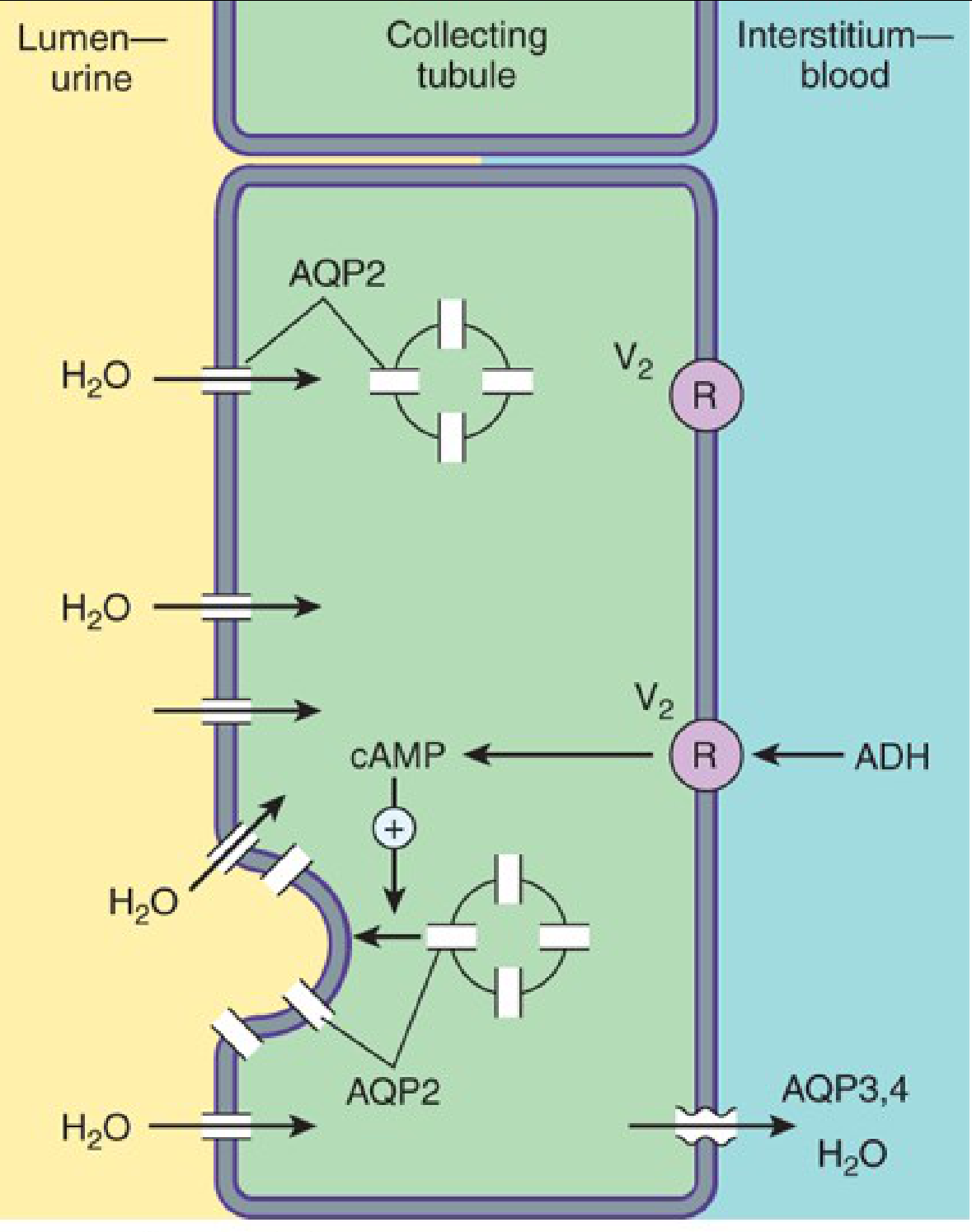
- ADH (AVP) is released from the posterior pituitary when plasma osmolality rises or blood volume falls.
- ADH binds to V2 receptors on the basolateral membrane of principal cells in the collecting duct.
- V2 is a Gs-coupled GPCR; binding activates adenylyl cyclase → raises intracellular cAMP.
- cAMP activates protein kinase A (PKA), which phosphorylates aquaporin-2 (AQP2) water channel vesicles.
- Phosphorylated AQP2 vesicles translocate and fuse with the apical (luminal) membrane.
- AQP2 in the apical membrane allows water to move from the tubular lumen into the cell, then exit via AQP3 and AQP4 on the basolateral side into the interstitium and blood.
- Result: concentrated urine, water retained.
"Vasopressin binding to V1 receptors activates the G-PLC-IP3 pathway... V2 receptors act via a Gs protein-coupled, cAMP-mediated process." - Goodman & Gilman's Pharmacological Basis of Therapeutics
How Tolvaptan Blocks This
- No V2 receptor activation → no rise in cAMP → PKA not activated → AQP2 channels stay in intracellular vesicles (not inserted into the apical membrane)
- Without AQP2 in the luminal membrane, the collecting duct becomes impermeable to water
- Water stays in the tubular lumen and is excreted as dilute urine
- This excretion of electrolyte-free water is called aquaresis
Why Only Water Is Lost (Not Electrolytes)
| Feature | Tolvaptan (aquaresis) | Loop/Thiazide diuretics |
|---|---|---|
| Target | V2 receptor (collecting duct) | NaK2Cl transporter / NaCl cotransporter |
| Sodium excretion | Minimal / none | Substantial |
| Potassium excretion | Minimal / none | Substantial |
| Water excretion | Yes (electrolyte-free) | Yes (with electrolytes) |
| Serum sodium effect | Raises serum [Na+] | May worsen hyponatremia |
- The V2 receptor only controls the water permeability of the collecting duct principal cells, via AQP2 trafficking.
- It has no direct role in sodium, potassium, or chloride transport.
- Na+ reabsorption is handled by separate transporters (ENaC channels, Na+/K+-ATPase) that are regulated by aldosterone, not vasopressin/V2.
- Therefore, blocking V2 removes only the water reabsorption signal, leaving all electrolyte transport mechanisms intact.
Clinical Uses
1. Hyponatremia (Euvolemic and Hypervolemic)
- SIADH (Syndrome of Inappropriate ADH secretion) - EMA approval specifically for this
- Heart failure with dilutional hyponatremia
- Hepatic cirrhosis with ascites and hyponatremia
- Initiate only in a hospital so sodium rise can be monitored
- Avoid fluid restriction initially - allow thirst to self-limit aquaresis
- Generally ineffective if serum creatinine >2.5 mg/dL
- Contraindicated in hypovolemic hyponatremia (would worsen volume depletion)
- Not to be used >30 days due to hepatotoxicity risk
2. Autosomal Dominant Polycystic Kidney Disease (ADPKD) - Jynarque
- Total kidney volume increased 2.8% per year with tolvaptan vs 5.5% per year with placebo
- eGFR decline reduced from 10.1 to 6.8 mL/min/1.73 m² over 3 years
- Also reduced kidney pain and urinary albumin excretion
Adverse Effects
| Effect | Cause |
|---|---|
| Polyuria, nocturia, polydipsia | Direct aquaretic effect (most common, does not diminish over time) |
| Thirst, dry mouth | Aquaresis with osmoreceptor activation |
| Hypernatremia | Over-correction of hyponatremia, especially if fluid restricted |
| Osmotic demyelination syndrome | If sodium corrects too rapidly (>10-12 mEq/24h) |
| Hepatotoxicity | Transaminase elevation in ~4.4% with ADPKD dosing; Jynarque has a REMS program |
Summary
Can u providee the book goodman and gilmans pharmacology book and other best books for pharmacology
Top Pharmacology Textbooks (with legitimate access options)
🏆 For Medical Students & Clinicians
| Book | Best For | Notes |
|---|---|---|
| Goodman & Gilman's The Pharmacological Basis of Therapeutics (14th Ed) | Deep mechanistic understanding, research | The gold standard reference |
| Katzung's Basic & Clinical Pharmacology (16th Ed) | Integrated basic + clinical, USMLE | Very widely used in med schools |
| Rang & Dale's Pharmacology (10th Ed) | Conceptual clarity, physiology-linked | Excellent for UK/European curricula |
| Lippincott Illustrated Reviews: Pharmacology | USMLE Step 1 prep, quick review | Great diagrams and mnemonics |
| Bertram Katzung's Pharmacology: Examination & Board Review | Board exams | Q&A format companion |
📚 For Pharmacists & Advanced Study
| Book | Best For |
|---|---|
| Stahl's Essential Psychopharmacology | Psychiatry/neuropharmacology |
| DiPiro's Pharmacotherapy: A Pathophysiologic Approach | Clinical decision-making |
| Basic & Clinical Pharmacology by Katzung | Most popular overall |
Where to Access Them Legally
- Your university/medical school library - most institutions have physical copies and digital access via ClinicalKey, AccessMedicine, or similar
- AccessMedicine (McGraw-Hill) - accessmedicine.mhmedical.com - Goodman & Gilman and Katzung are both available here with institutional login
- ClinicalKey (Elsevier) - Rang & Dale and many others
- Google Books - free preview of many chapters
- Open Library / Internet Archive - archive.org - older editions available for free borrowing (legal digital lending)
- PubMed / NCBI Bookshelf - ncbi.nlm.nih.gov/books - some pharmacology texts are freely available
What I Can Do Instead
- Answer any pharmacology question directly from these books with exact citations
- Explain drug mechanisms, uses, adverse effects, and interactions
- Summarize entire drug classes or topics
- Compare drugs side by side