
Can gall bladder stone remove by medicine?
gallbladder stone dissolution medicine ursodiol treatment 2024
gallbladder stones ultrasound
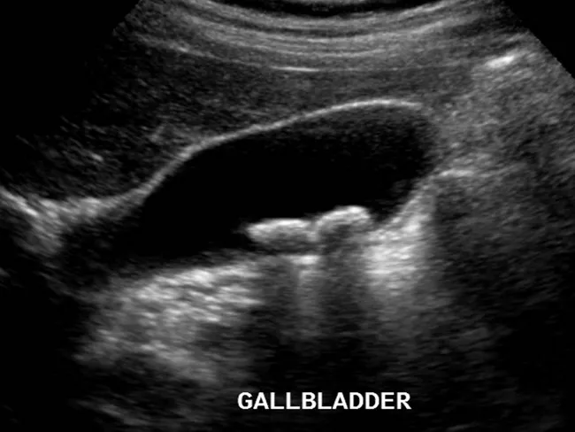
Ultrasound examination of the gallbladder using grayscale B-mode ultrasonography (transabdominal approach) demonstrates two echogenic foci within the gallbladder lumen. Both features are dependent on the gallbladder, implying intraluminal calculi rather than extrinsic objects. Each lesion casts a posterior acoustic shadow, a hallmark of gallstones, and their appearance distinguishes calcified cholesterol stones from gallbladder polyps, which typically lack shadowing. The stones measure over 2 millimeters in diameter, consistent with cholelithiasis and enabling reliable detection by ultrasound. The gallbladder wall appears without significant thickening, and there is no overt pericholecystic fluid evident on this image, arguing against acute cholecystitis at the time of imaging. The study highlights the classic sonographic criteria for gallstone disease: echogenic mobile calculi with clean shadowing, gravity-dependent location, and recognition of potential sludge in the gallbladder lumen. This imaging feature set supports clinical decisions regarding symptomatic biliary colic, risk stratification for complications, and surgical planning such as cholecystectomy. In differential considerations, gallbladder polyps or sludge can mimic stones; however, shadowing and mobility aid differentiation. Overall, ultrasound remains the initial modality of choice for suspected biliary pathology due to safety, accessibility, and high diagnostic yield. Correlative clinical data and laboratory tests further refine management decisions. Guiding patient-specific treatment options.
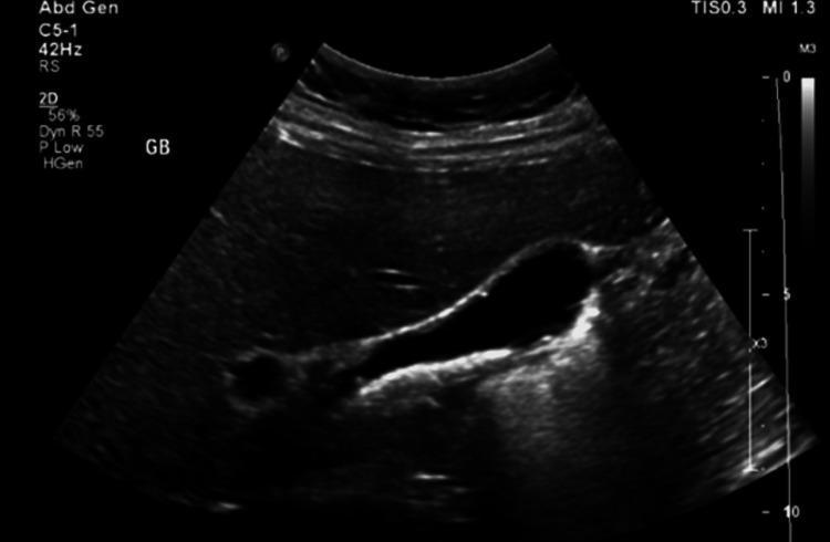
This diagnostic image is a 2D grayscale abdominal ultrasound focusing on the gallbladder (labeled 'GB'). The gallbladder is visualized as an anechoic (black) fluid-filled sac. Within the dependent portion of the gallbladder lumen, there are multiple hyperechoic (bright) mobile structures consistent with cholelithiasis (gallstones). These stones demonstrate posterior acoustic shadowing, a hallmark diagnostic feature where the sound waves are blocked by the dense stones. The gallbladder wall appears thin and regular, without obvious evidence of wall thickening, pericholecystic fluid, or other signs of acute cholecystitis. In the clinical context of situs inversus totalis, this ultrasound identifies the gallbladder positioned on the left side of the upper abdomen. This visual serves as a primary diagnostic tool for evaluating biliary colic and symptomatic cholelithiasis.
Can Gallbladder Stones Be Removed by Medicine?
The Only Approved Medicine: Ursodeoxycholic Acid (UDCA)
"Oral dissolution therapy (e.g., ursodeoxycholic acid, 15 mg/kg/day) may be considered in patients who have cholesterol gallstones, but it completely dissolves stones in only 40% of patients, and stones recur in up to 50% of patients within 5 years after therapy is stopped. Lifelong therapy may be necessary."
Who Can Benefit from Medicine?
| Requirement | Why it matters |
|---|---|
| Cholesterol stones (not pigment/mixed) | UDCA only dissolves cholesterol - does not work on pigment or calcified stones |
| Small stones (< 20 mm diameter) | Larger stones rarely dissolve |
| Non-calcified stones (radiolucent on X-ray) | Calcified stones don't respond |
| Functioning gallbladder | The gallbladder must be visualized on ultrasound |
| Mild or no symptoms | Symptomatic/complicated disease needs surgery |
| Patient refuses or cannot have surgery | UDCA is a surgical alternative, not a first choice |
What UDCA Cannot Do
- Cannot dissolve pigment stones (black or brown) - these are made of bilirubin, not cholesterol
- Cannot dissolve calcified (hard) stones
- Cannot work when the gallbladder is non-functioning
- Cannot prevent recurrence reliably - up to 50% of stones return within 5 years
- Takes 6 months to 2 years of daily treatment to work
- Cannot treat acute cholecystitis, cholangitis, or pancreatitis caused by gallstones
Other Non-Surgical Options (Very Limited Use)
- Extracorporeal shock wave lithotripsy (ESWL) - breaks stones into fragments using sound waves. Only suitable for a single stone 0.5-2 cm in diameter. Very few patients qualify, and recurrence rates are high.
- Methyl tert-butyl ether (MTBE) infusion - a solvent injected directly into the gallbladder to dissolve cholesterol stones. Rarely used due to technical difficulty and similar recurrence rates.
Why Surgery Is Preferred
"As recurrence rates of cholesterol gallstones are high following medical dissolution by UDCA, the role of medical therapy in gallstone patients is very limited."
- It is a one-time, definitive cure
- Recovery time is 1-2 weeks
- It eliminates all risk of future gallstone complications
- It is widely available and has a very low complication rate
Summary
| Treatment | Works for | Success Rate | Limitation |
|---|---|---|---|
| UDCA (ursodiol) | Small cholesterol stones, no/mild symptoms | ~30-40% complete dissolution | Takes 1-2 years; 50% stones return |
| ESWL | Single stone, 0.5-2 cm | Limited | Very few patients qualify |
| Surgery (lap. cholecystectomy) | All types and sizes | ~99% permanent cure | Requires anesthesia |
- Goldman-Cecil Medicine, International Edition
- Yamada's Textbook of Gastroenterology, 7th ed.
- Sleisenger and Fordtran's Gastrointestinal and Liver Disease