
examination of shoulder joint
shoulder joint examination clinical tests
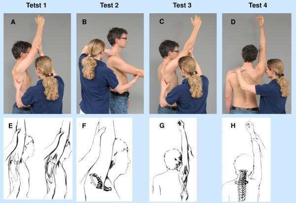
This composite educational graphic illustrates clinical mobility tests and biomechanical principles of the shoulder joint complex, specifically the Stenvers shoulder examination. Panels A-D are clinical photographs showing a practitioner performing manual assessments on a male subject. These include end-range flexion and abduction maneuvers with specific hand placements on the scapula, humerus, and acromioclavicular region to evaluate joint play and movement quality. Panels E-H provide corresponding anatomical diagrams. E compares lateral scapular positioning at end-range flexion between normal (left) and restricted (right) states, showing increased protraction in the latter. F details clavicular motion, contrasting normal posterior/dorsocranial trajectories (bold) with restricted movement patterns. G illustrates scapular posterior tilting during the terminal phase of flexion, and H depicts the associated motion of the cervicothoracic junction and spinal column. The content is designed for musculoskeletal physical therapy and orthopedic education, focusing on scapulohumeral rhythm, clavicular kinematics, and the identification of restricted range of motion in shoulder pathology.
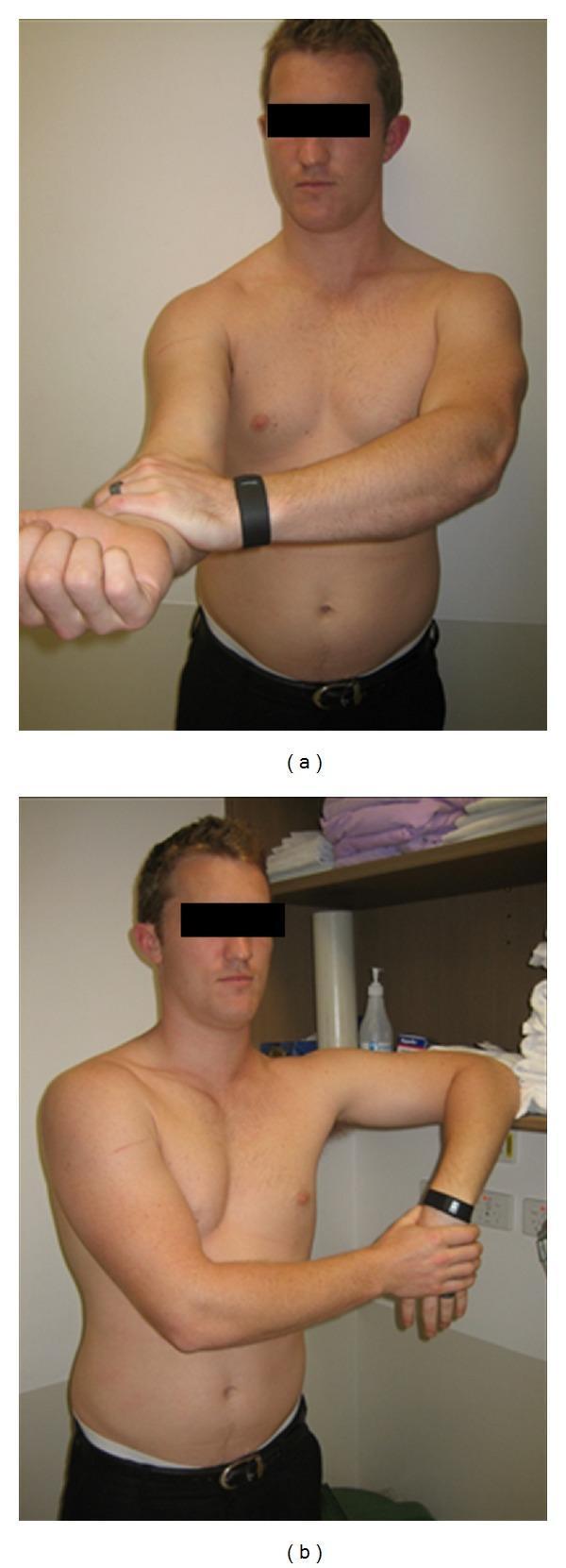
Clinical photograph showing two specific orthopedic physical examination maneuvers performed for shoulder pathology. Image (a) demonstrates Speed's test, which evaluates the long head of the biceps tendon; the participant has the right arm extended anteriorly with the forearm supinated, while the left hand applies downward resistance at the distal forearm/wrist. Image (b) illustrates a modification of the Hawkins-Kennedy impingement test, used to assess for subacromial impingement; the participant's right arm is flexed to 90 degrees at the shoulder with the elbow also flexed to 90 degrees, and the arm is internally rotated. These images represent telerehabilitation or self-administered physical examination techniques where the patient uses their own limbs or surrounding environment to recreate provocative orthopedic tests. The anatomical focus is the glenohumeral joint and associated musculotendinous structures. Educational context includes musculoskeletal assessment, physical therapy, and telemedicine clinical reasoning.
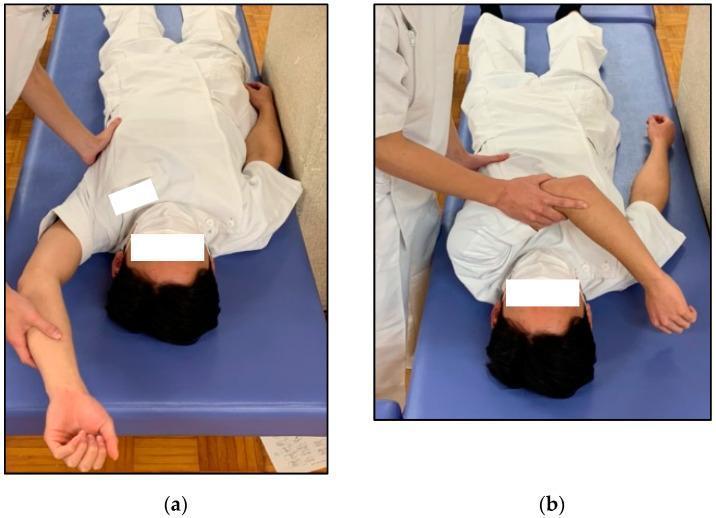
Clinical procedural photograph demonstrating physical examination techniques for assessing posterior shoulder tightness. The image is divided into two panels (a and b), showing a patient in the supine position on an examination table. In panel (a), the Combined Abduction Test (CAT) is shown: the examiner uses one hand to stabilize the lateral border of the scapula while the other hand passively abducts the patient's arm toward the head to check for restricted range of motion. In panel (b), the Horizontal Flexion Test (HFT) is depicted: the examiner maintains scapular stabilization while passively adducting the patient's arm across the torso toward the contralateral shoulder. These maneuvers are typically used in sports medicine and orthopedics to identify posterior capsule or muscle tightness, which are clinical indicators often associated with shoulder impingement or throwing-related overhead injuries. The examiner's hand placement is critical in both tests to prevent compensatory scapular motion, ensuring the assessment reflects true glenohumeral joint flexibility.
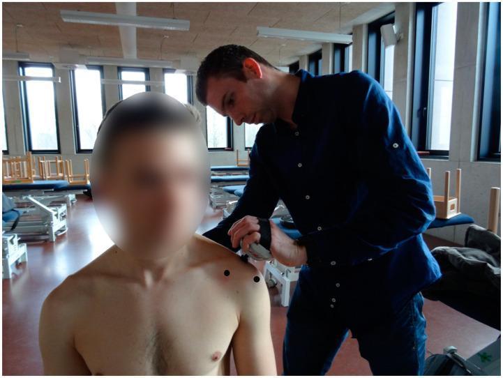
A clinical photograph depicting a physical examination of the shoulder joint in a medical or rehabilitative setting. An examiner is shown using a digital pressure algometer to quantify the pressure pain threshold (PPT) at the acromioclavicular (AC) joint of a seated patient. Three specific anatomical landmarks on the anterior and superior shoulder are indicated by black circular markers: the coracoid process, the AC joint, and the anterolateral subacromial area. The image demonstrates a standardized diagnostic procedure often used in the assessment of musculoskeletal conditions such as adhesive capsulitis (frozen shoulder) or subacromial impingement syndrome. The focus is on the correct placement of the algometer and the topographical identification of myofascial or articular trigger points for clinical evaluation of pain sensitivity and joint pathology.
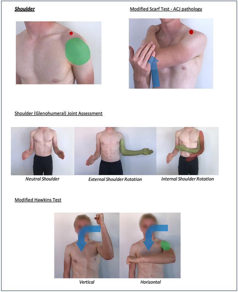
Educational clinical photograph compilation illustrating physical examination maneuvers for shoulder assessment. The content is organized into four main sections: 1. Shoulder (Basics): Highlights the deltoid region with a green overlay and a red dot indicating the acromioclavicular joint (ACJ). 2. Modified Scarf Test: Demonstrates cross-body adduction to assess ACJ pathology, featuring a blue directional arrow and a red dot at the ACJ. 3. Glenohumeral Joint Assessment: Displays the patient in neutral, external rotation (highlighted in green for normal range of motion), and internal rotation (highlighted in red for restricted or abnormal motion). 4. Modified Hawkins Test: Shows two variations for impingement assessment—a vertical approach with the arm abducted to 90 degrees and internally rotated, and a horizontal approach with the arm adducted across the midline. Blue arrows indicate the direction of force and rotation. This resource uses a color-coding convention where green overlays typically represent normal findings or anatomical target zones, and red signifies potential pathology or restricted range of motion. It is designed for clinical training and telemedicine frameworks.
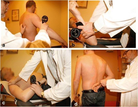
A composite of four clinical photographs demonstrating the clinical assessment of passive range of motion (PROM) of the shoulder joint. The series illustrates standardized physical examination techniques: (a) measurement of glenohumeral abduction with the patient standing and the examiner stabilizing the scapula; (b) assessment of external rotation with the patient supine and the shoulder in 90 degrees of abduction; (c) measurement of internal rotation in the supine position; and (d) functional assessment of internal rotation with the 'hand behind back' maneuver. In panels a, b, and c, a gravity-referenced inclinometer (Plurimeter-V) is utilized to objectively quantify the joint angles. The inclinometer is shown placed along the humerus, distal to the shoulder joint, to capture the angular displacement from the vertical or horizontal reference. This image serves as an educational guide for orthopedic and physical therapy practitioners in the assessment of shoulder pathologies such as adhesive capsulitis or rotator cuff disorders.
Examination of the Shoulder Joint
Preliminary
- The patient must be stripped to the waist and examined in good daylight
- Always compare both sides
- Observe the patient's attitude as they enter the clinic - this alone can suggest the diagnosis
- Pain arising from the shoulder joint is felt anterolaterally; referred pain can come from the cervical spine, heart, mediastinum, or diaphragm
A. INSPECTION (Look)
1. Attitude of the Limb
- Fracture clavicle: patient supports the flexed elbow of the injured side with the other hand
- Anterior dislocation of shoulder: patient supports the flexed elbow, arm held in slight abduction and external rotation
2. Deformity and Swelling
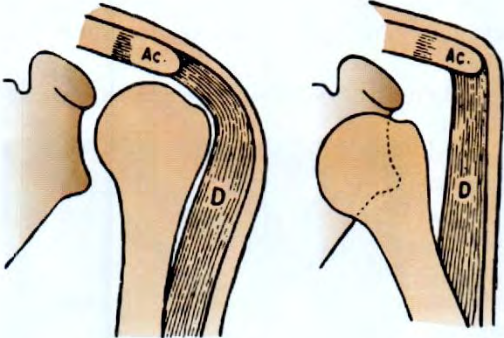
- Flattening / loss of roundness below the acromion = dislocation of shoulder (the greater tuberosity is displaced medially, losing the normal shoulder contour)
- The roundness of the shoulder is formed by the outward projection of the greater tuberosity beyond the acromion (AC) and the thickness of the deltoid (D):
- Swelling at middle-third clavicle or lateral one-third/medial two-third junction = fracture clavicle
- Undue prominence at acromial end of clavicle = AC joint dislocation
- Swelling at sternal end of clavicle = sternoclavicular dislocation
- Swelling in deltopectoral groove + prominent acromion = subcoracoid dislocation
- Drooping shoulder + lengthening of arm = fracture neck of scapula
3. Contour
- Wasting of deltoid (after shoulder dislocation due to axillary nerve injury)
- Wasting of rotator cuff muscles (supraspinatus, infraspinatus, subscapularis, teres minor)
4. Scars
- Anterior scar = deltopectoral approach
- Lateral scar = deltoid-splitting approach or lateral arthroscopic portals
- Posterior scars = arthroscopic portal sites
B. PALPATION (Feel)
1. Clavicle
- Stand behind the patient (seated on stool)
- Place hands on sternal ends of both clavicles simultaneously
- Palpate both clavicles from medial to lateral - any break in the line or abnormal prominence = fracture
- Check sternoclavicular joint (sternal end displaced anteriorly in dislocation) and AC joint (acromial end subluxates upward)
2. Upper End of Humerus
- Palpate acromion processes of both sides, then slide fingers down to the greater tuberosity
- Disappearance of greater tuberosity + loss of resistance = dislocation of shoulder (Fig. 13.5)
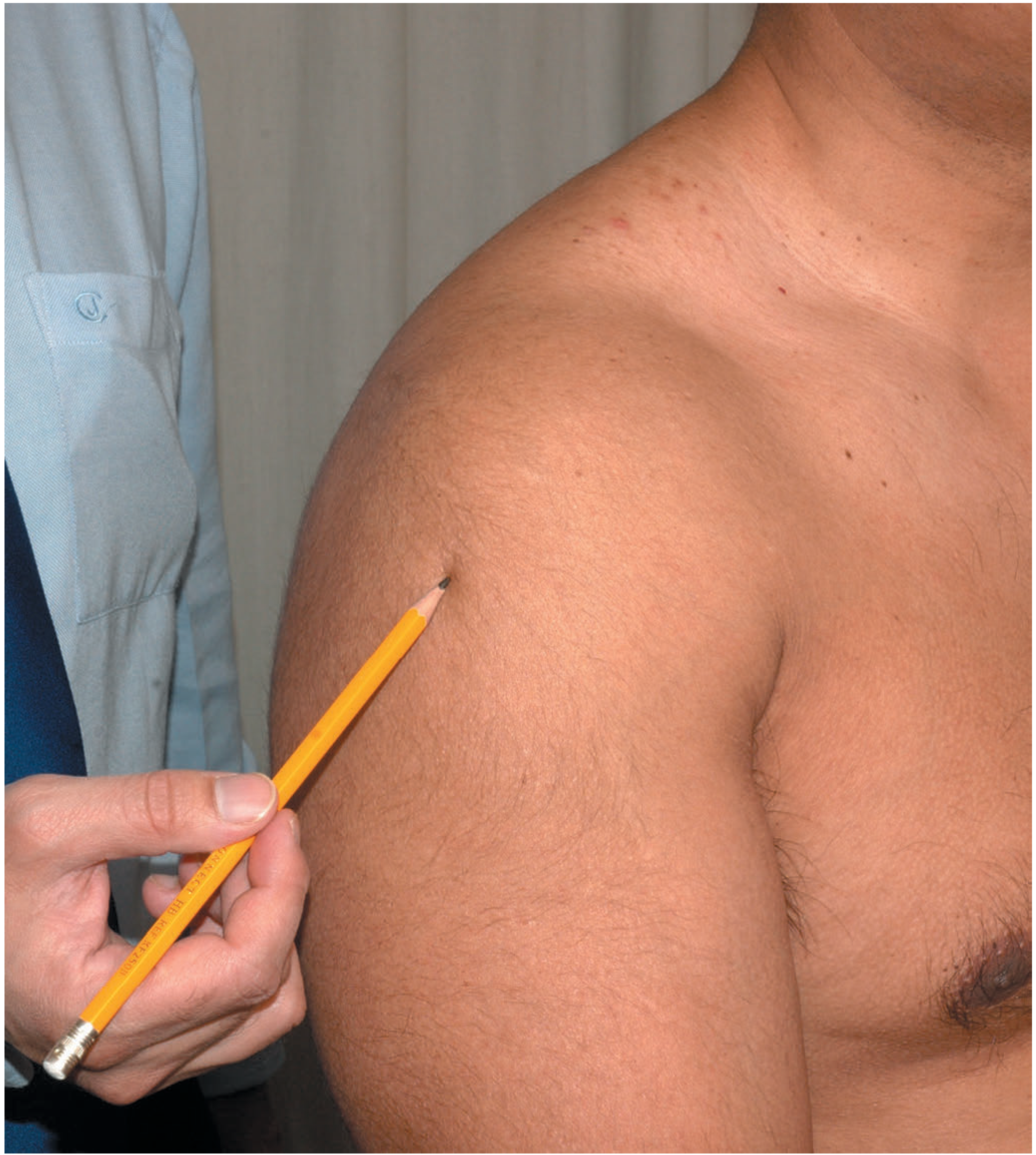
- Bimanual palpation of humeral head through the deltoid and axilla - the head is better felt from the axilla
3. Scapula
- Spine of scapula and acromion process are easily palpable
- Vertebral border, inferior angle, and coracoid process (½ inch below clavicle at medial 2/3rd and lateral 1/3rd junction) can be palpated
- Fracture of neck of scapula: tenderness medial to glenoid cavity + crepitus with axial pressure upward through flexed elbow; arm droops
4. Three Bony Points
- Coracoid process
- Acromial end of clavicle
- Greater tuberosity of humerus
5. Skin Sensation
- Test sensation over the regimental badge area (upper lateral arm) - loss indicates axillary nerve damage following shoulder dislocation
C. MEASUREMENTS
1. Length of the Arm
- Measured from the angle of the acromion to the lateral epicondyle of the humerus
- Note: the angle of the acromion is more posteriorly placed than the longitudinal axis of the arm
- Shortened in: subcoracoid dislocation, fracture neck of humerus, fracture shaft of humerus
- Lengthened in: subglenoid dislocation (head pushes humerus down)
2. Hamilton's Ruler Test
- A ruler placed along the lateral arm normally cannot touch both the acromion and the lateral epicondyle simultaneously, because the greater tuberosity pushes the ruler away
- In shoulder dislocation, the greater tuberosity is displaced medially - the ruler can touch both points simultaneously = positive test
D. MOVEMENTS (Move)
| Movement | Normal Range | How to Test |
|---|---|---|
| Forward flexion | 0-180° | Raise arms in front to touch ceiling, elbows extended |
| Extension | 0-30° | Extend both arms behind the body |
| Abduction | 0-180° | Raise arms sideways until fingers point to ceiling |
| Adduction | - | Touch other shoulder tip |
| Internal rotation | Up to T7-T9 | Touch back with dorsum of hand and raise up the back |
| External rotation | 0-40° | Elbows bent to 90°, rotate forearms outward |
E. SPECIAL TESTS
Impingement / Rotator Cuff Tests
| Test | Technique | Significance |
|---|---|---|
| Painful Arc test | Active abduction 60°-120° causes pain; pain-free above and below | Subacromial impingement or rotator cuff tendinitis |
| Neer's impingement sign | Passive forward flexion > 90° | Pain indicates impingement syndrome |
| Hawkins test | Passive forward flexion 90° + internal rotation | Pain indicates impingement |
| Jobe's test (Empty can) | Resisted pronation with forward flexion 90° in scapular plane | Pain/weakness indicates supraspinatus lesion |
| Drop-arm test | Examiner places arm in forward flexion in plane of scapula then releases | Inability to maintain position indicates supraspinatus tear |
| Lift-off (Gerber's) test | Arm in internal rotation behind back | Inability to lift arm off back = subscapularis lesion |
| Belly-push test | Elbow held anterior with abduction pressure | Inability to hold elbow forward = subscapularis lesion |
| Bear-hug test | Patient's hand on opposite shoulder; examiner attempts to lift hand | Inability to maintain contact = subscapularis lesion |
| Horn-blower sign | Resisted maximal external rotation with abduction 90° | Pain/weakness = infraspinatus/teres minor lesion |
Instability Tests
| Test | Technique | Significance |
|---|---|---|
| Apprehension test | Patient supine/standing; elbow 90°, shoulder abducted 90°, externally rotate | Apprehension (not just pain) = anterior instability |
| Relocation test | As above but apply posterior force on humeral head | Relief of apprehension = anterior instability confirmed |
| Load-and-shift test | Anterior/posterior force on humeral head | Degree of translation reflects laxity |
| Sulcus sign | Inferior traction with arm at side | Inferior subluxation groove below acromion = inferior/multidirectional instability |
| Jerk test | Posterior force with arm adduction and forward flexion | "Clunk" = posterior subluxation |
Labrum / Biceps Tests
| Test | Technique | Significance |
|---|---|---|
| O'Brien (Active Compression) test | 10° adduction, 90° forward flexion, maximal pronation then supination | Pain with pronation relieved by supination = SLAP lesion |
| Speed's test | Resisted forward flexion, elbow extended, forearm supinated | Pain = bicipital tendinopathy |
| Crank test | Full abduction, humeral axial loading, rotation | Pain/clunk = SLAP lesion |
| Yergason's test | Resisted supination, elbow at 90° | Pain in bicipital groove = biceps tendon pathology |
AC Joint
| Test | Technique | Significance |
|---|---|---|
| Scarf test (Cross-body adduction) | Bring arm horizontally across chest to opposite shoulder | Pain at AC joint = AC joint pathology |
Dislocation-Specific Tests
- Dugas' test: In dislocation of shoulder, the patient cannot touch the opposite shoulder tip with the hand of the affected side while keeping the arm against the chest wall. After reduction, this becomes possible - confirms successful reduction.
Frozen Shoulder vs Glenohumeral OA
- Both show global restriction of glenohumeral movement
- OA: radiographic changes, possible crepitus
- Frozen shoulder (adhesive capsulitis): painful then stiff phases, preserved bone architecture
F. NEUROLOGICAL ASSESSMENT
- Motor: test deltoid abduction (limited by pain in acute injuries, so not reliable acutely)
- Sensory: test regimental badge area (upper lateral arm) - this is the reliable test in the acute setting
Summary Box (Bailey & Love)
| Component | What to Assess |
|---|---|
| Look (standing) | Front - asymmetry, deformity; Side - muscle wasting; Back - wasting, scapular position |
| Look (supine) | Skin, scars, soft tissues, deformity |
| Feel | Palpate entire shoulder girdle from sternum to scapula |
| Move | Flexion/extension, abduction/adduction, internal/external rotation |
| Special tests | Impingement (painful arc, Jobe's, Hawkins); Instability (apprehension, relocation, sulcus); Rotator cuff; AC joint; Frozen shoulder vs OA |