
Allergic rhinitis
"allergic rhinitis"[MeSH Terms] AND treatment
allergic rhinitis nasal pathophysiology diagram
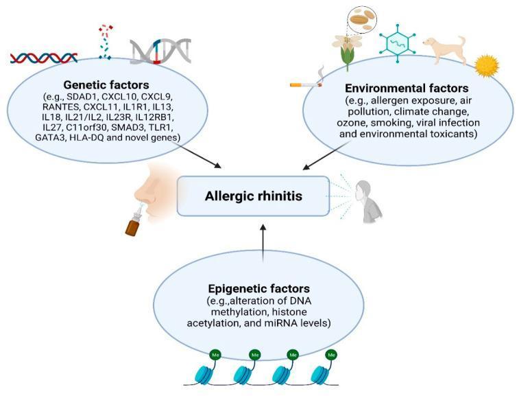
A pathophysiology diagram illustrating the multifactorial etiology of Allergic Rhinitis (AR). The graphic is organized into three major thematic bubbles converging on a central 'Allergic rhinitis' node, which is accompanied by icons of a nasal spray and an affected human profile. 1. Genetic Factors: Depicts a DNA double helix and lists specific susceptibility genes including SDAD1, Various Interleukins (IL1R1, IL13, IL18, IL21/IL2, IL23R, IL12RB1, IL27), Chemokines (CXCL9, CXCL10, CXCL11, RANTES), and others like SMAD3, GATA3, and HLA-DQ. 2. Environmental Factors: Features icons representing cigarette smoke (smoking), a dog (pet dander), a flower (pollen), a virus (infection), and the sun (climate change/ozone), alongside air pollution and toxicants. 3. Epigenetic Factors: Highlights mechanisms such as DNA methylation, histone acetylation, and miRNA level alterations, visually represented by chromatin structures with 'Me' (methylation) tags. This educational infographic summarizes the complex interplay between hereditary predisposition, external triggers, and gene-expression modifications in the pathogenesis of respiratory allergic disease.
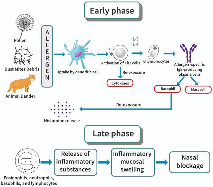
This pathophysiology diagram illustrates the two-phase immunological response in allergic rhinitis. The 'Early Phase' section details the sensitization process, starting with allergens such as pollen, dust mite debris, and animal dander being uptaken by dendritic cells. This triggers the activation of Th2 cells, which release cytokines and stimulate B lymphocytes via IL-3 and IL-4. These B cells differentiate into allergen-specific IgE-producing plasma cells that bind to basophils and mast cells. Upon allergen re-exposure, these sensitized cells undergo degranulation, leading to immediate histamine release. The 'Late Phase' section depicts the subsequent inflammatory cascade occurring hours later, characterized by the recruitment and infiltration of eosinophils, neutrophils, basophils, and lymphocytes. This influx results in the release of inflammatory substances, causing inflammatory mucosal swelling and culminating in clinical nasal blockage. The diagram serves as an educational tool for understanding Type I hypersensitivity and the transition from acute mast cell-mediated symptoms to chronic inflammatory tissue changes in the nasal mucosa.
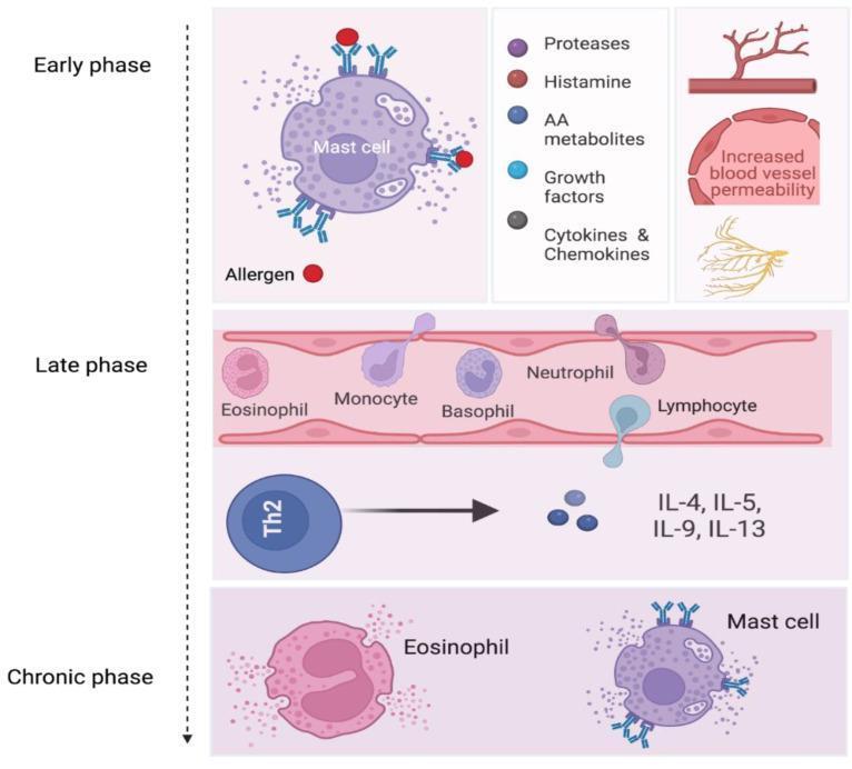
A pathophysiology diagram illustrating the three stages of allergic rhinitis: Early phase, Late phase, and Chronic phase. The 'Early phase' section depicts a mast cell activated by allergens binding to IgE on its surface receptors, triggering the release of inflammatory mediators including proteases, histamine, arachidonic acid (AA) metabolites, growth factors, and cytokines/chemokines. Associated vascular changes such as increased blood vessel permeability and nerve stimulation are shown. The 'Late phase' section demonstrates leukocyte recruitment and extravasation through a blood vessel wall, featuring an eosinophil, monocyte, basophil, neutrophil, and a migrating lymphocyte. It also shows a Th2 cell secreting interleukins IL-4, IL-5, IL-9, and IL-13. The 'Chronic phase' section illustrates the persistent presence and coexistence of degranulating eosinophils and mast cells in the inflamed tissue. This diagram provides a clinical overview of the immunological cascade and cellular interactions involved in allergic airway inflammation and tissue remodeling.
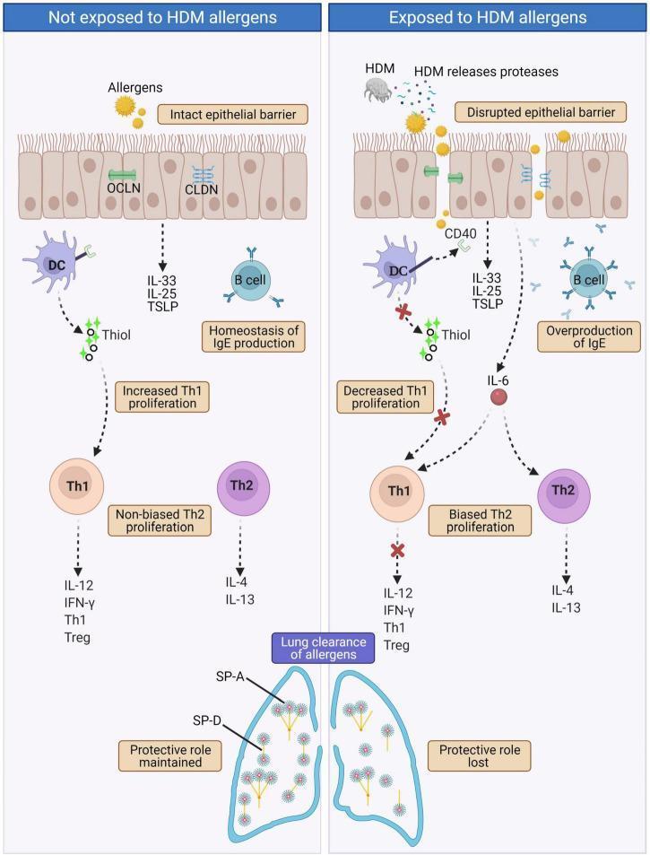
This pathophysiology diagram illustrates the immune response in Allergic Rhinitis (AR) due to House Dust Mite (HDM) exposure. The comparison chart depicts two states: 'Not exposed' (homeostasis) and 'Exposed to HDM allergens' (pathological state). In the normal state, an intact epithelial barrier with occludin (OCLN) and claudin (CLDN) tight junctions prevents allergen infiltration, resulting in balanced Th1/Th2 proliferation and IgE homeostasis. Following HDM exposure, HDM-released proteases disrupt tight junctions, allowing allergens to penetrate the barrier. Key downstream immunological changes include: 1) Dendritic cell (DC) surface CD40 cleavage leading to decreased Thiol production and suppressed Th1 proliferation; 2) Increased IL-6 secretion causing biased Th2 proliferation and production of IL-4 and IL-13 cytokines; 3) B cell overproduction of IgE; and 4) Cleavage of pulmonary surfactants SP-A and SP-D in the lungs, compromising allergen clearance. The diagram uses signaling pathway arrows and labels to demonstrate how HDM proteases facilitate allergic sensitization and mucosal inflammation through physical barrier disruption and Th2-mediated immune deviation.
"allergic rhinitis"[MeSH Terms] AND "immunotherapy"[MeSH Terms]