
Microscope and endoscope in ent in detail according to standard ENT proticol and recent articles in simplified manner
endoscope microscope ENT otolaryngology comparison
endoscopic ear surgery vs microscopic otology outcomes
endoscopic sinus surgery nasal endoscopy protocol
endoscopic tympanoplasty cholesteatoma outcomes meta-analysis
PMID: 39786316
PMID: 39582367
endoscopic ear surgery ENT operating microscope comparison
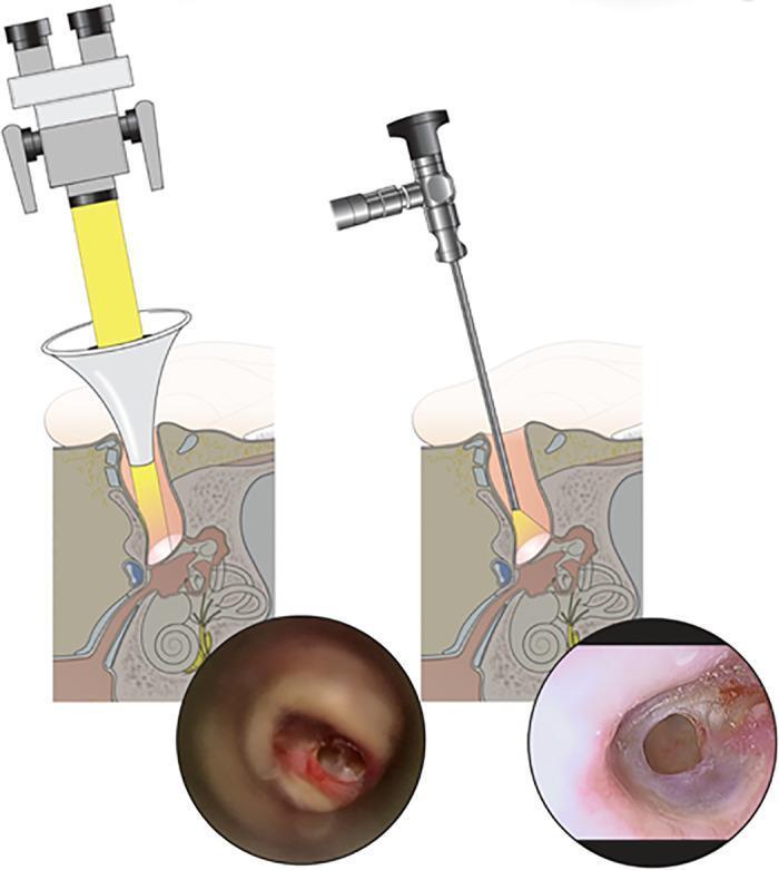
A comparison of microscopic versus endoscopic transmeatal approaches for visualizing the tympanic membrane. The left side features a diagram of an operating microscope directing a wide beam of light through an ear speculum, paired with a microscopic otoscopic photograph. The photograph shows a subtotal tympanic membrane perforation with restricted visualization of the anterior margins due to the linear line-of-sight constraints of the microscope. The right side displays a diagram of a rigid endoscope inserted deep into the external auditory canal, paired with a wide-angle endoscopic photograph. This view provides superior clarity and a comprehensive, circular perspective of the perforation edges, including the anterior-inferior quadrant. This educational visual demonstrates the clinical advantages of endoscopic ear surgery (EES) in achieving a wider field of view and enhanced detail of middle ear structures compared to traditional microscopic techniques.
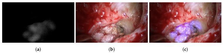
This surgical endoscopic comparison chart demonstrates the use of multispectral imaging in identifying middle ear pathology. The image is divided into three panels: (a) a spectrally segmented view under 420 nm illumination, appearing as a grayscale intensity map that highlights the spatial distribution of a cholesteatoma against a dark background; (b) a standard RGB broad-band illumination view showing the intraoperative surgical field, characterized by reddish soft tissue, white osseous structures, and the presence of pale, keratinous debris; and (c) an augmented spectral RGB view. In this final panel, the data from the spectral segmentation is overlaid onto the clinical photograph, highlighting the cholesteatoma tissue with a distinct bluish-purple hue. This comparison illustrates how multispectral imaging can differentiate between benign bone and pathological cholesteatoma during otological surgery. The anatomical context involves the mastoid cavity and middle ear, focusing on the identification and removal of recurrent cholesteatoma to assist surgical guidance and ensure complete resection.
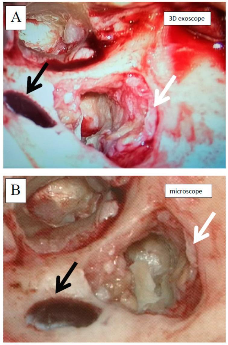
This medical comparison consists of two intraoperative images (A and B) showing the same surgical field during a cholesteatoma surgery in the left ear via a retroauricular approach. The images demonstrate an antromastoidectomy where the cholesteatoma has been partially removed. Image A is captured using a high-definition 3D exoscope, while Image B is captured using a standard operating microscope. In both images, a white arrow indicates the whitish, glistening cholesteatoma matrix, and a black arrow identifies the partially exposed sigmoid sinus, which appears as a dark, oval structure. The comparison highlights differences in visual perception between the two modalities: Image A shows more saturated red tones and a higher degree of visual interference from intraoperative bleeding, which appears to coat the tissue surfaces more intensely. Image B (microscope) exhibits a more natural, paler color palette with increased clarity of underlying bony structures and less perceived obscuration by blood. This serves as an educational comparison of visualization technologies in otolaryngology.
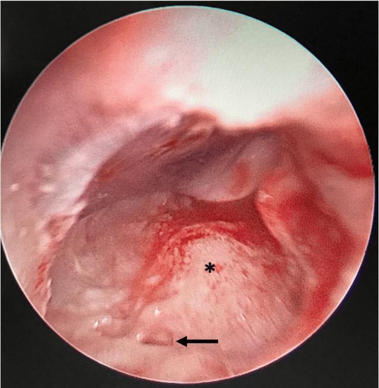
This clinical image presents an intraoperative endoscopic view of the human middle ear, obtained via a posterior tympanotomy. The field of view reveals the anatomical landmarks critical for otologic surgery, specifically cochlear implantation. A prominent, rounded, pinkish-white structure marked with an asterisk (*) represents the promontory, which corresponds to the basal turn of the cochlea. The surrounding mucosal tissue is erythematous and vascularized. A black horizontal arrow points to a localized region of bone covering the expected location of the round window niche, identifying an atretic round window plate. This finding is of high clinical significance as it indicates a congenital anomaly that complicates surgical access to the scala tympani. The image demonstrates the utility of otoendoscopy in identifying subtle middle ear pathologies that may be difficult to visualize using a standard operating microscope during mastoidectomy.
nasal endoscopy FESS functional endoscopic sinus surgery rigid endoscope
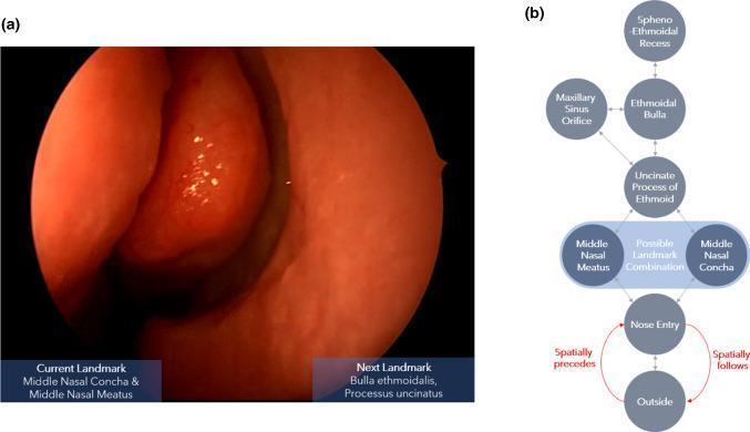
This composite educational graphic illustrates navigational landmarks and logic for Functional Endoscopic Sinus Surgery (FESS). Image (a) is a real-world clinical photograph captured during nasal endoscopy, showing a close-up view of the internal nasal anatomy. The image identifies the middle nasal concha (turbinate) and the middle nasal meatus, characterized by a healthy, reddish-orange mucosal surface with a moist, smooth texture. Image (b) is a clinical flowchart/algorithm detailing the spatial transitions between anatomical landmarks encountered during the procedure. The diagram uses a hierarchical structure to show connectivity between nodes such as the 'Outside', 'Nose Entry', 'Middle Nasal Meatus/Concha', 'Uncinate Process of Ethmoid', 'Ethmoidal Bulla', 'Maxillary Sinus Orifice', and 'Spheno-Ethmoidal Recess'. Red arrows define directional semantic relations, specifically 'spatially precedes' and 'spatially follows', which indicate the relative depth or inward/outward movement of the endoscope within the nasal cavity. This resource is designed to train surgical navigation prediction models by mapping visual anatomical landmarks to procedural pathways.
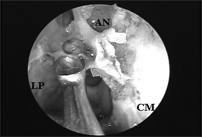
This procedural clinical photograph displays an endoscopic view of the right nasal cavity during a functional endoscopic sinus surgery (FESS). The image was captured using a 4mm 45-degree rigid endoscope. Key anatomical landmarks are labeled: the lamina papyracea (LP) on the lateral wall, the middle turbinate (CM) medially, and the agger nasi cell (AN) superiorly. The agger nasi cell is positioned such that it obstructs the direct endonasal visualization of the frontal sinus ostium, demonstrating a common anatomical variant that complicates frontal recess access. A surgical instrument is visible in the foreground, positioned near the lamina papyracea, indicating active manipulation for dissection or clearance of the obstructive ethmoidal cells. This image serves as an educational tool for identifying paranasal sinus anatomy and understanding the surgical challenges posed by agger nasi pneumatization in managing frontal sinus drainage.
Microscope and Endoscope in ENT - A Comprehensive Guide
Overview
PART 1: THE OPERATING MICROSCOPE IN ENT
What It Is
- Ear surgery (the primary domain - tympanoplasty, mastoidectomy, stapedotomy, cochlear implantation)
- Laryngeal microsurgery (microlaryngoscopy - vocal cord lesions)
- Salivary gland / parotid microdissection
Key Technical Features
| Feature | Detail |
|---|---|
| Magnification | 6x to 40x (typical operating range: 10-16x) |
| Light | Coaxial illumination (same axis as line of sight) |
| Working distance | 200-400 mm |
| Vision | True binocular/stereoscopic (3D depth perception) |
| Both hands | FREE for instrumentation |
| Field | Limited to line-of-sight (straight-ahead view only) |
Advantages of the Microscope
- Two-handed surgery - the single most important advantage. Suction in one hand, instrument in the other. Critical for bleeding control and fine dissection.
- True 3D stereoscopic depth perception - essential for delicate work near facial nerve and ossicular chain.
- Superior magnification - fine detail recognition of structures like the annulus, round window, facial nerve sheath.
- Less thermal risk to middle ear structures.
- Greater stability - mounted arm, no instrument fatigue.
- Excellent ergonomics for long cases (surgeon sits comfortably).
- Established learning curve - most ENT surgeons are trained in microscopic technique.
Limitations of the Microscope
- Sinus tympani (posterior mesotympanum)
- Anterior epitympanum
- Protympanic recess
- Facial sinus recess
- Anterior tympanic membrane perforation edges
PART 2: THE ENDOSCOPE IN ENT
Types Used in ENT
A. Rigid Endoscope (Hopkins Rod-Lens System)
| Diameter | Length | Angle | Application |
|---|---|---|---|
| 4 mm | 180 mm | 0° | Standard otologic/sinus surgery |
| 3 mm | 110-140 mm | 0° | Endoscopic ear surgery (EES) |
| 2.7 mm | 110 mm | 30°/70° | Angled views - sinus tympani, epitympanum |
| 4 mm | 180 mm | 30° | FESS (Messerklinger technique) |
| 4 mm | 180 mm | 45°/70° | FESS - frontal recess, posterior ethmoid |
B. Flexible Endoscope (Fiberoptic / Chip-on-Tip)
- Nasopharyngoscopy (nasopharynx, adenoids)
- Laryngoscopy (vocal cords, supraglottis, subglottis)
- DISE (Drug-Induced Sleep Endoscopy) - for obstructive sleep apnea evaluation
- Transnasal esophagoscopy
PART 3: ENDOSCOPE VS MICROSCOPE - THE DEBATE
| Parameter | Operating Microscope | Rigid Endoscope (EES) |
|---|---|---|
| Depth perception | 3D binocular (superior) | 2D monocular (improving with 3D scopes) |
| Hands free | Both hands free | One hand holds scope, one hand works |
| Hidden areas (sinus tympani etc.) | NOT visible | Excellent visualization |
| Field of view | Narrow (line of sight) | Wide angle (panoramic) |
| Bleeding control | Easy (two hands) | Difficult |
| Thermal risk | None | Real - tip heating can reach 49-51°C |
| External incision | Often needed | Usually NOT needed |
| Bone removal | More required | Minimal |
| Mastoid dissection | Often needed | Avoided in EES |
| Learning curve | Established / shorter | Longer, needs lab practice |
| Cost | Standard | Higher (camera system, HD screen) |
| Ergonomics | Good | More demanding (arm fatigue) |
PART 4: APPLICATIONS BY SUBSPECIALTY
4.1 OTOLOGY (Ear Surgery)
The Endoscopic Ear Surgery (EES) Concept
- Assessment of retraction pockets - serial photodocumentation to guide conservative vs surgical management
- Evaluation of cholesteatoma extent in blind spots
- Assessment of ossicular continuity through a perforation (outpatient, can use local anaesthesia)
- Endoscopy through myringotomy - unexplained conductive hearing loss, perilymphatic fistula, occult cholesteatoma
- Assess round window patency before intratympanic therapy (Ménière's, SSNHL)
- Otitis media with effusion - ventilation tube insertion
- Chronic otitis media - myringoplasty (type I tympanoplasty)
- Retraction pockets and atelectasis
- Cholesteatoma:
- Transcanal exclusively: attic / Prussak space / mesotympanic disease
- Combined approach: antral or mastoid cell involvement
- Otosclerosis - endoscopic stapedotomy (avoids need to drill external canal wall to see anterior stapes crus)
- Ossicular chain reconstruction / ossiculoplasty
- Middle ear tumors: glomus tympanicum, adenoma, osteoma
- Cochlear implantation - endoscope-assisted
- Lateral skull base: cholesterol granuloma of petrous apex, acoustic neuroma in IAC fundus (newer applications)
Protocol: Endoscopic vs Microscopic in Cholesteatoma Surgery
- Perform as much dissection as possible under the microscope (two-handed, safe)
- Use the endoscope at critical junctures - especially when matrix is adherent in sinus tympani or epitympanum
- End every cholesteatoma case with endoscopic inspection of all recesses to check for residual disease
- If disease is limited to attic, facial recess, or sinus tympani - endoscope-assisted surgery may eliminate the need for mastoidectomy
- Second-look procedures can often be done as transcanal endoscopic cases - avoiding postauricular re-incision
Safety Protocol for Thermal Injury
- Use light source at 40-50% intensity only
- Regularly clean the tip - blood and debris act as insulators and concentrate heat
- Limit contact time of tip with middle ear structures
- Studies found Hopkins rod (3mm) produced heating equivalent to a 44°C caloric test at the lateral semicircular canal. A 5mm burn was documented on the medial middle ear wall in one study.
- Newer LED cold-light sources reduce thermal risk significantly
4.2 RHINOLOGY (Nose and Sinuses)
Nasal Endoscopy Protocol (Diagnostic - Clinic)
- Preparation: Topical decongestant (oxymetazoline) + local anaesthetic (lignocaine spray or pledgets) applied 5-10 minutes prior
- Pass 1 (0° scope): Along the floor of the nasal cavity - assess inferior turbinate, floor, nasopharynx, Eustachian tube orifice, adenoids
- Pass 2: Between middle turbinate and septum - assess middle meatus, uncinate process, hiatus semilunaris, ethmoid bulla, maxillary sinus ostium
- Pass 3: Above middle turbinate - assess olfactory cleft, superior turbinate, sphenoethmoidal recess, sphenoid ostium
- Document: septal deviation, turbinate hypertrophy, polyps, discharge, mucosal edema, masses
FESS (Functional Endoscopic Sinus Surgery)
- Uncinectomy (remove uncinate process)
- Middle meatal antrostomy (open maxillary sinus ostium)
- Anterior ethmoidectomy (open anterior ethmoid cells)
- Posterior ethmoidectomy if needed
- Sphenoidotomy (open sphenoid sinus)
- Frontal recess dissection / Draf procedure (frontal sinusotomy)
- 0° for uncinectomy and maxillary antrostomy
- 30°/45° for frontal recess
- 70° for posterior frontal recess and sphenoethmoidal recess
4.3 LARYNGOLOGY
Rigid Laryngoscopy / Videostroboscopy
- Sustained "ee" at most comfortable pitch and loudness (several repetitions)
- Phonation on inhalation
- Glide midrange to high (sustain high note)
- Glide midrange to low (sustain low note)
- Quiet "ee" / Loud "ee"
- Locked mode (assess regularity)
- CAPE-V standard sentences: "The blue spot is on the key again," "How hard did he hit him?" etc.
- Connected speech, singing tasks as needed
Microlaryngoscopy (Microscope + Laryngoscope)
- High magnification (10-25x) for precise cold-steel or CO2 laser dissection
- Both hands free for microsurgical instruments (cup forceps, microflap technique)
- Essential for vocal fold polyps, nodules, Reinke's edema, papillomatosis, early glottic carcinoma
PART 5: RECENT EVIDENCE (2022-2026)
Endoscopic vs Microscopic Ear Surgery - What Does the Evidence Say?
- Operating time: Endoscopic was significantly shorter (mean difference -22 min)
- Air-bone gap closure: Comparable between the two groups (no significant difference)
- Post-operative pure tone audiogram: Equivalent
- Surgical success: Similar
- Additional advantages of EES: Better visualization, fewer supplemental incisions, excellent ergonomics, less post-op pain
- Conclusion: Endoscopic ossiculoplasty is a reasonable alternative to microscopic ossiculoplasty
- Endoscopic group: shorter surgical time (69.8 min vs 88.9 min)
- Graft success rate: 90.6% (microscopic) vs 92.8% (endoscopic) - no significant difference
- Conclusion: Endoscopic ear surgery is a suitable approach for atelectatic otitis media
PART 6: CURRENT CONSENSUS AND PROTOCOL RECOMMENDATIONS
| Situation | Recommended Approach |
|---|---|
| Tympanoplasty (type I) - straightforward | Endoscopic (transcanal) preferred |
| Cholesteatoma - limited to tympanic cavity | Endoscopic transcanal |
| Cholesteatoma - mastoid/antrum involvement | Microscopic + endoscope-assisted |
| Cholesteatoma second-look | Transcanal endoscopic |
| Stapedotomy | Endoscopic (avoids anterior canal wall drilling) |
| Cochlear implantation | Microscope (primary), endoscope-assisted for round window |
| Microlaryngoscopy | Microscope (mandatory - two-handed surgery) |
| Diagnostic nasal endoscopy | Rigid 0°/30° (Hopkins rod) |
| FESS | Rigid endoscope 0°/30°/45°/70° |
| DISE (sleep apnea) | Flexible nasopharyngoscope |
| Vocal cord examination (clinic) | Rigid 70° telescope or flexible scope |
- European (Marchioni et al.): short 3mm endoscopes as primary
- Anglo-American: 4mm 0° as primary, angled scopes (30°/70°) for recesses
Summary
- The microscope gives 3D vision, two-handed control, and is indispensable for complex dissection and microlaryngoscopy
- The endoscope (rigid Hopkins rod) eliminates blind spots in the middle ear, enables minimally invasive transcanal surgery, and has largely replaced the microscope for nasal/sinus work (FESS)
- The two are complementary - most centers use both in the same operation
- Recent high-quality evidence (2024-2025 meta-analyses and RCTs) confirms endoscopic ear surgery provides equivalent audiological outcomes with shorter operating times and less post-operative pain
- The thermal safety of endoscopy demands strict protocol: low light intensity (40-50%), frequent tip cleaning, limited contact time