
Anatomical causes of recurrent pregnancy loss
I'll start by reading the medical library skill, then search for anatomical causes of recurrent pregnancy loss.
Reading File
Now I'll run parallel searches across the textbook library, PubMed, and web simultaneously.
Finding Sources
Finding Sources
Searching PubMed
"recurrent pregnancy loss" AND "uterine anomaly"
Good. Now let me read the key textbook sections in parallel.
Reading File
Reading File
Excellent content. Let me read more of both sections for detailed anatomical content.
Reading File
Reading File
I have rich content. Let me read the Berek & Novak anatomic section more carefully and get additional details.
Reading File
Now let me read the core Anatomic Abnormalities section from Berek & Novak that starts at line 1098.
Reading File
I now have comprehensive content from both textbooks. Let me compile the full answer.
Anatomical Causes of Recurrent Pregnancy Loss (RPL)
Anatomical abnormalities account for approximately 10-15% of cases of RPL, affecting both the uterine body and the cervix. These causes are broadly divided into congenital and acquired categories.
Overview of Uterine Development
The uterus forms via apposition of bilateral hollow Mullerian (paramesonephric) ducts. Three major disturbances lead to congenital anomalies:
- Failure of one or more Mullerian ducts to develop
- Abnormal fusion of the ducts
- Failure of uterine septal absorption
The prevalence of Mullerian anomalies in the general population ranges from 0.06% to 38% depending on the series; in patients with RPL specifically, it rises to approximately 6.3-16.7% (vs. 3.8-6.7% in controls). - Creasy & Resnik's Maternal-Fetal Medicine, p. 981
A. Congenital Anatomical Abnormalities
1. Uterine Septum (Septate Uterus) - Most Common and Strongest Association
- Results from incomplete resorption of the median septum after Mullerian duct fusion
- Carries the strongest association with RPL of all congenital anomalies
- Miscarriage risk may be as high as 60% in women with an intrauterine septum
- Losses most frequently occur in the second trimester, though first-trimester losses also occur
- Mechanism: if an embryo implants on the poorly vascularized fibromuscular septum, abnormal placentation results
- 3D ultrasound is the preferred diagnostic modality (distinguishes septate from bicornuate uterus); hysterosalpingography (HSG) and sonohysterography are also used
- Treatment: Hysteroscopic metroplasty; success rates of 60-90% (some series >90%) for subsequent live birth after surgical correction
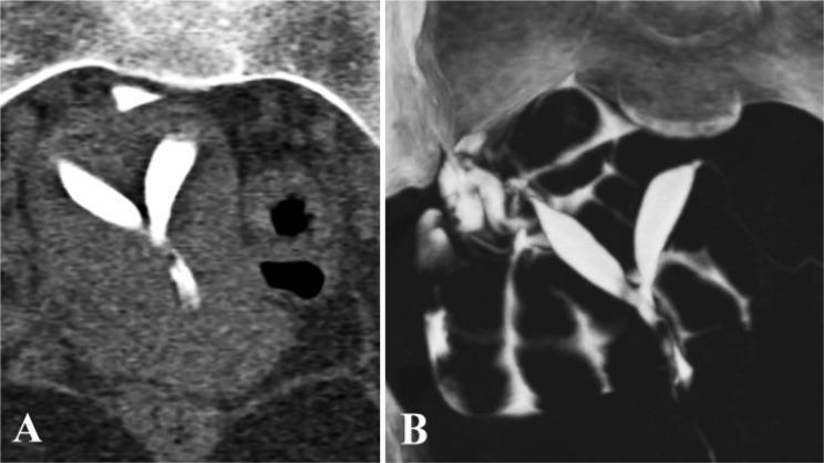
3D imaging of septate uterus: two spindle-shaped cavities separated by a midline incomplete septum; flat/convex fundal contour distinguishes septate from bicornuate uterus
2. Bicornuate Uterus
- Results from incomplete fusion of the two Mullerian ducts (defect of lateral fusion)
- Produces two uterine horns with a single cervix (partial) or two cervices (complete)
- Associated with second-trimester losses and preterm labor; weaker association with first-trimester RPL than septate uterus
- Diagnosis: 3D ultrasound or MRI (concave fundal contour on external surface)
- Surgical correction: Strassman metroplasty
3. Unicornuate Uterus
- Results from failure of one Mullerian duct to develop fully
- Only one functional uterine horn forms
- Associated with RPL, preterm labor, malpresentation, and intrauterine growth restriction
- May have a rudimentary (non-communicating or communicating) contralateral horn, which can itself be the site of ectopic pregnancy
4. Didelphys Uterus (Uterus Didelphys)
- Complete failure of Mullerian duct fusion
- Two separate uterine bodies, two cervices, and often a longitudinal vaginal septum
- Paradoxically, carries a better reproductive prognosis than other anomalies
- Associated with late-trimester losses and preterm labor
5. Arcuate Uterus
- Minimal midline indentation of the uterine fundus; considered a normal variant by most authorities
- Reproductive outcomes are similar to women with a normal uterine cavity
- Studies that exclude arcuate uterus show lower rates of Mullerian anomalies in RPL patients (3-7% vs. 6.3-16.7%) - Creasy & Resnik, p. 981
6. DES (Diethylstilbestrol) Exposure - Congenital
- Prenatal exposure to maternally ingested DES causes complex congenital uterine, cervical, and vaginal changes
- The most common anomaly is uterine hypoplasia ("T-shaped uterus")
- Contributes to first- and second-trimester losses, cervical incompetence, and premature labor
- Less common since DES was withdrawn from use (1971 in the USA), but daughters of exposed women remain at risk
7. Congenital Cervical Insufficiency
- Congenital weakness of the internal cervical os leads to painless dilation of the cervix in the second trimester
- Presents as characteristic second-trimester losses
- Can be primary (congenital) or secondary to prior cervical trauma
- Treatment: Prophylactic cervical cerclage (McDonald or Shirodkar technique)
8. Congenital Anomalies of the Uterine Arteries
- Aberrant arterial supply can adversely alter blood flow to the implanted blastocyst and developing placenta, contributing to early pregnancy loss - Berek & Novak's Gynecology, p. 1803
Molecular/Genetic Basis of Congenital Anomalies
HOX gene mutations play a role. HOXA10 expression is critical for implantation; HOXA10 and HOXA11 gene mutants show abnormal uterine/stromal development and reduced endometrial receptivity. Dysregulation of endometrial stromal tissue and epidermal growth factor receptor expression may explain why even anomalies with relatively normal cavity volumes predispose to pregnancy loss. - Creasy & Resnik, p. 981
B. Acquired Anatomical Abnormalities
1. Intrauterine Adhesions (Asherman Syndrome)
- Fibrotic bands and synechiae within the uterine cavity, most commonly following curettage after pregnancy (postabortal, postpartum), intrauterine infection, or surgery
- Endometrium overlying synechiae is inadequately vascularized, impairing implantation and placentation
- Spectrum from minor adhesions to complete obliteration of the cavity
- Diagnosis: Hysteroscopy (gold standard), HSG, sonohysterography
- Treatment: Hysteroscopic adhesiolysis
2. Uterine Leiomyomas (Fibroids)
Three locations, each with different clinical significance:
| Type | RPL Association |
|---|---|
| Submucosal (intracavitary) | Strongest association - distorts cavity, impairs implantation and placentation |
| Intramural (≥5 cm, distorting cavity) | Probable association; large intramural fibroids (≥5 cm) associated with pregnancy loss, removal may improve outcomes |
| Subserosal | Unlikely to affect RPL |
Note: ASRM and ESHRE both acknowledge the association between submucosal fibroids and RPL is controversial, with insufficient high-quality evidence. - Creasy & Resnik, p. 982
Mechanism: Endometrium overlying a submucous fibroid may be inadequately vascularized, promoting abnormal placentation.
3. Endometrial Polyps
- Focal overgrowths of endometrial glands and stroma
- Hypothetically impair implantation by occupying or distorting the endometrial cavity
- Association with RPL is controversial; evidence is limited
- Diagnosis: Sonohysterography, hysteroscopy
- Treatment: Hysteroscopic polypectomy
4. Chronic Endometritis
- Histopathologic finding of plasma cells in the endometrial stroma
- Identified by CD138 (syndecan-1) immunohistochemistry or H&E staining
- Possible (though controversial) cause of RPL
- Treated with antibiotics (most commonly doxycycline)
C. Cervical Factors
Cervical Incompetence (Cervical Insufficiency)
- Structural weakness of the cervix leading to painless, progressive dilation typically at 14-24 weeks gestation
- Classic presentation: painless second-trimester loss without preceding contractions
- Causes include prior cervical trauma (LEEP, cone biopsy, forceful dilation), congenital weakness, and DES exposure
- Diagnosis: Clinical (history-based); transvaginal ultrasound showing short cervix (<25 mm) or funneling
- Treatment: Cerclage (history-indicated, ultrasound-indicated, or exam-indicated)
D. Diagnostic Evaluation (ASRM/ESHRE Guidelines)
| Investigation | Preferred Modality |
|---|---|
| Uterine cavity assessment | 3D transvaginal ultrasound (preferred by ESHRE); can distinguish septate from bicornuate uterus |
| Alternative imaging | Sonohysterography, HSG |
| Complex anomalies | MRI |
| Definitive diagnosis & treatment | Hysteroscopy |
E. Prognosis After Correction
Women with corrected anatomical anomalies can expect a 60-90% subsequent live birth rate, with some series reporting >90% success after septum resection. - Berek & Novak's Gynecology, p. 1802
Summary Table
| Category | Condition | Key Feature | Trimester of Loss |
|---|---|---|---|
| Congenital | Septate uterus | Strongest RPL association; 60% miscarriage risk | 1st and 2nd |
| Congenital | Bicornuate uterus | Incomplete lateral fusion | 2nd, preterm |
| Congenital | Unicornuate uterus | One Mullerian duct fails | Any |
| Congenital | Didelphys uterus | Complete failure of fusion | 2nd, preterm |
| Congenital | DES hypoplasia | T-shaped uterus | 1st and 2nd |
| Congenital | Cervical insufficiency | Painless dilation | 2nd trimester |
| Acquired | Asherman syndrome | Post-curettage adhesions | 1st and 2nd |
| Acquired | Submucosal fibroid | Distorts cavity | 1st |
| Acquired | Endometrial polyp | Cavity distortion | 1st |
| Acquired | Chronic endometritis | Plasma cells in stroma | 1st |
Sources:
- Berek & Novak's Gynecology, Ch. 33 (Recurrent Pregnancy Loss), pp. 1801-1803
- Creasy & Resnik's Maternal-Fetal Medicine, Ch. 41, pp. 981-982
Recent meta-analytic evidence confirms obstetric complications from congenital Mullerian anomalies, including increased RPL risk - Panagiotopoulos et al., Obstet Gynecol 2022 (PMID 34856567).
Recurrent pregnancy loss
Asking for Preferences
What aspect of recurrent pregnancy loss would you like to focus on?
Everything
Quick answer: Recurrent pregnancy loss (RPL) is defined as 2 or more consecutive pregnancy losses. Its causes span genetic, anatomical, endocrine, immunological (especially antiphospholipid syndrome), thrombophilic, and unexplained categories. Workup includes karyotyping, uterine imaging, antiphospholipid antibodies, thyroid function, and glucose screening. Management is etiology-directed; many couples still achieve live birth even without a found cause.
Searching sources...
Finding Sources
Searching PubMed
"recurrent pregnancy loss" AND management
Searching Images
recurrent pregnancy loss causes etiology diagram
Now let me read the full RPL chapter from both major textbooks in parallel.
Reading File
Reading File
Excellent content. Now let me read the etiology continuation from Berek & Novak and the treatment/management sections from Creasy & Resnik in parallel.
Reading File
Reading File
Now let me get the treatment/management sections and the APS/thrombophilia/endocrine sections.
Reading File
Reading File
Now let me get the treatment/therapy section from Berek & Novak and APS/immunologic section from Creasy & Resnik.
Reading File
Reading File
Excellent. Let me get the APS/hematologic treatment section and the prognosis/unexplained RPL sections.
Reading File
Reading File
Now let me get the prognosis, APS treatment, and unexplained RPL sections.
Reading File
Reading File
I now have comprehensive content across all major sections. Let me fetch the RPL etiological factors diagram image and compile the full answer.
Loading Image
I now have everything needed for a comprehensive answer. Let me compile it.# Recurrent Pregnancy Loss (RPL) - Comprehensive Review
1. Definition
Traditional definition (ACOG): Three or more clinically recognized pregnancy losses before 20 weeks gestation, occurring in approximately 1 in 300 pregnancies (<1% of women).
Current definition (ASRM & ESHRE): Two or more pregnancy losses - adopted to facilitate earlier investigation, research, and psychological support.
Miscarriage affects 15-25% of clinically recognized pregnancies. Using sensitive hCG assays, actual loss after implantation is ~31%. After 2 losses, ~1% of pregnant women qualify for investigation. - Berek & Novak's Gynecology, p. 1792
2. Etiology - Overview
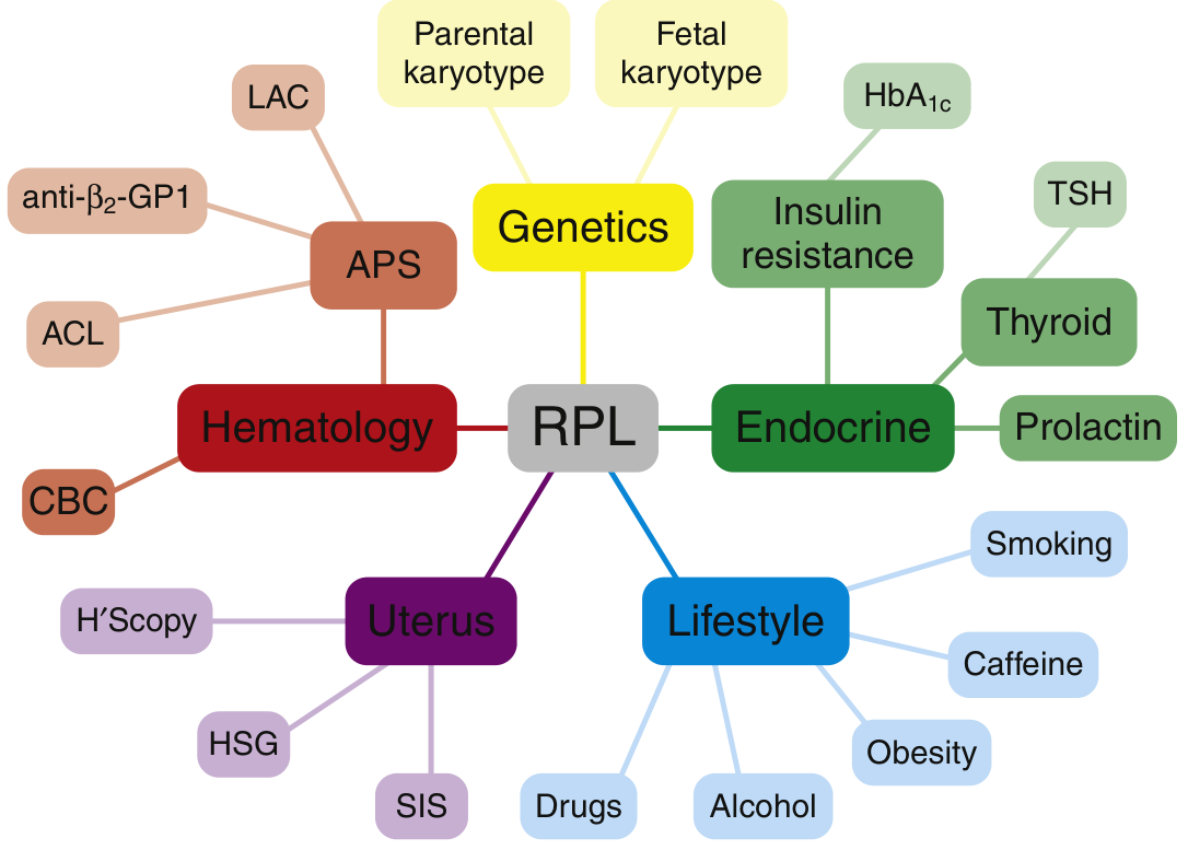
Figure: Factors contributing to RPL - Genetics, APS/Hematology, Endocrine, Uterine, and Lifestyle factors with corresponding investigations (from Creasy & Resnik's MFM)
| Cause | Approximate Proportion |
|---|---|
| Fetal aneuploidy (de novo) | 50-80% of individual losses |
| Unexplained | ~50% of couples |
| Antiphospholipid syndrome (APS) | ~15% |
| Anatomic abnormalities | 10-12% |
| Other identifiable causes | ~10% |
| Parental chromosomal abnormalities | 2-5% |
3. Etiology - Detailed
A. Genetic Factors
Fetal Aneuploidy (Most Common Cause of Individual Losses)
- At least 60% of preclinical and early clinical losses are from de novo fetal aneuploidy
- In women ≥35 years with RPL, chromosomal abnormalities account for >80% of losses
- Most common: autosomal trisomy (60-70%), monosomy X (~10-20%), polyploidy (~10-20%)
- Modern chromosomal microarray analysis (CMA) finds genetic abnormalities in 53.7% of miscarriage tissue samples
- Anembryonic pregnancies (blighted ova) are the typical presentation
- De novo aneuploidy is a confounding factor in RPL research - must be documented
Parental Chromosomal Rearrangements (3-5% of RPL)
- Types: balanced reciprocal translocations (most common), Robertsonian translocations, inversions, insertions, mosaicism
- Diagnosis: Metaphase karyotype with G-banding for both partners (NOT microarray - cannot detect balanced rearrangements)
- Robertsonian translocation: fusion of long arms of acrocentric chromosomes (13-15); unbalanced forms cause trisomies/monosomies → miscarriage
- ESHRE: parental karyotyping not routinely recommended unless history of genetically abnormal fetus/newborn; ASRM recommends it
- All patients with balanced rearrangements should receive genetic counseling
B. Anatomic Abnormalities (10-12% of RPL)
Congenital Mullerian Anomalies
Result from failure of Mullerian duct development, abnormal fusion, or failure of septal absorption.
| Anomaly | Mechanism | RPL Association |
|---|---|---|
| Septate uterus | Incomplete septal resorption | Strongest - up to 60% miscarriage risk; 2nd trimester losses most common |
| Bicornuate uterus | Incomplete lateral fusion | 2nd trimester loss, preterm labor |
| Unicornuate uterus | One Mullerian duct fails to develop | RPL, preterm labor, IUGR |
| Didelphys uterus | Complete failure of fusion | Better prognosis than others; late losses |
| DES hypoplasia (T-shaped) | Prenatal DES exposure | 1st and 2nd trimester loss, cervical incompetence |
| Arcuate uterus | Minor fundal indentation | Normal variant - outcomes similar to normal uterus |
Molecular basis: HOXA10 and HOXA11 gene mutations disrupt endometrial stromal development and implantation - Creasy & Resnik, p. 981
Acquired Uterine Anomalies
- Asherman syndrome (intrauterine adhesions): Post-curettage/infection; poorly vascularized endometrium over synechiae impairs placentation
- Submucosal fibroids: Distort cavity; strongest fibroid-RPL link; evidence controversial (ASRM, ESHRE)
- Intramural fibroids ≥5 cm: Associated with pregnancy loss; removal may improve outcomes
- Endometrial polyps: Controversial association
- Chronic endometritis: Plasma cells in stroma (CD138+); possible RPL cause
Cervical Factors
- Cervical insufficiency: Painless, progressive dilation at 14-24 weeks; 2nd trimester loss
- Causes: congenital weakness, prior cervical trauma (LEEP, cone biopsy), DES exposure
C. Immunologic Factors
Antiphospholipid Syndrome (APS) - ~15% of RPL; only proven immune cause
Diagnostic criteria (Sapporo/revised): At least one clinical + one laboratory criterion, antibodies present on ≥2 occasions ≥12 weeks apart.
Clinical (obstetric) criteria:
- ≥3 unexplained consecutive losses before 10th week
- ≥1 unexplained fetal death of morphologically normal fetus ≥10 weeks
- ≥1 premature birth of morphologically normal neonate <34 weeks from placental insufficiency/preeclampsia/eclampsia
Laboratory criteria (moderate-to-high titer):
- Lupus anticoagulant (LA)
- Anticardiolipin (aCL) IgG and/or IgM
- Anti-beta2-glycoprotein I (aβ2GPI) IgG and/or IgM
Mechanism: aPL antibodies activate complement, bind trophoblast phospholipids, cause placental thrombosis and infarction.
Cellular Immune Mechanisms (Under Investigation)
- TH1/TH2 imbalance: Successful implantation requires a TH2-dominant environment; shift toward TH1 inflammatory responses at the maternal-fetal interface may cause RPL
- Elevated TNF-α in early pregnancy correlates with RPL
- NK cell dysregulation: Investigated but not proven causative
- MHC class II genotypes may influence susceptibility
- Testing for TH1/TH2 cytokine dysregulation and NK cells in peripheral blood: NOT recommended (unproven clinical benefit)
D. Endocrine Abnormalities
| Condition | Association with RPL | Management |
|---|---|---|
| Thyroid disease | Hypothyroidism and anti-TPO antibodies associated with RPL, even subclinically | Levothyroxine for subclinical hypothyroidism; target trimester-specific TSH ranges |
| PCOS | ~50% miscarriage rate in PCOS (double general population); polycystic ovaries in most RPL on USS | Address comorbidities; insulin sensitizers controversial |
| Insulin resistance | 27% of RPL women vs 9.5% controls have insulin resistance | Metformin may reduce miscarriage rate in insulin-resistant women (55% → 15%) but insufficient trial evidence |
| Hyperprolactinemia | Small but significant association | Bromocriptine may increase live birth rate by 30% in hyperprolactinemic RPL women |
| Luteal phase deficiency | Controversial; no reproducible definition or standard | ASRM and ESHRE: screening/treatment NOT recommended |
| Decreased ovarian reserve | Low AMH/day-3 FSH predicts higher aneuploid embryos and miscarriage | Ovarian reserve testing useful; treatment individualized |
E. Thrombophilias (Inherited)
- Factor V Leiden (FVL): OR 2.4 for RPL; OR 10.7 for ≥2 second/third trimester losses; heterozygous FVL alone may not significantly increase first-trimester loss
- Prothrombin G20210A: Possible weak association
- MTHFR mutations (C677T, A1298C): Most experts now consider NOT associated with RPL
- Protein C, S, antithrombin deficiencies: Insufficient data
IMPORTANT: ACOG, ASRM, and ESHRE do NOT recommend routine thrombophilia screening for RPL unless there is a personal/family history of thrombosis. Evidence for treatment to prevent miscarriage is lacking (outside APS). - Berek & Novak, p. 1802
F. Male Factors
- Standard semen analysis parameters (count, motility, morphology) do NOT predict RPL
- Sperm DNA fragmentation: Increased fragmentation index correlates with RPL; 40% of men with normal semen analysis but partner RPL had sperm aneuploidy (by FISH)
- Mechanism: Likely contributes to embryo aneuploidy
- No established role in routine evaluation yet; more research needed
G. Infection
- Certain uterine infections (mycoplasma, ureaplasma, chlamydia) may contribute
- No single infectious agent is consistently proven to cause RPL
- Cervical cultures may be considered but not standard
H. Environmental and Lifestyle Factors
| Factor | Risk |
|---|---|
| Alcohol ≥5 drinks/week | Increased 1st trimester loss (8.9% vs 1.4%) |
| Heavy smoking >10 cigarettes/day | RR 1.2-3.4 for pregnancy loss; 25% rate at >20 cig/day |
| Second-hand smoke | 11% increased miscarriage rate |
| Cocaine use | Up to 38% early pregnancy loss |
| Methamphetamine | Higher loss rate (2.1 vs 1.2 lifetime average) |
| Obesity (BMI >30) | 31% miscarriage rate vs 18% for normal BMI; higher euploid miscarriage rate |
| Caffeine | Limit to <200 mg/day (most societies); association debated |
| Advanced maternal age | Major independent risk factor for aneuploidy |
4. Preconception Evaluation
Recommended Testing (ASRM/ESHRE Guidelines)
| Test | Purpose | Guideline Status |
|---|---|---|
| Parental karyotype (both partners) - metaphase G-banding | Detect balanced translocations | ASRM: Recommended; ESHRE: Not routine (individual risk) |
| Products of conception - CMA | Confirm aneuploidy; provide closure | ASRM: Recommended at each subsequent loss |
| Uterine cavity assessment | Detect anatomical causes | Both: Recommended; ESHRE prefers 3D TVS (can distinguish septate from bicornuate) |
| Antiphospholipid antibodies (LA, aCL IgG/IgM, aβ2GPI IgG/IgM) | Diagnose APS | Both: Recommended |
| TSH ± TPO antibodies | Thyroid disease | Both: Recommended (ESHRE adds TPO Ab) |
| HbA1c | Diabetes screening | ASRM: Recommended |
| Prolactin | Hyperprolactinemia | ASRM: Routine; ESHRE: Only if symptomatic |
Uterine Imaging Options
- 3D transvaginal ultrasound - preferred (can distinguish septate from bicornuate); noninvasive
- Sonohysterography (SIS) - best for polyps and adhesions
- Hysterosalpingography (HSG) - assesses tubal patency and cavity
- MRI - complex anomalies; only if 3D USS unavailable per ESHRE
- Hysteroscopy - gold standard diagnostic and therapeutic; proceeds after imaging suggests correctable anomaly
Tests with Unproven Utility (Under Investigation)
- Ovarian reserve (day-3 FSH, estradiol, AMH) - gaining support
- Anti-thyroid peroxidase antibodies (rapidly gaining support)
- Endometrial biopsy for chronic endometritis
Tests NOT Recommended
- Thrombophilia panel (FVL, prothrombin mutation, protein C/S, antithrombin, MTHFR, homocysteine) - unless personal/family VTE history
- TH1/TH2 cytokine testing
- NK cell testing (peripheral blood or endometrial)
- Antinuclear antibodies (ANA)
5. Management
A. Genetic Abnormalities
- Parental balanced translocation: Genetic counseling + options:
- Natural conception (expectant - still high chance of live birth)
- PGT-SR (preimplantation genetic testing for structural rearrangements) via IVF - selects embryos with balanced/normal chromosomes; reduces miscarriage rate but still investigational for some indications
- Donor gametes
- De novo aneuploidy: Expectant - favorable overall prognosis
B. Anatomic Anomalies
- Uterine septum: Hysteroscopic metroplasty - live birth rate improves from 61.5% → 81.3% with surgery (though no RCT; Cochrane found no randomized trials of metroplasty vs expectant management)
- Asherman syndrome: Hysteroscopic adhesiolysis - 71% live birth rate post-lysis
- Submucosal fibroids: Hysteroscopic myomectomy; significant reduction in subsequent miscarriage in one study
- Cervical insufficiency: Prophylactic cerclage (McDonald or Shirodkar); history-indicated, ultrasound-indicated, or exam-indicated
C. APS - The Only Proven Immune/Thrombophilic Treatment
Standard treatment for APS with RPL:
- Low-dose aspirin (LDA) 81 mg/day + Low molecular weight heparin (LMWH) (e.g., enoxaparin 40 mg SC daily) OR unfractionated heparin (UFH), started with positive pregnancy test, continued through pregnancy
- Live birth rate with treatment: 70-90% (vs ~30% without treatment in some series)
- Aspirin alone is NOT effective for APS-related RPL
For inherited thrombophilias without APS:
- Anticoagulation is based on individualized VTE risk, NOT miscarriage prevention (no RCT evidence supports anticoagulation to prevent miscarriage in non-APS thrombophilia)
D. Endocrine Management
- Thyroid disease: Levothyroxine to achieve trimester-specific TSH targets
- Hyperprolactinemia: Bromocriptine (dopamine agonist) - 30% increase in live birth rate
- PCOS/Insulin resistance: Metformin may be beneficial in insulin-resistant RPL women, but insufficient evidence for routine use
- Luteal phase defect: Progesterone supplementation remains controversial:
- Cochrane 2018: Possible reduction in miscarriage (RR 0.73)
- Large RCT (PRISM-type): Treatment with vaginal progesterone at positive pregnancy test did NOT improve outcomes in all RPL women
- Current evidence supports: Vaginal progesterone 400 mg twice daily in women with RPL + first-trimester bleeding in the current pregnancy (PROMISE trial reanalysis)
E. Progesterone Supplementation - Nuanced Position
- Vaginal micronized progesterone (Utrogestan) 400 mg twice daily or dydrogesterone 10 mg twice daily
- Most benefit: Women with RPL + first-trimester vaginal bleeding in current pregnancy
- NOT shown to benefit all RPL women as routine supplementation
- Earlier start (luteal phase) may be more beneficial - still under study
F. Immunologic Treatments
| Treatment | Evidence | Recommendation |
|---|---|---|
| Heparin + aspirin for APS | Strong - 70-90% live birth rate | Recommended |
| IVIg for unexplained RPL | Multiple small RCTs negative; Cochrane: no benefit | Not recommended |
| Corticosteroids (prednisolone) | Insufficient evidence; harms include preterm labor, GDM | Not routinely recommended |
| Leukocyte immunization therapy | Cochrane: no significant benefit; potential harm | Not recommended |
G. Anticoagulation for Non-APS
- Large RCTs show no benefit of aspirin alone or aspirin + heparin in unexplained RPL
- Aspirin in early pregnancy may even increase isolated spontaneous loss risk (conflicting data)
H. Lifestyle Modifications (All RPL patients)
- Smoking cessation (complete)
- Abstinence from alcohol during conception and pregnancy
- Avoid illicit substances
- Achieve healthy BMI (both underweight and obesity increase risk)
- Limit caffeine <200 mg/day
- Folic acid supplementation (at least 0.4-1.0 mg/day)
I. Psychological Support
- Risk of major depression is >2-fold increased after pregnancy loss
- Empathetic communication; acknowledge suffering
- Refer to support groups and counselors
- Self-help: meditation, yoga, exercise, biofeedback
- Remind patients that the majority will achieve a live birth
6. Postconception Management (Once Pregnant)
- Serial serum β-hCG monitoring
- Early transvaginal ultrasound (documentation of fetal cardiac activity = prognostic; live birth rate ~77% after cardiac activity confirmed at 5-6 weeks in unexplained RPL)
- Chromosome analysis of products of conception from any subsequent loss (chromosomal microarray preferred)
- Psychological support throughout
7. Prognosis
| Scenario | Live Birth Rate |
|---|---|
| After 4 prior losses (no treatment, no cause found) | Up to 60% |
| Corrected anatomical anomalies | 60-90% (some series >90% post-septum resection) |
| Corrected endocrine abnormality | >90% |
| APS on heparin + aspirin treatment | 70-90% |
| Parental cytogenetic etiology | 20-80% (highly variable by specific rearrangement) |
Key take-home: Even after several losses, a patient is more likely to carry her next pregnancy to term than to miscarry. Reminding patients of this is an invaluable therapeutic tool. - Berek & Novak, p. 1835
8. Unexplained RPL (~50% of couples)
- No identifiable cause found despite full workup
- Most likely reflects high background rate of de novo aneuploidy
- Management: Reassurance, supportive monitoring, lifestyle optimization, and psychological support
- No proven pharmacological intervention improves outcomes over placebo in well-designed RCTs
- Tender loving care (TLC) - supportive early pregnancy monitoring - has been shown to improve live birth rates in some studies (possibly up to 80% in supportive care clinics)
Sources:
- Berek & Novak's Gynecology, Ch. 33 (Recurrent Pregnancy Loss), pp. 1791-1835
- Creasy & Resnik's Maternal-Fetal Medicine, Ch. 41, pp. 978-988
- Australasian RPL Clinical Management Guideline 2024 Part I (PMID 38934264)
- Australasian RPL Clinical Management Guideline 2024 Part II (PMID 38934293)
- SOGC Guideline No. 464: RPL 2025 (PMID 41176277)
Apla positive
Finding Sources
Finding Sources
Searching PubMed
"antiphospholipid syndrome" AND pregnancy
Given the context (following the RPL discussion), this is about a pregnant patient with positive antiphospholipid antibodies (obstetric APS). Let me read the most relevant sections from Creasy & Resnik and Rheumatology text for management detail.
Reading File
Reading File
Let me get the remaining detail from the treatment table and Creasy & Resnik continuation.
Reading File
Reading File
I now have comprehensive detail. Let me compile the answer on managing an aPL (antiphospholipid antibody) positive patient.
Antiphospholipid Antibody (aPL) Positive - Diagnosis & Management
Given the context of our RPL discussion, "aPL positive" typically refers to a patient testing positive for one or more antiphospholipid antibodies. Here is a complete framework for interpreting and managing this result.
1. What Counts as "aPL Positive"?
Three antibodies are recognized under international consensus (Sydney/revised Sapporo) criteria:
| Antibody | Threshold for Significance |
|---|---|
| Lupus anticoagulant (LA) | Positive (functional clotting assay) - the strongest predictor of poor pregnancy outcome |
| Anticardiolipin (aCL) IgG/IgM | Moderate-to-high titer |
| Anti-β2-glycoprotein I (aβ2GPI) IgG/IgM | Moderate-to-high titer |
Critical rule: A single positive test is NOT diagnostic. Antibodies must be confirmed on two or more occasions at least 12 weeks apart to distinguish true APS from a transient positive (e.g., from infection or medication). - Creasy & Resnik's Maternal-Fetal Medicine, p. 984
2. Does aPL Positivity = Antiphospholipid Syndrome (APS)?
Not necessarily. Definite APS requires at least one laboratory criterion AND one clinical criterion:
Clinical (obstetric) criteria - any one of:
- ≥3 unexplained consecutive losses before 10 weeks gestation
- ≥1 unexplained death of a morphologically normal fetus ≥10 weeks gestation
- ≥1 premature birth before 34 weeks due to preeclampsia, eclampsia, or placental insufficiency
Clinical (thrombotic) criteria:
- Prior arterial or venous thrombosis
If a patient is aPL-positive but has no thrombosis history and no APS-defining obstetric history, she is simply an asymptomatic carrier - a distinct, lower-risk category.
3. Management by Risk Category
This is the key clinical decision point - management differs substantially by category:
| Category | Definition | Antenatal Management | Postpartum |
|---|---|---|---|
| Labs only (asymptomatic carrier) | aPL positive, no thrombosis, no APS-defining obstetric history | Consider low-dose aspirin (LDA) based on thrombotic risk profile; some clinicians treat, others don't | LDA ± prophylactic LMWH for 6 weeks, based on risk |
| Obstetric APS | aPL positive + meets obstetric clinical criteria, no thrombosis history | LDA + prophylactic-dose LMWH | LDA + prophylactic LMWH for 6 weeks |
| Thrombotic APS | aPL positive + prior arterial/venous thrombosis (± obstetric criteria) | LDA + therapeutic-dose LMWH | Warfarin indefinitely (lifelong anticoagulation) |
(Table adapted from Rheumatology, 2-Volume Set, p. 1297)
Key practical points:
- LDA alone is NOT sufficient for confirmed obstetric or thrombotic APS - it must be combined with heparin/LMWH
- Heparin/LMWH is generally started once pregnancy is confirmed and continued throughout gestation
- Anticoagulation continues at least 6 weeks postpartum given ongoing thrombotic risk in the puerperium
- With appropriate treatment, live birth rates of 50-90% have been reported (compared to much lower rates untreated)
4. Why aPL Matters Beyond Miscarriage
Antiphospholipid antibodies are associated with:
- Placental insufficiency
- Oligohydramnios
- Fetal growth restriction (IUGR)
- Fetal death
- Preeclampsia (often early-onset/severe)
- Maternal thrombosis (arterial or venous)
Importantly, the mother can be completely asymptomatic even while placental compromise is developing - this is why monitoring (growth scans, Dopplers, BP surveillance) matters even in "labs only" carriers being watched closely.
5. If Standard Treatment Fails (Refractory APS)
For women who lose the pregnancy or develop thrombosis despite LDA + heparin:
- Hydroxychloroquine - emerging evidence for benefit in improving outcomes, though not yet confirmed in prospective RCTs
- IVIg, corticosteroids, and plasmapheresis have been proposed but are not recommended by ACR guidelines due to insufficient evidence and side-effect burden
- These refractory/high-risk cases should be managed jointly with maternal-fetal medicine, hematology, and rheumatology
A note of caution from the literature: much of the historical trial evidence combining heparin + aspirin vs aspirin alone is heterogeneous and methodologically inconsistent - many trial subjects would not meet current strict APS criteria, so treatment effect sizes should be interpreted cautiously. - Creasy & Resnik, p. 984
6. If aPL Is Positive But Doesn't Meet Criteria
Patients with low-positive or indeterminate titers, or antibodies outside the three internationally recognized ones, do not meet definite APS criteria. Management here is genuinely uncertain ("vexing" per the literature) - there's no robust evidence-based protocol, and decisions should be individualized with specialist input.
7. Bottom Line - What to Ask Next
To give a more precise recommendation for your specific patient, it would help to know:
Asking for Preferences
What best describes this aPL-positive patient's situation?
This confirms Obstetric APS category (assuming the antibodies have been confirmed on two occasions ≥12 weeks apart and meet the obstetric clinical criteria, i.e., ≥3 losses <10 weeks, or ≥1 loss ≥10 weeks, or ≥1 preterm delivery <34 weeks from preeclampsia/placental insufficiency).
Management Plan for This Patient
Antenatal (throughout pregnancy):
- Low-dose aspirin (81 mg/day) - start as soon as pregnancy confirmed (ideally preconception or very early)
- PLUS prophylactic-dose LMWH (e.g., enoxaparin 40 mg subcutaneously once daily) - started once intrauterine pregnancy is confirmed
- Aspirin alone is not adequate for this category - the combination is what improves live birth rates (70-90% reported with treatment vs much lower untreated)
Monitoring during pregnancy:
- Serial growth ultrasounds (risk of fetal growth restriction from placental insufficiency)
- Umbilical artery Doppler studies, especially third trimester
- Close blood pressure surveillance and preeclampsia screening (aPL carries elevated preeclampsia risk, often early-onset)
- Early confirmation of fetal cardiac activity - reassuring prognostic sign (studies show ~77-86% of aPL-positive RPL patients who reach fetal cardiac activity on ultrasound go on to have that pregnancy continue past the point of prior losses)
Postpartum:
- Continue LDA + prophylactic LMWH for at least 6 weeks after delivery - thrombotic risk is highest in the puerperium even without prior thrombosis history
If she loses this pregnancy or shows breakthrough placental compromise despite treatment (refractory APS):
- Consider adding hydroxychloroquine (emerging evidence, not yet RCT-confirmed)
- IVIg, corticosteroids, plasmapheresis are proposed but not recommended by ACR due to weak evidence and side effects
- Refer for joint management with maternal-fetal medicine, hematology, and rheumatology
Important caveat: Confirm this patient truly meets criteria - a single positive lupus anticoagulant, aCL, or aβ2GPI test is not diagnostic. If retesting at ≥12 weeks was not yet done, this should happen before committing to long-term anticoagulation, since the diagnosis materially changes management (and heparin/LMWH is not without bleeding/HIT risk).
Sources: Creasy & Resnik's Maternal-Fetal Medicine, pp. 984; Rheumatology 2-Volume Set, pp. 1296-1297 (Table 145.4)
This is a shared conversation. Sign in to Orris to start your own chat.