
Humerus notes
Reading File
The humerus is the long bone of the upper arm, articulating with the scapula at the shoulder (glenohumeral joint) and with the radius/ulna at the elbow. Key structures include the head, anatomical neck, surgical neck (common fracture site), greater/lesser tubercles, bicipital groove, shaft, and distal condyles. The radial nerve spirals in the radial groove of the shaft.
Searching sources...
Finding Sources
Finding Sources
Good - found an anatomy atlas with a humerus diagram. Let me read the key anatomy sections in parallel.
Reading File
Reading File
Finding Sources
Excellent. Now let me read the Gray's Anatomy for Students sections on humerus in detail.
Reading File
Reading File
Reading File
Reading File
Now let me get the proximal humerus section from Gray's and fetch the key humerus diagram images.
Finding Sources
Loading Image
Loading Image
I now have all the core material needed. Let me also get the proximal humerus section from Gray's to cover the head, tubercles, and bicipital groove.
Finding Sources
Reading File
Reading File
Loading Image
I now have rich, detailed content from Gray's Anatomy for Students and the THIEME Atlas. Here are comprehensive humerus notes:
The Humerus
The humerus is the single long bone of the arm, articulating proximally with the scapula at the glenohumeral joint and distally with the radius and ulna at the elbow joint.
Proximal End
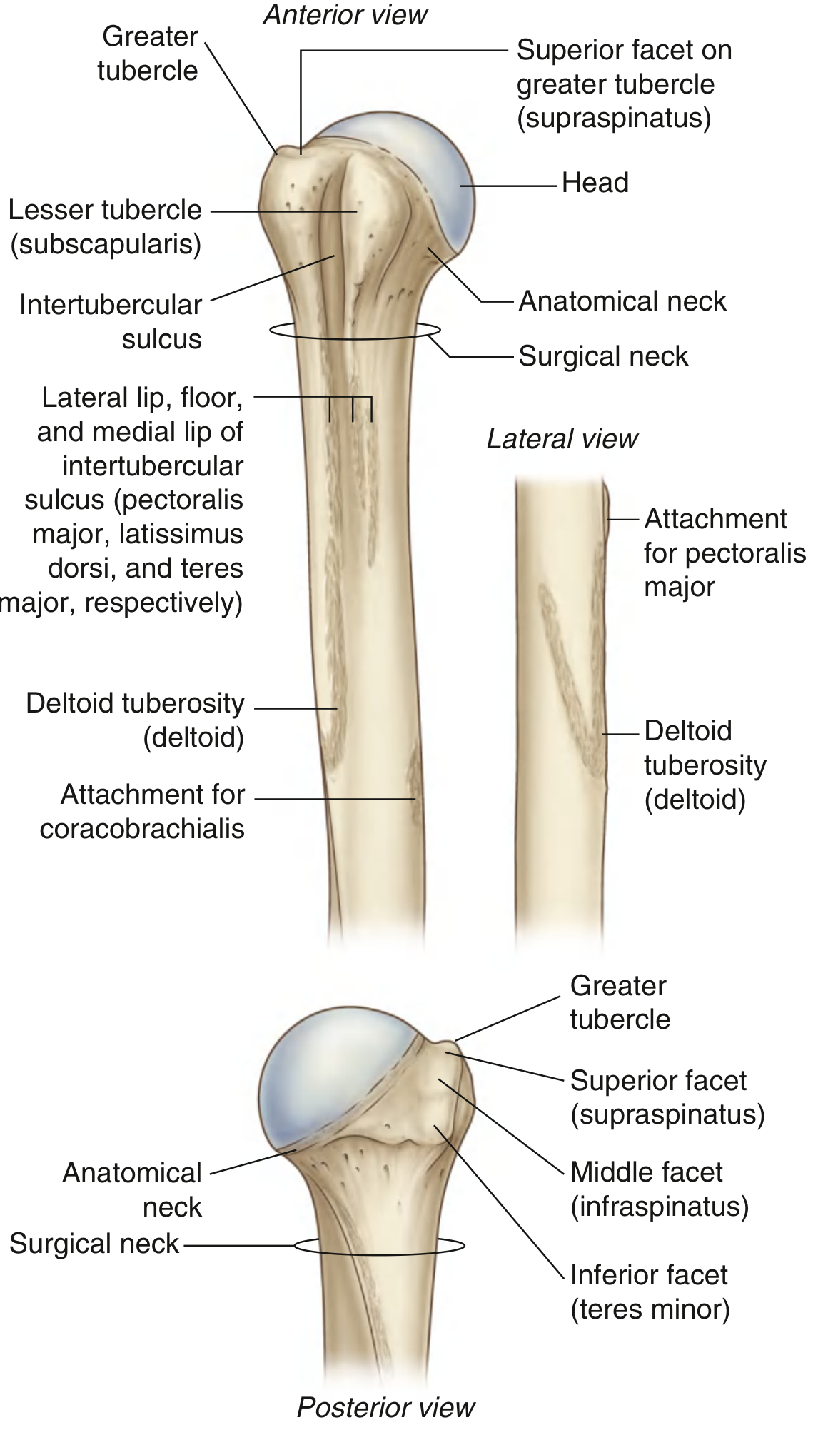
(Fig. 7.22, Gray's Anatomy for Students)
Head
- Half-spherical; projects medially and superiorly to articulate with the glenoid cavity of the scapula.
- Covered in hyaline cartilage.
Anatomical Neck
- Very short constriction immediately distal to the head, between the head and the tubercles laterally, and between the head and the shaft medially.
- Fractures here are rare because the bone is thick at this oblique angle.
Greater Tubercle (lateral)
Three facets on its superior and posterior surface - the "SIT" mnemonic for rotator cuff insertions:
| Facet | Muscle |
|---|---|
| Superior | Supraspinatus |
| Middle | Infraspinatus |
| Inferior | Teres minor |
Lesser Tubercle (anterior)
- Single impression for subscapularis attachment.
Intertubercular Sulcus (Bicipital Groove)
- Deep groove separating the lesser and greater tubercles; continues down the proximal shaft.
- Transmits the tendon of the long head of biceps brachii.
- Muscle attachments to the groove lips:
- Lateral lip - pectoralis major
- Floor - latissimus dorsi
- Medial lip - teres major
Surgical Neck
- The horizontal constriction between the expanded proximal humerus (head + tubercles) and the narrower shaft.
- Most common fracture site of the proximal humerus (especially in elderly patients falling on an outstretched arm).
- The axillary nerve and posterior circumflex humeral artery pass immediately posterior to the surgical neck through the quadrangular space - both are at risk in surgical neck fractures.
Shaft
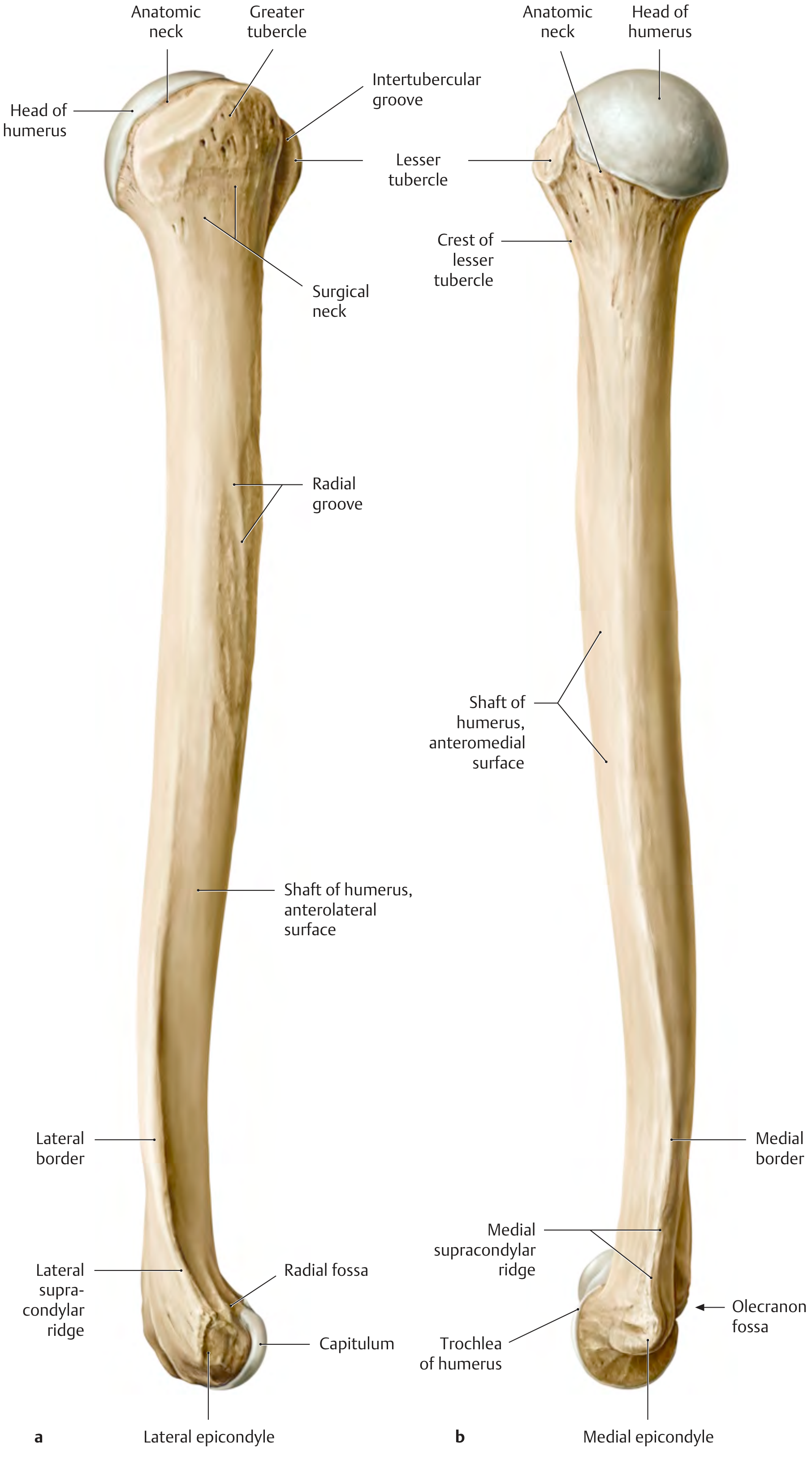
(THIEME Atlas of Anatomy)
Cross-sectional shape
Triangular in cross-section with:
- 3 borders: anterior, lateral, medial
- 3 surfaces: anterolateral, anteromedial, posterior
Key landmarks
| Feature | Location | Significance |
|---|---|---|
| Deltoid tuberosity | Lateral surface, midshaft | Insertion of deltoid muscle |
| Radial groove (spiral groove) | Posterior surface, passes diagonally | Houses the radial nerve and profunda brachii artery |
| Coracobrachialis attachment | Medial surface, mid-shaft | Thin vertical roughening |
Intermuscular septa
Attach to the medial and lateral borders, dividing the arm into anterior (flexor) and posterior (extensor) compartments.
Distal End
The Condyle (two articular parts)
| Part | Shape | Articulates with |
|---|---|---|
| Capitulum | Hemispherical, lateral, projects anteroinferiorly, NOT visible from posterior | Radius (head) |
| Trochlea | Pulley-shaped, medial, extends onto posterior surface, medial edge more pronounced | Ulna |
Epicondyles (non-articular, proximal to the condyle)
- Medial epicondyle - large, most palpable landmark on medial elbow; bears attachment for anterior compartment forearm muscles; the ulnar nerve passes posterior to it (palpable here; "funny bone")
- Lateral epicondyle - less pronounced; attachment for posterior compartment (extensor) forearm muscles
Supracondylar ridges
- Both medial and lateral borders expand distally into medial and lateral supraepicondylar ridges.
- The lateral ridge is more pronounced and roughened for posterior forearm muscle attachments.
Three Fossae (superior to the condyle)
| Fossa | Surface | Relation |
|---|---|---|
| Radial fossa | Anterior, above capitulum (least distinct) | Accommodates radial head in elbow flexion |
| Coronoid fossa | Anterior, above trochlea | Accommodates coronoid process of ulna in flexion |
| Olecranon fossa | Posterior, above trochlea (largest) | Accommodates olecranon in elbow extension |
Nerves Related to the Humerus
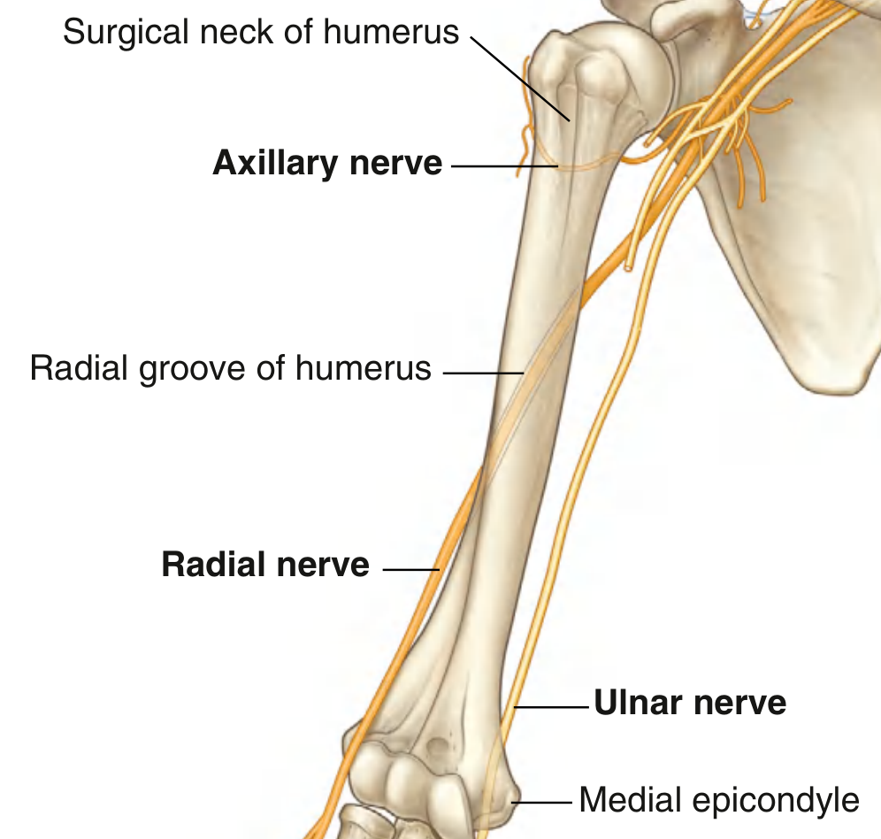
(Fig. 7.17, Gray's Anatomy for Students)
Three major nerves are directly related to the humerus:
| Nerve | Region | At-risk fracture |
|---|---|---|
| Axillary nerve (posterior cord) | Passes around surgical neck posteriorly through quadrangular space | Surgical neck fracture → loss of deltoid function + loss of sensation over "regimental badge" area |
| Radial nerve | Spirals diagonally in the radial groove (middle third of shaft) | Humeral shaft fracture → wrist drop (loss of wrist + finger extension) |
| Ulnar nerve | Passes posterior to the medial epicondyle | Medial epicondyle fracture → loss of intrinsic hand muscles, claw hand |
Blood Supply to the Humeral Head
- Anterior circumflex humeral artery - main supply to humeral head
- Posterior circumflex humeral artery - accompanies axillary nerve through quadrangular space
Fractures at the anatomical neck can disrupt both vessels, leading to avascular necrosis of the humeral head.
Torsion of the Humerus
- The proximal humerus is rotated relative to the distal end (torsion angle ~16° in adults, decreasing from ~60° at birth).
- Assessed by comparing the humeral head axis to the epicondylar axis.
- The decrease in torsion angle with growth correlates with repositioning of the scapulae.
Clinical Correlations
| Injury | Site | Nerve at risk | Deficit |
|---|---|---|---|
| Fall onto outstretched arm (elderly) | Surgical neck | Axillary nerve | Deltoid paralysis, loss of shoulder abduction |
| Humeral shaft fracture | Radial groove | Radial nerve | Wrist drop, loss of finger extension |
| Medial epicondyle fracture | Distal humerus | Ulnar nerve | Claw hand, loss of intrinsic muscles |
| Supracondylar fracture (children) | Distal humerus | Anterior interosseous nerve / brachial artery | "Danger zone" - can injure brachial artery |
Sources: Gray's Anatomy for Students (9780323934237); THIEME Atlas of Anatomy - General Anatomy and Musculoskeletal System (9781626237186); Campbell's Operative Orthopaedics 15th Ed 2026 (9780443117633)
This is a shared conversation. Sign in to Orris to start your own chat.