
Prompt- Create a super aesthetic, handwritten-style revision note page for the topic: dental caries • Make it look like neat exam notes written on a page (top view, clean layout, colorful headings, underlines, boxes). • Keep it crisp, high-yield, and exam-oriented. • Use clear headings and organize content like this (modify if needed for topic): ○ Definition ○ Etiology ○ Pathogenesis (flow/arrow style) ○ Clinical features ○ Radiographic features (if applicable) ○ Histopathology (if applicable) ○ Key features ○ Differential diagnosis ○ Management ○ Complications ○ Key points / last-minute revision box • Add simple diagrams or labeled sketches wherever helpful. • Highlight important keywords. • Include one memory trick / one-line summary
A top-view aesthetic handwritten-style dental exam revision notes page on white lined paper with a clean, colorful, organized layout. The page looks like neat student notes written with colored pens. Layout: TOP HEADER: Big colorful title "DENTAL CARIES" in teal bubble letters, with a small tooth sketch next to it. Below: pink underline. Small pink text: "Multifactorial bacterial disease of teeth" SECTION 1 [purple heading, underlined]: "DEFINITION" Handwritten text: "Dynamic, multifactorial bacterial disease → acid dissolution of calcified tooth tissue (enamel, dentin, cementum)" Orange box highlight: "DEMINERALIZATION > REMINERALIZATION" SECTION 2 [orange heading]: "ETIOLOGY — KEYES TRIAD" A neat Venn diagram with 3 overlapping circles drawn in pen: - Circle 1 (blue): "HOST (Tooth + Saliva)" - Circle 2 (green): "MICROORGANISM (S. mutans ★)" - Circle 3 (red): "SUBSTRATE (Sucrose ↑ risk)" Center overlap: "CARIES" in red Below circles: "+ TIME (4th factor) ← Newbrun addition" SECTION 3 [red heading]: "PATHOGENESIS" Arrow flowchart in handwriting: Sucrose intake → S. mutans metabolizes → Lactic acid produced → pH drops <5.5 → Hydroxyapatite dissolves → Enamel demineralized → Spreads to DEJ → Dentinal tubule invasion → Pulp exposure → Pulpitis/Abscess Key note starred: "★ pH <5.5 = critical pH for enamel dissolution" SECTION 4 [blue heading]: "CLINICAL FEATURES" Bullet points in neat handwriting: • Early: white spot lesion (reversible!) — initial enamel caries • Progressed: brown/black discoloration, cavitation • Sensitivity to sweet, hot, cold • Pain: starts intermittent → becomes spontaneous (pulpitis) • Pit & fissure caries (Class I) vs Smooth surface (Class II-V) Small tooth cross-section sketch labeled: Enamel / DEJ / Dentin / Pulp with arrow showing caries progression SECTION 5 [green heading]: "RADIOGRAPHIC FEATURES" • Bitewing X-ray = GOLD STANDARD for interproximal caries • Appears as RADIOLUCENCY (dark area) • Triangular radiolucency in enamel, widens at DEJ • Periapical X-ray → periapical abscess (dark halo at root tip) Classification box: Class I (occlusal) | II (posterior proximal) | III (anterior proximal) | IV (proximal + incisal) | V (gingival margin) SECTION 6 [purple heading]: "HISTOPATHOLOGY" Enamel caries zones (listed with arrows): → Zone 1: Translucent zone (deepest) — demineralization begins → Zone 2: Dark zone — remineralization attempts → Zone 3: Body of lesion — maximum demineralization → Zone 4: Surface zone — relatively intact Dentin caries zones: → Dead tracts → Turbid zone → Transparent zone → Subtransparent zone → Normal dentin Note: "Bacterial invasion of dentinal tubules → liquefaction foci" SECTION 7 [teal heading]: "KEY POINTS" Stars/bullets: ★ Most cariogenic organism: S. mutans (initiation); Lactobacilli (progression) ★ Most cariogenic sugar: SUCROSE ★ Fluoride: replaces OH⁻ in hydroxyapatite → fluorapatite → more acid resistant ★ Critical pH enamel = 5.5 | Dentin = 6.0-6.7 ★ Stephan curve: pH drop after sugar → recovers in 30-40 min SECTION 8 [orange heading]: "MANAGEMENT" Two columns: NON-OPERATIVE: Fluoride (topical/systemic) | Fissure sealants | Diet modification | Plaque control | Remineralizing agents (CPP-ACP) OPERATIVE: Amalgam/Composite filling | Glass ionomer cement | Pulp capping | Root canal treatment (RCT) | Crown Note: "White spot lesion = REVERSIBLE → no drilling needed!" SECTION 9 [red heading]: "COMPLICATIONS" Chain: Caries → Pulpitis (reversible → irreversible) → Pulp necrosis → Periapical abscess → Cellulitis → Osteomyelitis → Ludwig's angina / Cavernous sinus thrombosis SECTION 10 [yellow sticky note box in corner]: "⚡ LAST MINUTE BOX" • DMFT/dmft index = measure of caries • Streptococcus mutans = most imp organism • Sucrose = most cariogenic sugar • Fluoride optimal in water = 0.5-1.0 ppm • Bitewing X-ray = interproximal caries • White spot = earliest visible lesion • Stephan curve — know it! BOTTOM PINK BANNER: "🧠 MEMORY TRICK: 'SAD TOOTH' = Substrate + Acid + Demineralization → TIME → Organism + Oclusal vulnerability + Tubule invasion + Host factors" One-line summary in teal: "Caries = bacteria + sugar + time → acid → tooth destruction — prevent with fluoride!" Overall aesthetic: clean notebook paper look, multiple ink colors (teal, orange, red, purple, green), small tooth sketches, stars, underlines, boxes, arrows — looks like a topper's exam notes.
Generate
Top-view aesthetic handwritten-style dental exam revision notes page on white lined notebook paper. Clean, colorful, organized layout like a topper's neat handwritten study notes using colored pens and markers. Full page, dense with content, no blank spaces. ═══ TOP HEADER ═══ Large teal bubble-letter title: "DENTAL CARIES" with a cute hand-drawn tooth icon beside it. Pink double underline beneath. Coral/pink italic subtitle: "Multifactorial bacterial disease of calcified tooth tissues" ─── SECTION 1 [Purple marker heading + underline]: DEFINITION ─── Handwritten: "Dynamic, multifactorial, bacterial disease → acid dissolution of enamel, dentin & cementum" Orange highlighted box: "DEMINERALIZATION > REMINERALIZATION = CARIES" ─── SECTION 2 [Orange heading]: ETIOLOGY — KEYES TRIAD + TIME ─── Neat hand-drawn Venn diagram (3 overlapping circles): • Blue circle: "HOST (Tooth anatomy + Saliva)" • Green circle: "MICROORGANISM (S. mutans ★)" • Red circle: "SUBSTRATE (Sucrose)" • Center: "CARIES" in bold red Arrow pointing down: "+ TIME (4th factor — Newbrun)" Small note: "S. mutans = initiation | Lactobacilli = progression" ─── SECTION 3 [Red heading]: PATHOGENESIS (arrow flow) ─── Hand-drawn arrow chain in blue ink: Sucrose ingestion ➜ S. mutans ferments ➜ Lactic acid produced ➜ pH drops below 5.5 ➜ Hydroxyapatite dissolves ➜ Enamel demineralized ➜ Reaches DEJ ➜ Spreads laterally ➜ Dentinal tubule invasion (bacteria) ➜ Pulp exposure ➜ Pulpitis ➜ Abscess Star note in red: "★ Critical pH = 5.5 (enamel) | 6.0-6.7 (dentin)" Star note: "★ Stephan Curve: pH drops after sugar, recovers in ~30-40 min" ─── SECTION 4 [Blue heading]: CLINICAL FEATURES ─── Small hand-drawn tooth cross-section on right side labeled: Enamel / DEJ / Dentin / Pulp with a red arrow showing caries progression inward. Bullet points: • Earliest sign: White spot lesion (subsurface demineralization — REVERSIBLE ✓) • Progressed: brown/black discoloration, chalky appearance • Cavitation when enamel collapses • Pain: cold/sweet sensitivity → spontaneous throbbing pain (pulpitis) • Site predilection: pit & fissures > proximal surfaces > smooth surfaces • Rampant caries: nursing bottle caries in children (upper anteriors) ─── SECTION 5 [Green heading]: RADIOGRAPHIC FEATURES ─── • Bitewing X-ray = GOLD STANDARD for interproximal caries detection • Appears as RADIOLUCENCY (darker area on film) • Enamel caries: triangular radiolucency with apex toward DEJ • Dentin caries: wider base at DEJ, apex toward pulp (reversed triangle) • Periapical X-ray: periapical abscess = radiolucent halo at root apex Classification mini-table: Class I → Occlusal | Class II → Posterior proximal | Class III → Anterior proximal | Class IV → Proximal + incisal | Class V → Gingival margin (cervical) ─── SECTION 6 [Purple heading]: HISTOPATHOLOGY ─── Two side-by-side neat mini-columns: ENAMEL CARIES (deep to surface): ① Translucent zone — earliest, demineralization begins ② Dark zone — remineralization zone ③ Body of lesion — max mineral loss ④ Surface zone — relatively intact (protective) DENTIN CARIES (pulp outward): ① Subtransparent zone — normal ② Transparent zone — sclerotic ③ Turbid zone — bacterial invasion ④ Dead tracts — degenerated tubules Note boxed: "Bacteria invade dentinal tubules → liquefaction foci" ─── SECTION 7 [Teal heading]: MANAGEMENT ─── Two neat columns separated by dotted line: NON-OPERATIVE (Preventive): ✦ Topical fluoride (gel, varnish, mouthwash) ✦ Systemic fluoride — water fluoridation 0.5-1.0 ppm optimal ✦ Pit & fissure sealants ✦ Dietary counseling — reduce sucrose frequency ✦ Plaque control — brushing, flossing ✦ Remineralizing agents: CPP-ACP (Tooth Mousse) ✦ White spot lesion = NO drilling → fluoride remineralization OPERATIVE (Restorative): ✦ Amalgam / Composite resin filling ✦ Glass ionomer cement (releases fluoride ★) ✦ Indirect pulp capping → Direct pulp capping ✦ Root Canal Treatment (RCT) if pulp involved ✦ Crown placement post-RCT ─── SECTION 8 [Red heading]: COMPLICATIONS ─── Arrow chain: Caries → Pulpitis (reversible) → Pulpitis (irreversible) → Pulp Necrosis → Periapical Abscess → Cellulitis → Osteomyelitis → Ludwig's Angina / Cavernous Sinus Thrombosis (life-threatening!) ─── SECTION 9 [Orange heading]: DIFFERENTIAL DIAGNOSIS ─── • Dental fluorosis (white/brown spots — not cavitated, history of excess fluoride) • Amelogenesis imperfecta (developmental enamel defect, bilateral) • Dental erosion (acid from GERD/diet — smooth, no bacteria) • Attrition / Abrasion (mechanical wear, different surface pattern) • Hypocalcification (opaque white areas, non-progressive) ─── YELLOW STICKY NOTE BOX (bottom right corner) ─── "⚡ LAST MINUTE REVISION" ★ DMFT / dmft index = measures caries experience ★ Most cariogenic organism = S. mutans ★ Most cariogenic sugar = SUCROSE ★ Fluoride optimal = 0.5–1.0 ppm in water ★ Earliest lesion = WHITE SPOT ★ Bitewing X-ray = interproximal caries ★ Critical pH enamel = 5.5 ★ GIC releases fluoride — best in high-risk patients ★ Stephan curve — must know! ─── BOTTOM CORAL BANNER ─── "🧠 MNEMONIC: SNAP TIME = Substrate + No saliva + Acid + Plaque + Time + Infection + Microbial virulence + Enamel susceptibility" Teal one-liner: "Caries = Sugar + Bacteria + Time → Acid → Tooth destruction → Prevented by Fluoride!" Overall look: white lined paper background, multiple ink colors (teal, coral/orange, purple, green, red, blue), hand-drawn Venn diagram and tooth sketch, neat bullet points, stars, arrows, underlines, highlight boxes — exactly like colorful topper exam notes.
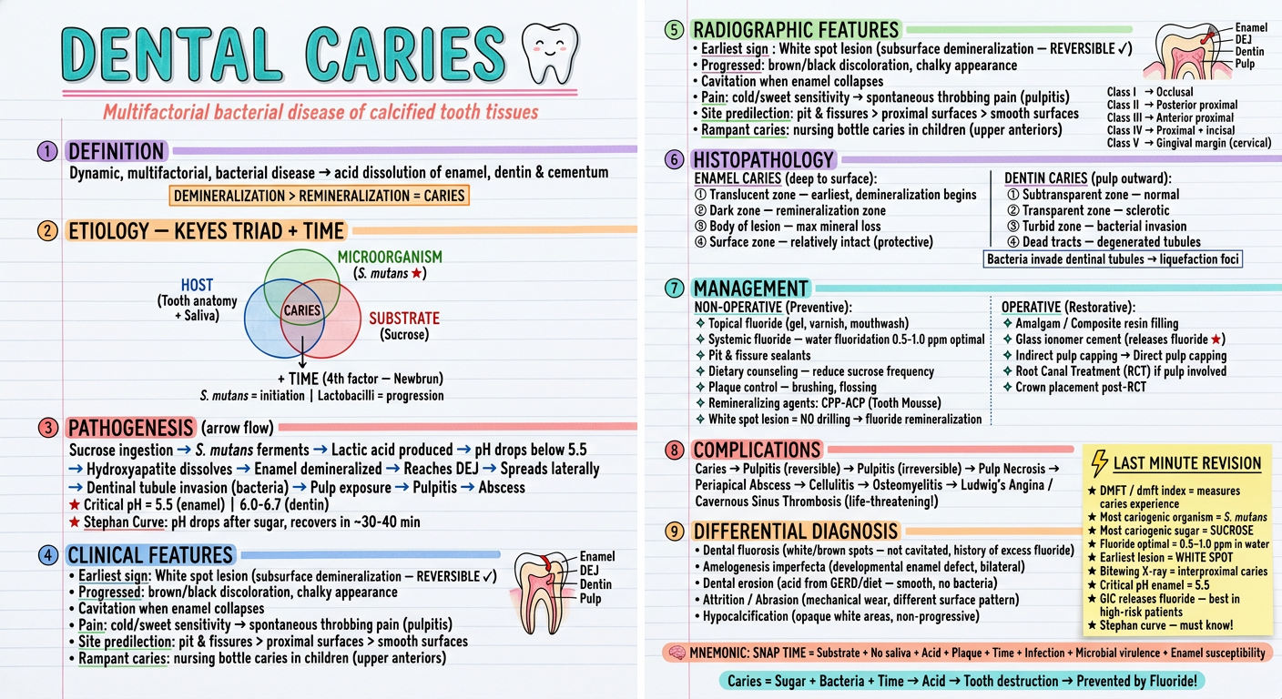
- Definition - with the key demineralization > remineralization concept highlighted
- Keyes Triad Venn diagram - Host + Microorganism + Substrate + Time (Newbrun's 4th factor)
- Pathogenesis flowchart - sucrose → acid → pH <5.5 → enamel dissolution → pulp
- Clinical features - white spot to cavitation, pain progression, rampant/nursing bottle caries
- Radiographic features - bitewing X-ray gold standard, Caries Class I-V classification
- Histopathology - 4 zones of enamel caries + dentin caries zones
- Management - non-operative (fluoride, sealants) vs. operative (filling, RCT)
- Complications chain - all the way to Ludwig's angina
- Differential diagnosis - fluorosis, erosion, amelogenesis imperfecta
- Last-minute sticky note - DMFT, critical pH, Stephan curve, GIC pearl
- Mnemonic + one-line summary at the bottom