
Management of dengue hemorrhagic fever. Management of dengue hemorrhagic fever.
"dengue hemorrhagic fever" AND management
dengue hemorrhagic fever management fluid resuscitation
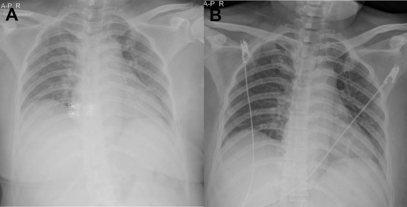
This diagnostic comparison presents two anterior-posterior (AP) chest X-rays (labeled A and B) tracking the progression of pulmonary findings in a patient with dengue hemorrhagic fever (DHF) and iatrogenic fluid overload. Image A shows significant bilateral perihilar haze and increased pulmonary vascular markings, consistent with hilar congestion and early pulmonary edema following aggressive fluid resuscitation. The heart silhouette is visible, and the peripheral lung fields remain relatively clear. Image B, taken after the reduction of intravenous fluids and management toward a negative fluid balance, demonstrates a marked resolution of the bilateral hilar congestion. The central lung fields appear clearer with more distinct bronchial markings. Both images show metallic ECG leads overlying the thorax and a central venous catheter. This comparison illustrates the clinical relevance of radiographic monitoring in managing plasma leakage and fluid therapy in DHF patients to prevent and resolve pulmonary congestion.
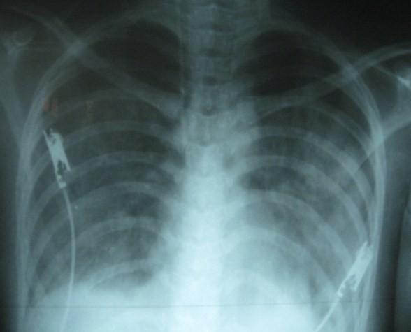
This diagnostic image is an anteroposterior (AP) chest X-ray demonstrating severe pulmonary edema and pleural effusion in a 19-year-old female patient with Dengue Shock Syndrome. The lung fields show bilateral, diffuse, fluffy opacities and increased interstitial markings, characteristic of pulmonary edema. There is significant blunting of the right costophrenic angle, indicating a right-sided pleural effusion. The cardiac silhouette appears moderately enlarged, and the normal clear visualization of pulmonary vasculature is obscured by interstitial and alveolar infiltrates. ECG electrodes and leads are visible on the chest wall, reflecting an acute clinical monitoring setting. This imaging serves as a critical educational example of 'third space' fluid accumulation and capillary leak syndrome secondary to viral hemorrhagic fever, illustrating the rapid progression of respiratory distress during fluid resuscitation in Dengue Shock Syndrome.
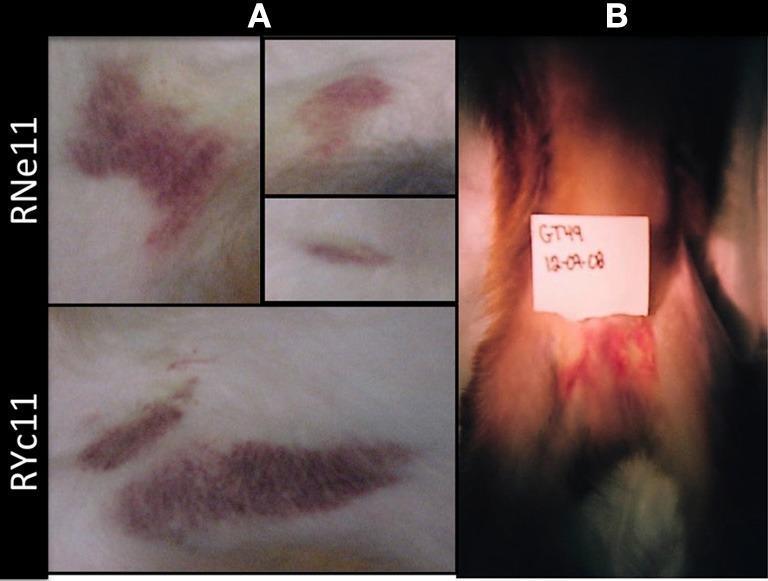
Clinical photograph presenting cutaneous manifestations of Dengue Virus 2 (DV2) infection in a rhesus macaque animal model, serving as a surrogate for human hemorrhagic fever pathology. The image is divided into two primary panels illustrating hematomas and ecchymoses. Panel A displays localized skin lesions on Indian rhesus macaques (labeled RNe11 and RYc11) at Day 7 post-infection. These lesions appear as dark reddish-purple, irregular patches (ecchymoses) and deeper, raised areas of blood accumulation (hematomas). The morphology ranges from diffuse, blotchy purpura to elongated, streak-like hemorrhagic patterns. Panel B shows a Chinese rhesus macaque (GT49) at Day 6 post-infection, highlighting localized hemorrhaging in the perineal region. This area exhibits a patchy distribution of erythema and subcutaneous bleeding against a background of light-colored fur. The visual data demonstrates the clinical spectrum of vasculopathy and coagulopathy associated with acute flavivirus infection, specifically modeling the capillary leak and hemorrhagic tendencies observed in human Dengue Hemorrhagic Fever (DHF).
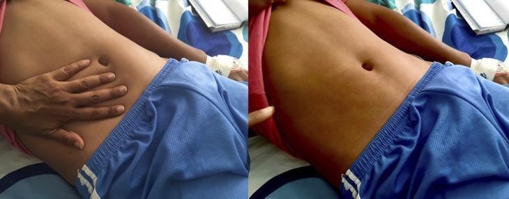
This clinical photograph displays a diffuse, erythematous maculopapular rash on the abdomen of a patient, characteristic of Dengue fever. The image is presented in a two-part sequence to demonstrate clinical blanching. On the left, a clinician's hand is shown applying direct pressure to the right side of the patient's abdomen. On the right, after the hand is removed, a clear, pale handprint-shaped outline remains where the pressure was applied, indicating that the redness fades (blanches) as blood is temporarily displaced from the superficial vessels. This finding confirms the rash is inflammatory rather than hemorrhagic (petechial/purpuric). The rash is widespread across the torso but typically spares the palms and soles in this clinical presentation. This image serves as a teaching tool for medical students and clinicians to distinguish between vascular inflammatory rashes and non-blanching hemorrhagic lesions in the context of viral hemorrhagic fevers and infectious diseases.
WHO dengue hemorrhagic fever management guidelines 2024 fluid therapy
Management of Dengue Hemorrhagic Fever (DHF)
Overview
- Fever lasting 2-7 days
- Hemorrhagic tendencies (positive tourniquet test, petechiae, ecchymoses, spontaneous bleeding)
- Thrombocytopenia (platelet count ≤ 100,000/mm³)
- Evidence of plasma leakage - rising hematocrit ≥20% from baseline, pleural effusion, ascites, or hypoproteinemia
WHO Grading of DHF
| Grade | Clinical Features | Lab |
|---|---|---|
| I | Fever + positive tourniquet test, evidence of plasma leakage | Platelets <100,000/mm³, Hct rise ≥20% |
| II | Grade I + spontaneous bleeding (epistaxis, black stools, gum bleeding) | Same |
| III | Grade II + circulatory failure (weak rapid pulse, pulse pressure ≤20 mmHg, hypotension, cold clammy skin, restlessness) | Same |
| IV | Grade III + profound shock - undetectable BP/pulse | Same |
Disease Phases - Key to Management
- Febrile phase (Day 1-3): High fever, myalgia, headache, retro-orbital pain. Minor bleeding possible. Manage symptomatically.
- Critical phase (Day 3-6, lasts 24-48 hours): Plasma leakage begins at defervescence. Hematocrit rises. This is when shock and hemorrhage occur. Intensive monitoring and fluid therapy required.
- Convalescent/reabsorption phase: Plasma reabsorption begins - watch for fluid overload. Bradycardia is common. Hematocrit falls due to dilution.
Management by Group
Group A - Dengue Without Warning Signs (Outpatient)
- Oral rehydration: ORS, fruit juice, electrolyte-containing fluids
- Paracetamol for fever; keep temperature below 39°C. Space doses ≥6 hours apart
- Avoid: Aspirin, NSAIDs (ibuprofen) - aggravate gastritis and bleeding; aspirin may cause Reye's syndrome
- Monitor daily: temperature, fluid intake/output, urine frequency, platelet count, hematocrit
- Return to hospital immediately if: no improvement, deterioration at defervescence, severe abdominal pain, persistent vomiting, cold extremities, restlessness, black stools/coffee-ground vomiting, no urine for >4-6 hours
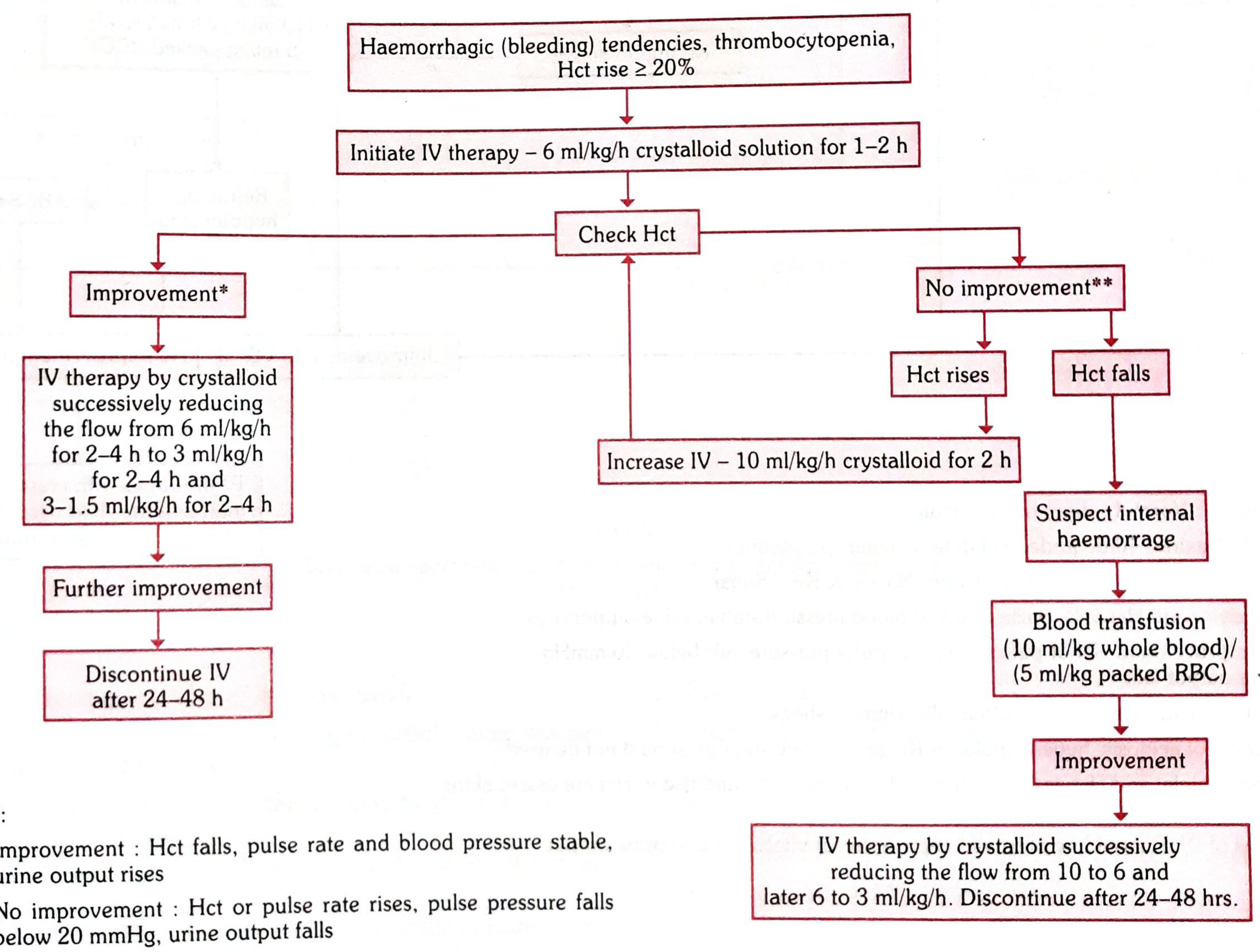
Group B - DHF Grades I and II (Hospitalize)
- Oral rehydration continued; IV fluids if vomiting or unable to tolerate orals
- Serial hematocrit every few hours - a rising Hct signals plasma leakage
- If Hct rises ≥20% from baseline: initiate IV isotonic crystalloid (normal saline or Ringer's lactate)
- Start at 6 ml/kg/h crystalloid for 1-2 hours
- If improvement (Hct falls, pulse/BP stable, urine output rises): reduce stepwise - 6 → 3 → 1.5 ml/kg/h over 6-8 hours; discontinue after 24-48 hours
- If no improvement (Hct rises, pulse pressure falls, urine output falls):
- Increase to 10 ml/kg/h crystalloid for 2 hours
- If Hct falls (suspect internal hemorrhage): blood transfusion 10 ml/kg whole blood or 5 ml/kg packed RBCs
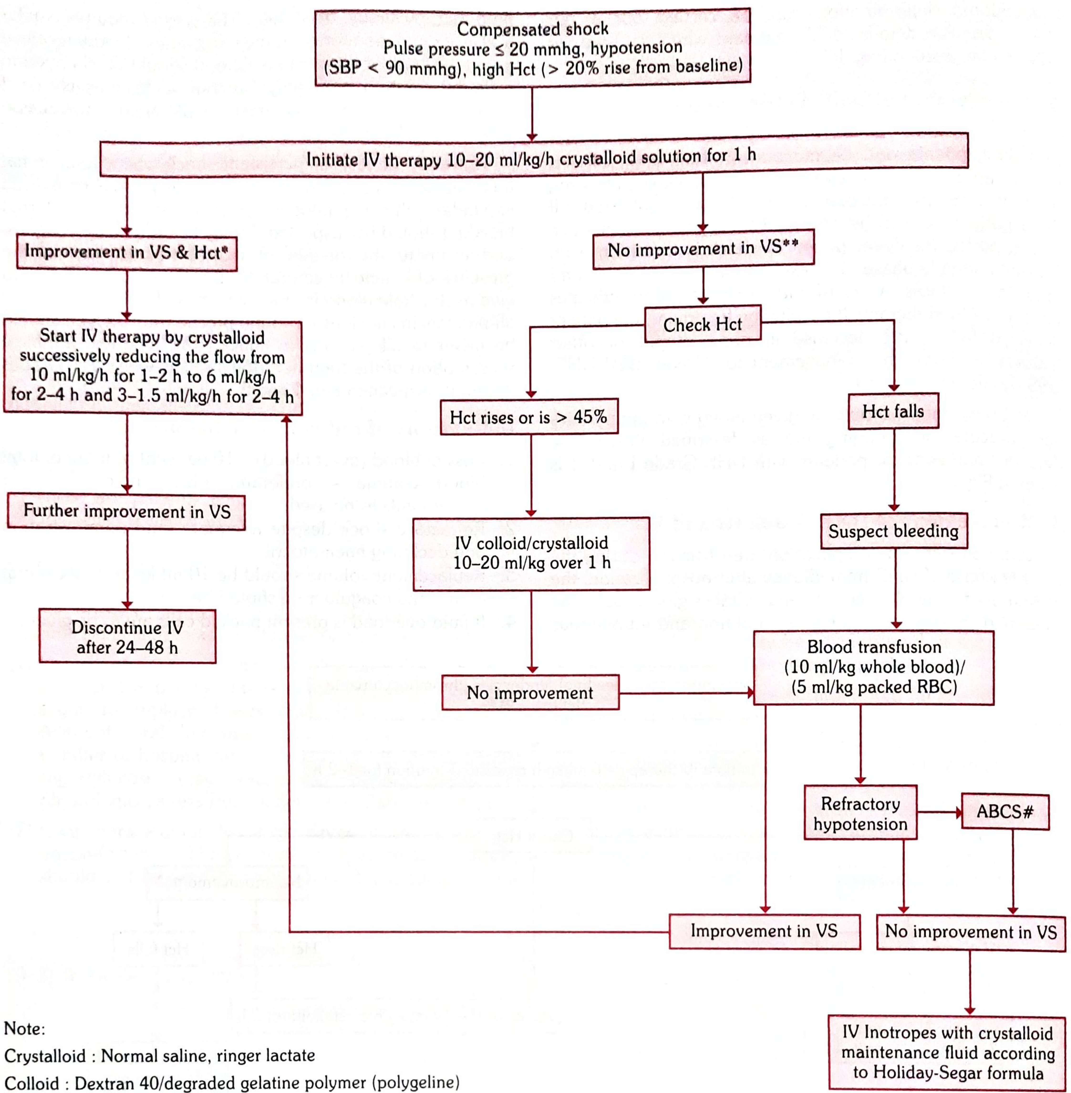
Group C - DHF Grades III and IV / DSS (Emergency Management)
- Start IV therapy immediately at 10-20 ml/kg/h crystalloid for 1 hour
- Oxygen for all patients in shock
- Monitor vital signs (VS) and Hct every 30-60 minutes
- Check hematocrit:
- Hct rises (>45%): Switch to colloid (Dextran 40 or polygeline) 10-20 ml/kg over 1 hour
- Hct falls: Suspect bleeding - give blood transfusion (10 ml/kg whole blood or 5 ml/kg packed RBCs)
- A - Acidosis
- B - Bleeding
- C - Calcium (electrolyte imbalance: Na⁺, K⁺)
- S - Sugar (hypoglycemia)
Fluid Therapy - Key Principles
| Principle | Detail |
|---|---|
| Preferred fluids | Isotonic crystalloids: Normal saline or Ringer's lactate |
| Avoid | Hypotonic fluids (risk of hyponatremia and worsening plasma leakage) |
| Colloids (Dextran 40) | Use when crystalloids fail and Hct remains elevated |
| Guide therapy by | Serial hematocrit trends + urine output (target >0.5 ml/kg/h) |
| Discontinue IV when | Patient enters convalescent phase, plasma leakage resolves |
| Watch for fluid overload | During reabsorption phase - falling Hct from dilution, pulmonary edema, ascites |
| High-volume IV | Associated with increased risk of severe dengue progression in recent studies |
Blood Product Transfusions
Red Cell Transfusion - Indications:
- Overt blood loss ≥10% of total blood volume
- Refractory shock despite adequate fluid administration with declining hematocrit
- Replace at 10 ml/kg body weight at a time; check coagulogram
- Packed cells if fluid overload is present
Platelet Transfusion - Indications:
- In general, no prophylactic platelet transfusion even at <20,000/mm³
- Prophylactic transfusion may be given at <10,000/mm³
- Prolonged shock with coagulopathy and abnormal coagulogram
- Systemic massive bleeding (in addition to red cell transfusion)
Monitoring Parameters
| Parameter | Frequency |
|---|---|
| Vital signs (BP, pulse, pulse pressure) | Every 1 hour in critical phase |
| Hematocrit | Every 4-6 hours (or more frequently in shock) |
| Urine output | Every 1-2 hours |
| Platelet count | Daily (or more often if declining rapidly) |
| Blood glucose | Regularly (tight glycemic control) |
| Coagulogram | If bleeding or refractory shock |
| Chest X-ray | If pleural effusion or respiratory distress suspected |
Specific Complications and Their Management
| Complication | Management |
|---|---|
| Severe bleeding/hemorrhage | Blood/blood product transfusion; identify source |
| Acute liver failure | Similar to ALF from other causes |
| Acute kidney injury | Judicious fluid management (target urine >0.5 ml/kg/h); early RRT if needed; CVVH preferred modality |
| ARDS/pulmonary edema | Reduce IV fluids; target negative fluid balance; oxygen/ventilatory support |
| Encephalopathy | Supportive; tight glycemic control; manage cerebral edema |
| Myocarditis | Inotropic support; avoid fluid overload |
| Electrolyte imbalance | Correct acidosis, hyponatremia, hypocalcemia, hypoglycemia |
Contraindicated/Avoid
- Aspirin and NSAIDs - increase bleeding risk; aspirin may precipitate Reye's syndrome
- Steroids - randomized studies show no benefit in dengue shock syndrome; not recommended
- Intramuscular injections - risk of hematoma at injection sites
- Hypotonic IV fluids - worsen plasma leakage physiology
- Excessive fluid - can cause pulmonary edema/fluid overload, especially in reabsorption phase
- Prophylactic platelet transfusion at modest thrombocytopenia (not indicated unless <10,000/mm³ or active bleeding)
Clinical Warning Signs Requiring Urgent Hospitalization
- Abdominal pain or tenderness
- Persistent vomiting
- Clinical fluid accumulation (ascites, pleural effusion)
- Mucosal bleeding
- Lethargy or restlessness
- Liver enlargement >2 cm
- Rising hematocrit concurrent with rapid fall in platelet count
- No urine for >4-6 hours
Criteria for Discharge
- Afebrile for ≥24 hours without antipyretics
- Return of appetite
- Visible clinical improvement
- Good urine output
- Minimum 2-3 days after recovery from shock
- No respiratory distress from pleural effusion or ascites
- Platelet count >50,000/mm³
Dengue Shock Syndrome (DSS) - Quick Reference Summary
| Step | Action |
|---|---|
| 1 | Rapid IV isotonic crystalloid 10-20 ml/kg/h for 1 hour + oxygen |
| 2 | Reassess VS and Hct after 1 hour |
| 3 | If improved: stepwise reduction of IV fluid rate |
| 4 | If not improved + Hct high: colloid/crystalloid bolus |
| 5 | If not improved + Hct falling: suspect bleeding → transfuse |
| 6 | Refractory shock: correct ABCS; consider inotropes |
| 7 | Once convalescent: reduce and stop fluids to prevent overload |
- Park's Textbook of Preventive and Social Medicine, pp. 293-298 (WHO Classification, fluid algorithms)
- Rosen's Emergency Medicine: Concepts and Clinical Practice, p. 2630 (Management overview)
- Red Book 2021 - Report of the Committee on Infectious Diseases, pp. 520-524 (Pediatric management, shock therapy)
- Creasy & Resnik's Maternal-Fetal Medicine, p. 1565 (Supportive care principles)
- Xu et al. (2024): High IV fluid volume associated with worse outcomes in hospitalized dengue patients
- StatPearls - Dengue Fever (NCBI): Cornerstone is judicious fluid therapy with isotonic crystalloids