
Paraneoplastic leukemoid reaction in a patient with stage 4 lung cancer small cell cancer
leukemoid reaction small cell lung cancer paraneoplastic
PMID: 36149442
G-CSF ectopic production small cell lung cancer leukocytosis prognosis
paraneoplastic leukemoid reaction small cell lung cancer G-CSF mechanism treatment prognosis 2022 2023 2024
Paraneoplastic Leukemoid Reaction in Stage 4 Small Cell Lung Cancer (SCLC)
Definition
Epidemiology and Context in SCLC
- Paraneoplastic syndromes affect up to 8-20% of all lung cancer patients, and SCLC is the subtype most commonly associated with paraneoplastic phenomena overall. - Fishman's Pulmonary Diseases and Disorders, p. 2062
- Among patients with newly diagnosed lung cancer, approximately 14.5% have leukocytosis, and ~20% of those with leukocytosis have a solid malignancy as the underlying cause. Frontiers Oncol, 2019
- True leukemoid reactions (WBC >50,000) are much rarer and documented predominantly in case reports. SCLC, along with squamous cell carcinomas of the lung, head and neck, kidney, and bladder, are the tumors most often implicated.
Pathophysiology: Ectopic G-CSF / GM-CSF Secretion
| Cytokine | Effect |
|---|---|
| G-CSF (granulocyte colony-stimulating factor) | Drives massive neutrophilic leukocytosis; most implicated |
| GM-CSF (granulocyte-macrophage CSF) | Contributes to mixed leukocytosis |
| IL-6 | Contributes to thrombocytosis and acute-phase response |
- SCLC and other lung tumors may directly secrete G-CSF as an ectopic hematopoietic growth factor, driving WBC counts that can exceed 100,000/µL (and in extreme cases up to 178,000/µL). - Goldman-Cecil Medicine, p. 3136
- The mechanism is analogous to the therapeutic use of recombinant G-CSF: tumor-produced G-CSF bypasses normal feedback regulation, causing uncontrolled marrow stimulation.
- Hematologic paraneoplastic syndromes (including leukocytosis) develop through tumor-derived cytokines activating progenitor cells in the bone marrow. - Fishman's, p. 2062
- The course of hematologic paraneoplastic syndromes parallels tumor activity - they worsen with progression and resolve (or improve) with tumor response to treatment.
Two Overlapping Mechanisms in Lung Cancer
- Pure paraneoplastic (ectopic G-CSF): Tumor secretes G-CSF → peripheral neutrophilia with left shift, no leukoerythroblastic morphology, bone marrow not invaded.
- Leukoerythroblastic reaction (myelophthisis): Metastasis to bone marrow → left-shifted leukocytosis + nucleated red blood cells + teardrop-shaped red cells (dacrocytes) + thrombocytosis. Stage 4 SCLC may cause both simultaneously. - Goldman-Cecil, p. 3136
Diagnosis: Excluding Other Causes
| Differential Diagnosis | Distinguishing Feature |
|---|---|
| Infection / sepsis | Fever, cultures, CRP/PCT elevation |
| Acute leukemia / CML | Peripheral blast count, BCR-ABL mutation, bone marrow biopsy |
| Myeloproliferative neoplasm | JAK2 V617F mutation (present in ~50% of essential thrombocytosis; absent in reactive causes) |
| Corticosteroid-induced | History of steroid use (causes demargination) |
| G-CSF/GM-CSF therapy (iatrogenic) | Drug history |
- Complete blood count with differential - look for left shift, toxic granulation, absence of blasts
- Peripheral blood smear - nucleated RBCs, teardrop cells suggest myelophthisis
- Bone marrow biopsy - to exclude primary hematologic malignancy and detect marrow metastases
- Genomic/molecular testing - in cancer patients with hyperleukocytosis, genomic testing can discriminate myeloproliferative neoplasms from a paraneoplastic leukemoid reaction. - Goldman-Cecil, p. 3136
- Serum G-CSF level (if available) - elevated in ectopic production
- JAK2 V617F mutation - negative in reactive/paraneoplastic leukocytosis
Clinical Features and Complications
- WBC may range from mildly elevated to >100,000/µL; extreme cases (>100,000) are associated with leukostasis complications (pulmonary hypoxia, CNS symptoms)
- Thromboembolic risk is elevated: paraneoplastic leukemoid reactions can cause arterial thrombosis (reported in lung adenocarcinoma, PMID 33051200), DIC, and microvascular occlusion
- The reaction may precede recognition of the cancer, serving as a herald of occult malignancy or disease progression
- Fever may occur from cytokine release, mimicking infectious leukocytosis
Prognosis
- The 2022 German case report (PMID 36149442) described an adenocarcinoma patient with WBC up to 178,000/µL who died in neutropenia after just 2 cycles of immunochemotherapy, only ~8 weeks after initial presentation.
- Paraneoplastic leukocytosis is associated with advanced disease and worse clinical outcomes. - Fishman's, p. 2067 (by analogy with thrombocytosis data)
- The degree of leukocytosis may correlate with tumor burden and proliferative activity; the NLR elevation (from G-CSF-driven neutrophilia with relative lymphopenia) is an established poor prognostic marker in lung cancer.
- In stage 4 SCLC, the baseline prognosis is already poor (median OS ~9-12 months with systemic therapy); a paraneoplastic leukemoid reaction likely signals a more aggressive tumor phenotype.
Treatment
| Approach | Details |
|---|---|
| Systemic chemotherapy | First-line for extensive-stage SCLC: etoposide + carboplatin (or cisplatin), with or without atezolizumab/durvalumab (PD-L1 inhibitor) |
| G-CSF avoidance | Do NOT add prophylactic G-CSF - this would dramatically worsen the leukemoid reaction |
| Monitoring for leukostasis | If WBC >100,000/µL with symptoms: consider leukapheresis as a bridge to systemic therapy |
| Treat complications | Anticoagulation if thrombosis present; avoid hyperviscosity |
| Anti-G-CSF approaches | Investigational - specific G-CSF/MDSC-targeting strategies are not yet clinically established |
Key Clinical Pearls for SCLC Stage 4
- Do not give G-CSF with chemotherapy in a patient who already has paraneoplastic leukocytosis - it is contraindicated and dangerous.
- The WBC trend mirrors tumor response - falling WBC with treatment indicates tumor control; rising WBC may herald progression before imaging changes.
- Exclude CML definitively (BCR-ABL/FISH) - WBCs in the 50,000-200,000 range can look identical on the peripheral smear.
- Bone marrow biopsy serves the dual purpose of excluding a primary hematologic process AND staging (detecting SCLC marrow metastases).
- Serum G-CSF measurement, when available, is the most direct evidence of ectopic production.
- The syndrome carries a poor prognosis and may complicate chemotherapy delivery due to associated immunosuppression (MDSC expansion) and infection risk.
- Goldman-Cecil Medicine (International Edition), p. 3136 - Nonhematologic Malignancy / Leukocytosis
- Fishman's Pulmonary Diseases and Disorders, p. 2062-2067 - Extrapulmonary Syndromes Associated with Lung Tumors
- Murray & Nadel's Textbook of Respiratory Medicine, p. 4353 - Musculoskeletal/Hematologic Paraneoplastic Effects
- Harrison's Principles of Internal Medicine 22E, Ch. 98 - Paraneoplastic Syndromes: Endocrinologic/Hematologic
- Heusinger et al. 2022, PMID 36149442 - Paraneoplastic hyperleukocytosis in lung cancer (WBC 178k/µL)
- Niu et al. 2024, PMID 38344203 - Paraneoplastic leukemoid reaction treated with Lorlatinib
- Frontiers Oncol 2019 - G-CSF/MDSC/NLR paradigm in NSCLC
What is paraneoplastic syndrome
Paraneoplastic Syndrome
Definition
- The local or distant spread (metastasis) of the tumor
- The elaboration of hormones typical to the tissue of tumor origin
- Complications of treatment
Why They Matter Clinically
- Earliest manifestation of occult cancer - the syndrome may appear before the tumor is diagnosed, serving as a diagnostic clue to find an underlying malignancy
- Clinically significant in their own right - they can cause severe morbidity and even be lethal if untreated
- Can mimic metastatic disease - potentially confounding staging and treatment decisions
Underlying Mechanisms
| Mechanism | How It Works | Examples |
|---|---|---|
| Ectopic hormone / peptide secretion | Tumor cells produce biologically active hormones not normally made by that tissue type | ACTH by SCLC → Cushing syndrome; ADH by SCLC → SIADH; PTHrP by squamous cell cancer → hypercalcemia |
| Immunological / autoimmune | Tumor expresses antigens shared with normal tissues; immune response directed against tumor cross-reacts with and damages normal tissue | Anti-Hu antibodies in SCLC → peripheral neuropathy; Lambert-Eaton myasthenic syndrome |
Classification by System
1. Endocrinopathies (most common category)
| Syndrome | Associated Cancer | Causal Mechanism |
|---|---|---|
| Cushing syndrome | Small cell lung carcinoma (SCLC), pancreatic carcinoma, neural tumors | Ectopic ACTH or ACTH-like peptide |
| SIADH (hyponatremia) | SCLC, intracranial neoplasms | Ectopic ADH (vasopressin) |
| Hypercalcemia | Squamous cell carcinoma of lung, breast, renal carcinoma, adult T-cell leukemia | PTHrP, TGF-α, TNF, IL-1 |
| Hypoglycemia | Fibrosarcoma, ovarian carcinoma | Insulin or insulin-like substance (IGF-2) |
| Polycythemia | Renal cell carcinoma, cerebellar hemangioma, hepatocellular carcinoma | Ectopic erythropoietin |
| Osteomalacia | Phosphaturic mesenchymal tumors | FGF-23 |
SCLC is responsible for the majority of ectopic endocrine syndromes. About 50% of Cushing syndrome cases from ectopic ACTH come from small-cell lung cancer. - Robbins Cotran, p. 263
2. Nerve and Muscle Syndromes (Neurological)
| Syndrome | Associated Cancer | Mechanism |
|---|---|---|
| Lambert-Eaton myasthenic syndrome | SCLC, thymoma | Autoantibodies against voltage-gated calcium channels (VGCCs) at neuromuscular junction |
| Paraneoplastic encephalomyelitis | SCLC | Anti-Hu (ANNA-1) antibodies |
| Limbic encephalitis | SCLC, testicular, ovarian | Anti-Hu, anti-NMDAR antibodies |
| Cerebellar degeneration | SCLC, ovarian, breast | Anti-Yo, anti-Hu antibodies |
| Peripheral sensory neuropathy | SCLC | Anti-Hu antibodies |
| Dermatomyositis / polymyositis | Bronchogenic and breast carcinoma | Immunologic |
3. Dermatological Syndromes
| Syndrome | Associated Cancer | Mechanism |
|---|---|---|
| Acanthosis nigricans (gray-black velvety skin patches) | Gastric, lung, uterine carcinoma | Secretion of EGF or other growth factors; in ~50% of adult cases with this finding, underlying cancer is present |
| Dermatomyositis | Bronchogenic, breast | Immunologic |
| Sweet syndrome (acute febrile neutrophilic dermatosis) | Hematologic malignancies | Cytokine-driven |
4. Osseous, Articular, and Soft Tissue Changes
| Syndrome | Associated Cancer | Mechanism |
|---|---|---|
| Hypertrophic pulmonary osteoarthropathy (HPO) | Bronchogenic carcinoma | Unknown; periosteal new bone formation + arthritis at ankles/knees/wrists |
| Digital clubbing | Bronchogenic carcinoma | Often associated with HPO |
5. Vascular and Hematologic Changes
| Syndrome | Associated Cancer | Mechanism |
|---|---|---|
| Trousseau phenomenon (migratory thrombophlebitis) | Pancreatic, lung, other cancers | Tumor mucins activate clotting cascade |
| Non-bacterial thrombotic endocarditis (NBTE) | Advanced cancers | Hypercoagulability |
| DIC | Acute promyelocytic leukemia, prostatic carcinoma | Tumor procoagulants |
| Red cell aplasia | Thymoma | Immunologic |
| Leukemoid reaction | Lung, kidney, head/neck cancers | Ectopic G-CSF/GM-CSF production |
| Polycythemia | Renal, hepatic, cerebellar tumors | Ectopic erythropoietin |
Which Cancers Are Most Commonly Associated?
- Lung cancer (especially SCLC) - the single most common cancer associated with paraneoplastic syndromes overall
- Breast cancer
- Ovarian cancer
- Thymoma
- Lymphomas and leukemias
General Principles of Management
- Treat the underlying tumor - this is the most effective approach; most endocrine and hematologic paraneoplastic syndromes parallel tumor activity and resolve with tumor response
- Symptomatic management of the specific syndrome (e.g., fluid restriction for SIADH, bisphosphonates for hypercalcemia, IVIg/plasmapheresis for neurological syndromes)
- Immunosuppression for autoimmune/neurological syndromes (steroids, IVIg, rituximab)
- Neurological paraneoplastic syndromes are an exception - they frequently do NOT improve with tumor treatment because the immune-mediated nerve damage is often irreversible
A key principle: the course of endocrine and hematologic paraneoplastic syndromes mirrors tumor activity. Neurological syndromes often do not. - Fishman's Pulmonary Diseases and Disorders, p. 2062
- Robbins, Cotran & Kumar - Pathologic Basis of Disease, pp. 244-277 (Paraneoplastic Syndromes)
- Robbins & Kumar Basic Pathology, pp. 539-555
- Harrison's Principles of Internal Medicine 22E, Ch. 98 - Paraneoplastic Syndromes: Endocrinologic/Hematologic
- Fishman's Pulmonary Diseases and Disorders, p. 2062 - Extrapulmonary Syndromes with Lung Tumors
- Murray & Nadel's Textbook of Respiratory Medicine, p. 4345
What is Charcot Marie disease
Charcot Marie Tooth disease foot deformity pes cavus peripheral neuropathy
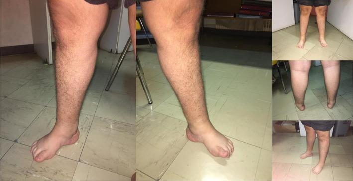
This clinical photograph montage displays the lower extremities of a patient with Charcot-Marie-Tooth (CMT) disease type 1C, caused by a LITAF gene variant. The images reveal characteristic marked distal muscle atrophy, particularly involving the calves and intrinsic foot muscles, resulting in a 'stork leg' appearance relative to the more preserved proximal thighs. Bilateral pes cavus is clearly visible, indicated by abnormally high longitudinal arches of the feet. There is evidence of muscle wasting around the ankles and feet, making the bony structures more prominent. Additional clinical findings shown include an ingrown nail on the right hallux. The visualization supports the diagnosis of a hereditary motor and sensory neuropathy, focusing on the musculoskeletal manifestations of distal peripheral nerve degeneration. The content is suitable for medical education regarding neurogenetics and clinical neurology physical examination findings.
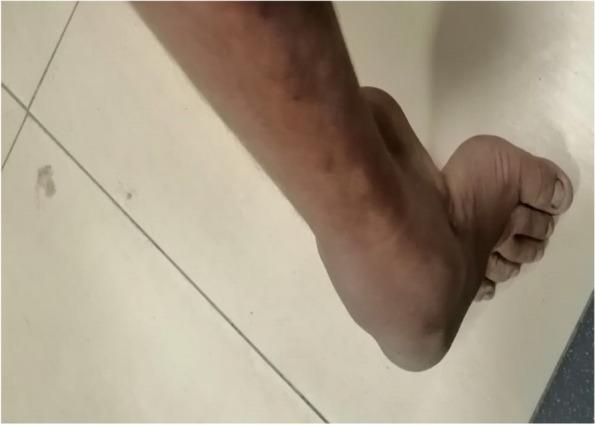
This clinical photograph displays the distal lower limb and foot of a patient, demonstrating classic physical manifestations of Charcot-Marie-Tooth (CMT) disease, specifically CMT Type 2. The image highlights a pronounced pes cavus (high-arched foot) deformity, which is a hallmark clinical sign of hereditary motor and sensory neuropathies. There is significant visible muscle atrophy in the distal leg, particularly involving the peroneal and tibial musculature, giving the lower leg a 'stork leg' or 'inverted champagne bottle' appearance. Additional structural changes include apparent clawing of the toes. These findings illustrate chronic, progressive distal muscle wasting and orthopedic deformities resulting from axonal degeneration. The clinical context associates these features with a homozygous GDAP1 gene mutation, reflecting an autosomal recessive inheritance pattern typical of certain CMT subtypes.
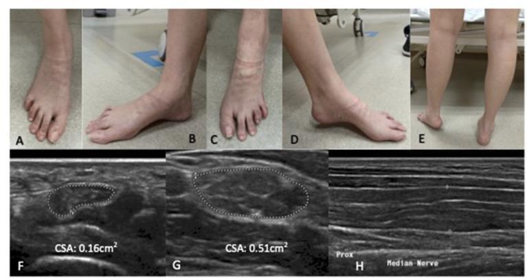
This composite educational image features clinical photographs and diagnostic ultrasound scans related to hereditary peripheral neuropathy, specifically Charcot-Marie-Tooth disease. Panels A-E consist of clinical photographs of a patient's lower extremities. Panels A-D demonstrate bilateral foot deformities characterized by mild pes cavus (elevated plantar arches), claw toes, and symmetrical wasting of the intrinsic foot muscles. Panel E shows a posterior view of the lower legs, highlighting relatively preserved calf muscle bulk. Panels F-H present high-resolution peripheral nerve ultrasound findings of the median nerve. Transverse sections (F and G) illustrate significant nerve hypertrophy with increased cross-sectional areas (CSA); image F shows a CSA of 0.16 cm² at the carpal tunnel, while image G shows a more pronounced enlargement of 0.51 cm² at the elbow, indicating diffuse nerve thickening typical of demyelinating neuropathies. Panel H provides a longitudinal view of the median nerve, demonstrating a thickened fascicular pattern. The visual content serves as a pedagogical reference for correlating physical findings like intrinsic muscle atrophy and foot architecture changes with sonographic evidence of diffuse neural enlargement.
Charcot-Marie-Tooth Disease (CMT)
Definition
Epidemiology
- Prevalence: 1 in 2,500 - making it the most common inherited peripheral neuropathy
- Affects approximately 37 per 100,000 people across all forms
- ~10% of cases arise from spontaneous (de novo) mutations; the rest are inherited
- Clinical severity varies enormously - some patients have severe symptoms in childhood, others only mild symptoms late in life
Pathophysiology
| Mechanism | CMT Type | What Is Damaged |
|---|---|---|
| Demyelination | CMT1, CMT4, CMTX | Myelin sheath (Schwann cell proteins) - nerve conduction velocity severely slowed |
| Axonal degeneration | CMT2 | Axon itself - conduction velocity near-normal but amplitude reduced |
Genetic Classification
| Type | Inheritance | Gene / Locus | Mechanism | Key Features |
|---|---|---|---|---|
| CMT1A (most common, ~50% of all CMT) | Autosomal dominant | PMP22 duplication (chr 17p11.2) | Overexpression of peripheral myelin protein 22 - demyelination | Onset 1st-2nd decade; rarely wheelchair-dependent |
| CMT1B (<5%) | Autosomal dominant | MPZ (chr 1q22-23) | Myelin protein zero mutation | Variable severity |
| CMT1X (10-20%) | X-linked | GJB1 / Connexin 32 (Xq13.1) | Gap junction protein - demyelination | Males more severely affected than females |
| CMT2A (most common CMT2, ~30% of CMT2) | Autosomal dominant | MFN2 / Mitofusin 2 (chr 1p36.22) | Mitochondrial fusion protein - axonal | Earlier onset, more severe - wheelchair in 20s, may have optic atrophy |
| CMT2B | Autosomal dominant | RAB7 (chr 3q21) | Endosomal trafficking - axonal | Prominent sensory loss, foot ulcerations |
| CMT4 | Autosomal recessive | Multiple genes | Demyelinating | Early onset, more rapidly progressive |
- Goldman-Cecil Medicine, p. 2292; Bradley and Daroff's Neurology in Clinical Practice, p. 2674
The PMP22 duplication (CMT1A) accounts for roughly 50% of all CMT cases globally. The most common CMT2 mutation is MFN2 (mitofusin 2 gene) - a mitochondrial fusion protein expressed in peripheral nerves.
Clinical Features
Characteristic Triad:
- Distal weakness - foot drop, difficulty walking, problems with fine hand movements (fastening buttons, turning keys, writing)
- Distal muscle wasting - classic "inverted champagne bottle" or "stork leg" appearance (thin calves with preserved thighs)
- Sensory loss - predominantly distal, affects proprioception and vibration sense more than pain/temperature
Orthopaedic Deformities:
- Pes cavus (high-arched foot) - hallmark finding
- Hammer toes / claw toes
- Cavovarus foot deformity (most common in CMT1; planovalgus in CMT2)
- Scoliosis and hip dysplasia in some patients
Neurological Signs:
- Absent or reduced ankle jerks (areflexia distally)
- Enlarged/palpable peripheral nerves (especially in CMT1 demyelinating forms)
- Distal loss of proprioception and spinal ataxia
- Poor balance (difficulty skating, walking on uneven surfaces)
- Children often present as slow runners with impaired balance
Upper Limb:
- Fine hand movement impairment appears later
- Ankle-foot orthoses (AFOs) frequently required by the third decade
- Most patients remain ambulatory throughout life (CMT1A)
Clinical photographs of CMT
Diagnosis
1. Clinical Assessment
- Claw toes + high arches + thin legs + poor balance + unsteady gait
- Positive family history
- Absent ankle jerks
- Distal sensory deficits
2. Nerve Conduction Studies (NCS) / EMG
- CMT1 (demyelinating): Severely slowed motor NCV (<38 m/s in the forearm) with prolonged distal latencies; uniform slowing across all nerve segments
- CMT2 (axonal): Near-normal NCV but reduced amplitude of nerve action potentials
- CMT type 2 may have normal velocity - EMG shows decreased amplitude, not velocity
3. Genetic Testing
- CMT1A PMP22 duplication testing by FISH or MLPA is the first-line genetic test (covers ~50% of cases)
- If negative, next-generation sequencing of CMT gene panels
- Genetic testing allows precise subtype identification and prognosis prediction
4. Nerve Biopsy (rarely needed)
- CMT1: "onion bulb" formations - repeated cycles of demyelination and remyelination
- CMT2: axonal loss, no onion bulbs
Distinguishing CMT from Idiopathic Pes Cavus
| Feature | CMT | Idiopathic Pes Cavus |
|---|---|---|
| Family history | Often positive | Usually absent |
| Weakness | Present | Absent |
| Sensory deficits | Present | Absent |
| Absent ankle jerks | Yes | No |
| EMG/NCS abnormalities | Yes | Normal |
| Bilateral | Usually | Variable |
Management
1. Physical and Occupational Therapy
- Stretching, strengthening of proximal muscles
- Balance training
- Occupational therapy for fine motor tasks
2. Orthotics
- Ankle-foot orthoses (AFOs) - most common intervention, corrects foot drop and improves gait stability
- Custom footwear to accommodate foot deformities
3. Surgical Interventions (orthopaedic)
- Cavovarus foot reconstruction - most common surgical procedure
- Plantar fascia release
- Osteotomies (Dwyer calcaneal osteotomy for hindfoot varus)
- Tendon transfers (tibialis posterior transfer to correct foot drop)
- Arthrodesis (triple fusion) for rigid severe deformity
- Scoliosis surgery if significant spinal deformity develops
- CMT1A patients rarely need wheelchairs; CMT2A (MFN2) patients often become non-ambulatory in their 20s - Campbell's Operative Orthopaedics, p. 4898-4900
4. Medications to Avoid
- Vincristine, taxanes, amiodarone, metronidazole - can dramatically worsen CMT neuropathy
- Statins - use with caution
- Patients must always carry a list of neurotoxic drugs to avoid
5. Emerging Therapies
- Gene therapy and antisense oligonucleotide (ASO) approaches targeting PMP22 overexpression in CMT1A are in early clinical trials
- No approved disease-modifying agents as of 2026
Prognosis
- CMT is slowly progressive but rarely life-threatening
- Most patients remain ambulatory throughout life, particularly CMT1A
- CMT2A (MFN2 mutations) has a worse prognosis - wheelchair dependence in the 20s is common
- CMT2C involves vocal cord, intercostal, and diaphragmatic weakness - can shorten life expectancy
- Life expectancy is normal in most forms
- Goldman-Cecil Medicine (International Edition), p. 2292-2302
- Bradley and Daroff's Neurology in Clinical Practice, pp. 2667-2732 - CMT Types 1 and 2 (full genetic classification)
- Campbell's Operative Orthopaedics 15th Ed 2026, pp. 4853-4905 - CMT Disease and Cavovarus Foot