
LIVER FUNCTION TESTS AND ANAESTHETIC IMPLICATIONS
liver function tests anaesthesia perioperative hepatic
liver function tests interpretation hepatocellular cholestatic pattern
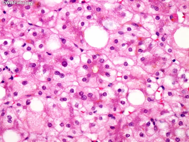
This is a liver biopsy prepared for light microscopy displaying features of parenchymal injury associated with long-term total parenteral nutrition (TPN). The hepatic parenchyma shows prominent fatty change (steatosis) with hepatocytes containing lipid vacuoles of variable size, compatible with macrovesicular and microvesicular steatosis. In addition, there is evidence of cholestasis with inspissated bile pigments accumulating within canaliculi and small intrahepatic ducts, a pattern that can mimic bile duct obstruction. The combination of steatosis and cholestatic injury is characteristic of PN-associated liver disease and can progress to ductular reaction, fibrosis, and ultimately cirrhosis if PN exposure persists. Damaged hepatocytes may display cytoplasmic ballooning and eosinophilic change, reflecting hepatocellular injury. The density of lipid droplets and bile plugs contributes to a heterogeneous lobular appearance. Clinically, this morphological spectrum mirrors the reported evolution of TPN-induced hepatic injury: steatosis in early stages, cholestasis with cholangiolar proliferation over time, and fibrotic remodeling in advanced disease. This histology is critical to distinguish PN-related liver injury from alternative etiologies such as biliary obstruction, viral hepatitis, or drug-induced cholestasis. It underscores the need for multidisciplinary management including PN modification, enteral stimulation, and monitoring of hepatic function tests to mitigate progression to cirrhosis.
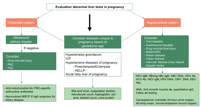
Summary : This flowchart outlines the diagnostic approach to evaluating abnormal liver tests in pregnancy, distinguishing between cholestatic and hepatocellular patterns, and detailing subsequent investigations and considerations based on findings. flowchart: # Nodes : • Start node: "Evaluation abnormal liver tests in pregnancy" (rectangle, top center) • Branch node: "Cholestatic pattern" (rounded rectangle, left) • Branch node: "Hepatocellular pattern" (rounded rectangle, right) • Process node: "Ultrasound without doppler" (rectangle, under cholestatic pattern) • Decision node: "If negative" (rectangle, under ultrasound) • Consideration node: "Consider: - Drug induced injury - PBC - PSC" (rectangle, under decision node) • Test node: "Anti-mitochondrial Ab; PBC-specific antinuclear antibodies - Noncontrast MRCP if high suspicion for biliary disease" (rectangle, bottom left) • Central process node: "Consider diseases unique to pregnancy based on gestational age" (rectangle, center) • List node: "Hyperemesis gravidarum ICP Hypertensive diseases of pregnancy - Preeclampsia/Eclampsia - HELLP Acute fatty liver of pregnancy" (rectangle, under central process node) • Test node: "Bile acid level, coagulation studies, reticulocyte count, haptoglobin, uric acid, platelet count, urine protein" (rectangle, bottom center) • Consideration node: "Consider: - Viral hepatitis - Autoimmune hepatitis - Drug induced liver injury - NASH/ASH - Wilson disease - Celiac disease - Vascular disease (may require Doppler study)" (rectangle, under hepatocellular pattern) • Test node: "HAV IgM, HBsAg, HBc IgM, HBV DNA, HDV Ab, HCV Ab, HEV IgM, HSV DNA, CMV DNA, EBV Abs ANA, Anti smooth muscle ab, quantitative IgG, Celiac ab testing Ceruloplasmin (consider 24-hour urine copper, slit lamp exam, nonceruloplasmin bound copper)" (rectangle, bottom right) # Connectors : • Downward arrows from "Evaluation abnormal liver tests in pregnancy" to both "Cholestatic pattern" and "Hepatocellular pattern", and to "Consider diseases unique to pregnancy based on gestational age". • Downward arrow from "Cholestatic pattern" to "Ultrasound without doppler". • Downward arrow from "Ultrasound without doppler" to "If negative". • Downward arrow from "If negative" to "Consider: - Drug induced injury - PBC - PSC". • Downward arrow from "Consider: - Drug induced injury - PBC - PSC" to "Anti-mitochondrial Ab; PBC-specific antinuclear antibodies - Noncontrast MRCP if high suspicion for biliary disease". • Downward arrow from "Consider diseases unique to pregnancy based on gestational age" to "Hyperemesis gravidarum ICP Hypertensive diseases of pregnancy - Preeclampsia/Eclampsia - HELLP Acute fatty liver of pregnancy". • Downward arrow from "Hyperemesis gravidarum..." to "Bile acid level, coagulation studies, reticulocyte count, haptoglobin, uric acid, platelet count, urine protein". • Downward arrow from "Hepatocellular pattern" to "Consider: - Viral hepatitis - Autoimmune hepatitis - Drug induced liver injury - NASH/ASH - Wilson disease - Celiac disease - Vascular disease (may require Doppler study)". • Downward arrow from "Consider: - Viral hepatitis..." to "HAV IgM, HBsAg, HBc IgM, HBV DNA, HDV Ab, HCV Ab, HEV IgM, HSV DNA, CMV DNA, EBV Abs ANA, Anti smooth muscle ab, quantitative IgG, Celiac ab testing Ceruloplasmin (consider 24-hour urine copper, slit lamp exam, nonceruloplasmin bound copper)". # Layout : • The flowchart is organized in a top-down manner, starting with a central evaluation node, splitting into three main branches: cholestatic pattern (left), hepatocellular pattern (right), and pregnancy-specific diseases (center). • Each branch contains sequential steps, with the left and right branches including further diagnostic considerations and specific tests. • The central branch focuses on pregnancy-specific conditions and associated laboratory investigations. # Analysis : • The flowchart provides a systematic approach to evaluating abnormal liver tests in pregnancy, emphasizing the importance of pattern recognition (cholestatic vs. hepatocellular) and gestational age. • It guides clinicians through appropriate imaging, laboratory tests, and disease considerations, ensuring comprehensive assessment and targeted workup based on initial findings. • The structure allows for exclusion of common etiologies before considering rare or pregnancy-specific conditions.
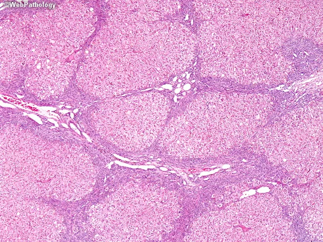
Light microscopy of a paraffin-embedded liver biopsy stained with Hematoxylin and Eosin (H&E) reveals advanced cirrhosis in a patient on long‑term total parenteral nutrition (TPN). Hepatic architecture is distorted by regenerative nodules embedded in a dense fibrous framework; thick fibrous septa traverse the parenchyma and connect portal tracts, producing bridging fibrosis. Within the nodules, hepatocytes are variably damaged, showing cytoplasmic rarefaction and isolated ballooning degeneration. A prominent ductular reaction is seen at the interface between residual hepatocytes and fibrous bands, reflecting biliary-type hepatoportal repair. The overall pattern is consistent with TPN‑associated liver disease comprising steatosis (historically described with TPN) and evolving portal‑to‑parenchymal scarring toward cirrhotic transformation. No discrete mass lesions are identified. This histomorphology supports a diagnosis of cholestatic/hepatocellular injury driven by prolonged parenteral nutrition, with potential clinical consequences including portal hypertension, synthetic dysfunction, and progression to decompensation if TPN exposure persists. Clinically, these findings should be correlated with liver function tests, bilirubin elevation, alkaline phosphatase, and gamma‑glutamyl transferase; imaging and nutrition management should focus on reducing TPN exposure when possible. The pattern must be differentiated from other causes of cirrhosis, including alcoholic liver disease, viral hepatitis, and biliary cholestasis. Educational use includes recognition of ductular reaction and nodular cirrhosis.
Liver Function Tests and Anaesthetic Implications
1. Overview of the Liver - Anaesthetic Relevance
- Portal vein: 75% of hepatic blood flow (nutrient-rich, relatively deoxygenated)
- Hepatic artery: 25% of blood flow - each vessel contributes ~50% of oxygen delivery
- Synthesis of all clotting factors (except Factor VIII) and albumin
- Drug metabolism (Phase I - cytochrome P450; Phase II - conjugation)
- Glucose homeostasis, ammonia detoxification (urea cycle)
- Bile production for fat-soluble vitamin absorption
2. Liver Function Tests: Classification and Interpretation
2.1 Hepatocellular Injury Markers
| Test | Source Tissue | Normal Range | Significance |
|---|---|---|---|
| ALT (SGPT) | Predominantly liver | 7-56 U/L | Most specific for hepatocellular injury |
| AST (SGOT) | Liver, cardiac + skeletal muscle, kidney, brain | 10-40 U/L | Less specific; also elevated in MI, rhabdomyolysis |
| LDH | Multiple tissues | 140-280 U/L | Hepatitis, infarction, trauma |
- AST:ALT ratio >2:1 - strongly suggests alcoholic liver disease
- Mild elevation (<3x ULN): NAFLD, chronic viral hepatitis, medication-induced
- Moderate elevation (3-20x ULN): Acute viral hepatitis
- Markedly elevated (>1000 U/L): Ischaemic hepatitis, acetaminophen toxicity, fulminant hepatitis
2.2 Cholestatic (Canalicular) Markers
| Test | Significance |
|---|---|
| Alkaline Phosphatase (ALP) | Most sensitive for hepatic metastases; also elevated in bone disease, pregnancy, childhood growth |
| GGT (γ-glutamyl transferase) | Corroborates hepatic source of elevated ALP; elevated in 70% of chronic alcoholics; induced by warfarin, barbiturates, phenytoin |
| 5'-Nucleotidase | Liver-specific isoenzyme; confirms hepatic source of ALP elevation |
| Bilirubin (total, direct, indirect) | Conjugated (direct) bilirubin rises in cholestasis and hepatocellular disease |
2.3 Hepatic Synthetic Function Tests
| Test | Normal Range | Significance |
|---|---|---|
| Serum Albumin | 35-50 g/L | Half-life 15-20 days; reflects chronic hepatic dysfunction; also affected by nutrition, renal losses |
| Prothrombin Time (PT)/INR | INR < 1.2 | Reflects Factors II, VII, IX, X (vitamin K-dependent) + V; sensitive to acute changes |
| Serum Ammonia | <50 µmol/L | Rises when >80% of liver tissue is destroyed (urea cycle failure) |
| Fibrinogen | 2-4 g/L | May be normal in quantity but dysfunctional (dysfibrinogenaemia in 70-80% of cirrhotics) |
2.4 Six Major Diagnostic Patterns (Henry's Laboratory Medicine)
| Pattern | ALT/AST | ALP/GGT | Bilirubin | Albumin/PT | Likely Diagnosis |
|---|---|---|---|---|---|
| Acute hepatitis | ↑↑↑ | Normal/mild ↑ | Mild ↑ direct | Normal early | Viral, toxic, ischaemic hepatitis |
| Chronic hepatocellular | ↑ (mild-moderate) | Normal/mild ↑ | Variable | ↓, PT prolonged | Cirrhosis, chronic hepatitis |
| Cholestatic | Mild ↑ | ↑↑ ALP + GGT | ↑↑ direct | Normal until late | Biliary obstruction, PBC, PSC |
| Infiltrative/metastatic | Normal/mild ↑ | ↑↑ ALP | Normal | Normal | Hepatic metastases, granulomas |
| Synthetic failure | Variable | Variable | ↑ | ↓↓ Albumin, ↑↑ PT | Cirrhosis, ALF |
| Isolated hyperbilirubinaemia | Normal | Normal | ↑ unconjugated | Normal | Gilbert syndrome, haemolysis |
3. Perioperative Risk Stratification in Liver Disease
3.1 Child-Turcotte-Pugh (CTP) Score
- Child's A: Perioperative mortality ~10% (acceptable surgical risk)
- Child's B: Perioperative mortality ~30-50% (significant risk; optimise first)
- Child's C: Perioperative mortality ~82% (elective surgery contraindicated)
3.2 MELD Score (Model for End-Stage Liver Disease)
MELD = 3.78 × ln[bilirubin mg/dL] + 11.2 × ln[INR] + 9.57 × ln[creatinine mg/dL] + 6.43
- Score range: 6-40
- MELD < 11: Low postoperative mortality - acceptable surgical risk
- MELD 12-20: Intermediate risk; consider optimisation
- MELD ≥ 20 (or Child's C): High mortality risk; elective procedures contraindicated until after liver transplantation
Key point from Barash's Clinical Anesthesia: "The perioperative risk of patients with end-stage liver disease depends more on the operative site and the degree of liver impairment than the anesthetic technique."
4. Anaesthetic Implications of Abnormal LFTs
4.1 Drug Pharmacokinetics in Liver Disease
- Hypoalbuminaemia increases free fraction of highly protein-bound drugs (e.g., thiopentone, diazepam, propofol)
- This increases pharmacodynamic effect for a given dose
- Ascites and peripheral oedema increase Vd for hydrophilic drugs
- Leads to reduced plasma concentrations but prolonged duration of action
- Reduced biotransformation of drugs dependent on hepatic enzymes
- Prolongs action of opioids (fentanyl, morphine), benzodiazepines, local anaesthetics
- Halothane hepatitis: trifluoroacetylated liver proteins formed from halothane, enflurane, isoflurane, and desflurane can trigger immune-mediated hepatic injury
- Bypasses hepatic first-pass metabolism
- Oral bioavailability of drugs (e.g., propranolol, midazolam) is markedly increased
- Nitrogenous waste enters systemic circulation - precipitates encephalopathy
4.2 Specific Drug Considerations
| Drug Class | Implications |
|---|---|
| Inhalational agents | All volatile agents reduce hepatic blood flow; halothane greatest risk of hepatotoxicity (halothane hepatitis); isoflurane preferred as it best preserves hepatic blood flow |
| Propofol | Safe; undergoes extrahepatic metabolism also; useful as TIVA in severe liver disease |
| Thiopentone | Increased free fraction due to hypoalbuminaemia; prolonged effect; reduce dose |
| Opioids | Significantly prolonged half-life in cirrhosis; morphine and pethidine avoided (active metabolite accumulation); fentanyl preferred but with caution |
| Benzodiazepines | Avoid long-acting (diazepam); may precipitate encephalopathy; use with caution |
| Succinylcholine | Prolonged action if plasma pseudocholinesterase is significantly reduced by liver disease |
| Atracurium/Cisatracurium | Hoffman elimination (plasma-based, non-hepatic); agents of choice in severe liver disease |
| Vecuronium/Rocuronium | Hepatic clearance; prolonged action; use with caution |
| NSAIDs | Avoid - reduce renal blood flow and precipitate hepatorenal syndrome |
| Paracetamol (acetaminophen) | Main cause of acute liver failure in overdose; use lower doses and with caution |
4.3 Coagulopathy: Assessment and Management
- Elevated von Willebrand factor compensates for low platelet count
- Thrombin generation may be normal or even elevated in cirrhosis
- Increased FVIII (not produced by hepatocytes)
- Reduced Factors II, V, VII, IX, X, XI
- Reduced Protein C and Antithrombin III (actually pro-thrombotic)
- Thrombocytopenia (30-64% of cirrhotics): primarily from splenic sequestration in portal hypertension
- Dysfibrinogenaemia in 70-80% of cirrhotics
- Standard PT/INR overestimates bleeding risk in cirrhosis
- Thromboelastography (TEG) or ROTEM better guide transfusion in bleeding cirrhotic patients
- Vitamin K infusion can correct coagulopathy of cholestatic disease (fat-soluble vitamin malabsorption), but NOT hepatocellular dysfunction
- Platelet transfusion indicated for active bleeding with count <56,000/mm³ (minimum needed for near-normal thrombin generation); not for prophylaxis alone
- In cholestatic disease with cholestasis: parenteral vitamin K corrects deficiency in Factors II, VII, IX, X, Protein C, and S
4.4 Cardiovascular Implications
- Decreased systemic vascular resistance (SVR)
- Increased cardiac output (CO)
- Relative hypotension
- Mechanism: excess nitric oxide and splanchnic vasodilation
- Hypotension during induction is common and exaggerated
- Volatile agents and neuraxial blockade worsen SVR reduction
- "Cirrhotic cardiomyopathy" - systolic dysfunction may be unmasked by TIPS (transjugular intrahepatic portosystemic shunt) which increases preload
4.5 Respiratory Implications
- Hepatopulmonary syndrome: intrapulmonary shunting causing hypoxaemia; V/Q mismatch; platypnoea (dyspnoea worse when upright)
- Portopulmonary hypertension: elevated PVR; significant perioperative risk
- Ascites: impairs respiratory mechanics; increased aspiration risk; patient may not tolerate supine position
- Requires modified rapid sequence induction consideration
4.6 Renal Implications
- Hepatorenal syndrome (HRS): intense renal vasoconstriction in setting of portal hypertension; functional renal failure
- Oliguria and rising creatinine in a cirrhotic are a medical emergency
- Avoid nephrotoxic agents: aminoglycosides, NSAIDs, contrast agents
- Albumin is indicated for HRS-AKI in conjunction with splanchnic vasoconstrictors (terlipressin)
- Bilirubin >4 mg/dL or creatinine >1 mg/dL + spontaneous bacterial peritonitis: albumin infusion prevents renal impairment
4.7 Hepatic Encephalopathy
- Precipitated by sedatives, opioids, benzodiazepines, constipation, GI bleeding, infection
- All CNS depressants dramatically worsen encephalopathy
- Grade III-IV encephalopathy: cerebral oedema risk 25-75%
- Avoid N2O (increases intracranial pressure in encephalopathy)
5. Preoperative Optimisation in the Patient with Liver Disease
- Treat active infection (SBP, etc.) before elective surgery
- Minimise vasoactive infusions - allow baseline haemodynamics to stabilise
- Optimise central blood volume and renal status - correct dehydration or volume overload
- Minimise ascites - therapeutic paracentesis; administer albumin 6-8 g per litre drained
- Improve encephalopathy - lactulose, rifaximin; restrict precipitants
- Correct coagulopathy - use TEG/ROTEM to guide targeted therapy; avoid blanket FFP prophylaxis
- Avoid oesophageal instrumentation if varices present or suspected
- Acute alcoholic hepatitis (high perioperative mortality)
- Acute liver failure
- Acute viral hepatitis
- MELD ≥ 20 or Child's C (unless surgery is life-saving)
6. Halothane Hepatitis and Anaesthetic Agent Hepatotoxicity
- Type I (mild, transient): Subclinical AST/ALT elevation after any halogenated volatile agent; due to Zone 3 hepatocyte hypoxia; self-limiting
- Type II (immune-mediated halothane hepatitis): Rare (1:35,000 exposures); occurs after repeated halothane exposure; trifluoroacetyl (TFA) protein adducts act as neoantigens; massive hepatocellular necrosis; mortality 50-75%
- All halogenated agents (halothane, enflurane, isoflurane, desflurane) form TFA protein adducts, but to differing degrees
- Halothane highest risk; desflurane and isoflurane very low risk
- Prior volatile agent hepatitis is a contraindication to future halogenated agents
7. Acute Liver Failure (ALF) - Anaesthetic Management
- Cerebral oedema is the leading cause of death (75% incidence in Grade IV encephalopathy)
- Targets: control ICP; maintain CPP; osmotherapy (hypertonic saline/mannitol); avoid hypercapnia
- Coagulopathy management: FFP and platelets as needed but guided by TEG/ROTEM
- Renal support: haemofiltration often required
- Haemodynamic instability: vasopressor support; avoid hypotension (reduces hepatic perfusion further)
8. Summary Table: LFT Abnormality and Anaesthetic Concerns
| LFT Abnormality | Anaesthetic Implication |
|---|---|
| ↑↑ ALT/AST | Hepatocellular damage; assess severity; avoid hepatotoxic agents |
| ↑ ALP + GGT | Cholestasis; malabsorption of fat-soluble vitamins; correct with parenteral Vit K if cholestatic |
| ↓ Albumin | Increased free drug fraction; ↑ sensitivity to protein-bound anaesthetics; ascites risk |
| ↑ PT/INR | Impaired synthesis; coagulopathy; use TEG; avoid regional block in severe cases |
| ↑ Bilirubin | Risk of renal tubular damage (jaundice surgery risk); nausea/vomiting management |
| ↑ Ammonia | Encephalopathy risk; avoid CNS depressants; avoid N2O; titrate opioids very carefully |
| ↑ Creatinine (+ liver disease) | HRS; nephrotoxin avoidance; albumin + vasoconstrictors |
| Thrombocytopenia | Splenic sequestration; platelet transfusion only for active bleeding or count <56,000 |
- Barash, Cullen, and Stoelting's Clinical Anesthesia, 9th Edition - Chapter 46 (Hepatic Function)
- Henry's Clinical Diagnosis and Management by Laboratory Methods - Chapter 9 (LFTs)
- Schwartz's Principles of Surgery, 11th Edition - Liver Function Tests
- Sleisenger and Fordtran's Gastrointestinal and Liver Disease - Surgical Candidacy
- Recent review: Espinosa et al., "Anaesthesia evaluation and perioperative strategies in liver disease patients with cardiohepatic syndrome," Rev Esp Anestesiol Reanim, 2025 [PMID: 40147631]