
Chronic Lower back pain in an 30 year old male . No history of trauma . What could be the reason and treatment
"low back pain" AND "young adults" AND management
"low back pain" AND treatment
chronic low back pain causes anatomy spine disc herniation
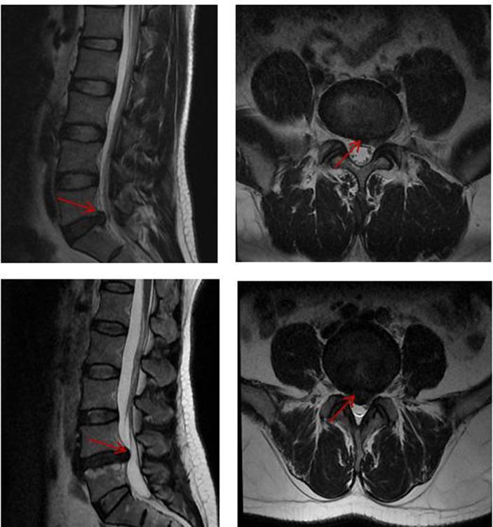
This diagnostic image displays four T2-weighted magnetic resonance imaging (MRI) views of the lumbar spine, specifically highlighting Lumbar Disc Herniation with Chronic Pain (LDHCP). The left panels show sagittal sections where the lumbar vertebrae and intervertebral discs are visible. Red arrows point to posterior disc protrusions at the L4-L5 or L5-S1 levels, demonstrating a loss of disc height and focal extrusion of nuclear material into the spinal canal. The right panels provide axial cross-sections at the level of the herniation. These views clearly show the herniated disc material (red arrows) compressing the thecal sac and narrowing the available space for the cauda equina nerve roots. The surrounding paraspinal musculature and facet joints are also visible. This imaging is characteristic of radiculopathy and chronic low back pain syndromes, used clinically to correlate structural spinal pathology with neurological symptoms and functional impairment.
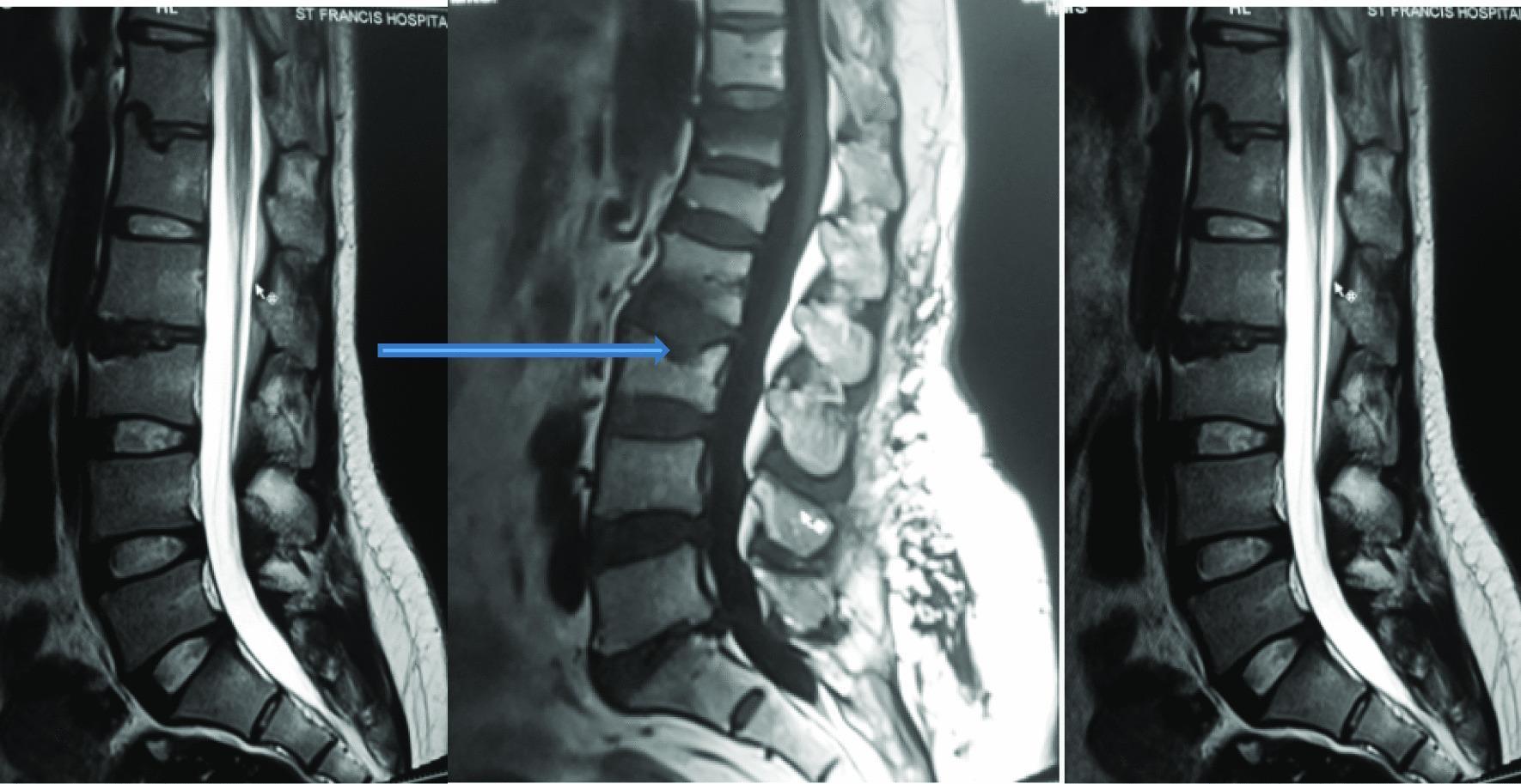
This diagnostic image consists of T2-weighted sagittal MRI sequences of the lumbar spine, demonstrating degenerative changes. A key finding is intervertebral disc desiccation at the L2/L3 level, characterized by a visible loss of high signal intensity (darkening) compared to the hyperintense signal of adjacent hydrated discs. Additionally, the image illustrates Schmorl's nodes at the L1 and L3 vertebral bodies. A blue arrow highlights a Schmorl's node, which appears as a focal, rounded indentation or herniation of the intervertebral disc material through the vertebral endplate into the spongy bone of the vertebral body. The spinal cord and conus medullaris are visible, with the cerebrospinal fluid appearing bright on this T2-weighted sequence. The images provide a clinical demonstration of common lumbar spinal pathologies, specifically focusing on disc degeneration and endplate herniation, which are relevant in the diagnostic evaluation of chronic low back pain.
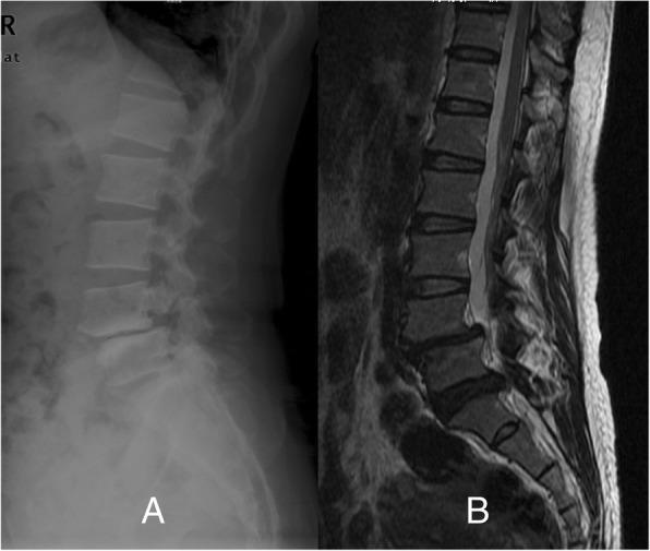
This composite diagnostic image displays a lateral X-ray (A) and a sagittal MRI (B) of the lumbar spine. The lateral radiograph demonstrates an anterior displacement of the L4 vertebral body relative to L5, characteristic of spondylolisthesis. There is evidence of spondylolysis, visible as a lucent defect in the pars interarticularis of the L4 vertebra. The sagittal MRI (B) provides a detailed view of the soft tissues and spinal canal, highlighting a significant disc protrusion at the L5/S1 level. This herniation causes mechanical compression of the thecal sac and likely impacts the descending L5 and S1 nerve roots. The imaging illustrates the mechanical instability of the mid-lumbar spine combined with secondary degenerative disc disease and neural foraminal/canal stenosis. These findings are clinically relevant for evaluating chronic low back pain, radiculopathy, and dermatomal sensory deficits in the lower extremities.
Chronic Lower Back Pain in a 30-Year-Old Male (No Trauma)
Why Diagnosis Is Difficult
Likely Causes (Differential Diagnosis)
1. Nonspecific / Mechanical LBP (Most Common)
- Pain from discs, facet joints, sacroiliac joints, ligaments, muscles, or fascia
- Worsened by activity, relieved by rest
- Often linked to poor posture, sedentary lifestyle, weak core muscles, obesity
- Pain processing changes in the dorsal horn, thalamus, and limbic system perpetuate chronicity even without ongoing tissue damage - Family Medicine 9e
2. Intervertebral Disc Disease
- Disc herniation (L4-L5 or L5-S1 most common): Can cause referred pain into buttocks/thighs and radiculopathy (sciatica)
- Disc degeneration: Loss of disc height, desiccation - common even in young adults
3. Ankylosing Spondylitis (Inflammatory - Key "No Trauma" Consideration)
- Classically affects young adult men (20s-30s); strongly associated with HLA-B27
- Presents with insidious onset, morning stiffness >1 hour, improvement with activity (unlike mechanical pain which worsens with activity)
- Back pain that awakens from sleep is a distinguishing feature
- Responds to NSAIDs - this response itself is considered a diagnostic clue
- Radiographs show sacroiliitis; late "bamboo spine" - Adams & Victor's Neurology, Goldman-Cecil Medicine
- Age of onset <40
- Insidious onset
- Morning stiffness >1 hour
- Improves with exercise, not with rest
- Night pain/awakening in second half of sleep
4. Spondylolysis / Spondylolisthesis
- Stress fracture of the pars interarticularis (spondylolysis) - common in young active men
- Forward slip of one vertebra on another (spondylolisthesis)
- Can present without a single trauma event
- X-ray and MRI are diagnostic
5. Degenerative Disc Disease with Schmorl's Nodes
- Disc desiccation and endplate herniation (Schmorl's nodes) visible on MRI, even in 30-year-olds
6. Sacroiliac (SI) Joint Dysfunction
- One of the more underdiagnosed sources of "low back" pain, especially in young men
- Pain typically below L5, may radiate to the posterior thigh
7. Other Causes to Rule Out ("Red Flags")
| Red Flag | Possible Cause |
|---|---|
| Night sweats, weight loss, fever | Spinal infection (discitis, osteomyelitis), malignancy |
| Bowel/bladder dysfunction, saddle anesthesia | Cauda equina syndrome - surgical emergency |
| Pain at rest, not relieved by any position | Malignancy (primary or metastatic) |
| Morning stiffness, peripheral arthritis, uveitis | Spondyloarthropathy (AS, psoriatic arthritis) |
Investigations
- First 6 weeks: No imaging needed unless red flags are present - Family Medicine 9e
- If red flags or inflammatory features: MRI lumbar spine, pelvic X-ray (sacroiliitis), HLA-B27, ESR, CRP
- Functional assessment: Ask about occupation, posture, activity level, sleep quality, and psychosocial factors
- Nerve tension signs: Straight leg raise (SLR) for radiculopathy
Treatment
Step 1 - Nonpharmacologic (FIRST LINE - All Guidelines Agree)
| Modality | Evidence |
|---|---|
| Therapeutic exercise (core strengthening + flexibility) | Strong evidence; network meta-analysis (PMID 38035307) confirms superiority over passive treatments |
| Aerobic exercise (150 min/week moderate intensity) | Improves mood, function, and pain perception |
| Physical therapy / Manual therapy | NICE-recommended alongside exercise |
| Patient education & self-management | Reduces visits and costs; improves outcomes |
| Weight loss (if overweight) | Reduces mechanical load |
| Cognitive Behavioral Therapy (CBT) | Effective for centralized pain/psychosocial component; recent meta-analysis supports Cognitive Functional Therapy |
| Acupuncture | NICE-endorsed as an option |
Step 2 - Pharmacologic (Add if Nonpharmacologic Insufficient)
| Drug | Notes |
|---|---|
| NSAIDs (ibuprofen, naproxen, diclofenac) | First-line pharmacotherapy; moderate evidence for analgesia; use at lowest effective dose |
| Acetaminophen | Evidence is NOT compelling for chronic LBP - the rheumatology texts now consider it ineffective |
| Duloxetine (SNRI) | Second-line option; modest benefit for chronic nonspecific LBP |
| Tricyclic antidepressants (amitriptyline) | Useful especially when sleep is disrupted or centralized pain is suspected |
| Tramadol | Weak opioid; short-term use only |
| Opioids | NOT superior to NSAIDs; associated with significant harms; should be avoided or used only in severe disabling pain with careful risk-benefit assessment - Kelley's Textbook of Rheumatology |
| Muscle relaxants (cyclobenzaprine, methocarbamol) | Short-term use for muscle spasm only |
If ankylosing spondylitis is confirmed, NSAIDs remain first-line and biologic TNF inhibitors (adalimumab, etanercept) or IL-17 inhibitors (secukinumab) are used if NSAIDs fail.
Step 3 - Multidisciplinary Rehabilitation
- Multidisciplinary pain program (combining physical, psychological, and occupational therapy)
- Cognitive Functional Therapy - targets the biopsychosocial drivers of pain
- Interventional procedures: Epidural steroid injections (if radiculopathy), SI joint injections, medial branch blocks for facet pain
- Surgery: Only for specific indications (large disc herniation with progressive neurological deficit, instability, cauda equina)
Key Takeaway
Drug which can be given for generalized anxiety in a 55 year old male patient with hypertension and diabetes