
Teach me Prostate cancer and Robotic Prostatectomy
"robotic prostatectomy"[MeSH Terms] AND "prostate cancer"[MeSH Terms]
"radical prostatectomy" AND "robotic" AND outcomes
Prostate Cancer & Robotic Prostatectomy: A Complete Guide
PART 1: PROSTATE CANCER
1. Epidemiology & Risk Factors
| Factor | Details |
|---|---|
| Age | Rare before 50; incidence rises steeply after 60 |
| Race | African-American men have highest incidence and mortality worldwide |
| Family history | 2-3x risk if first-degree relative affected; BRCA2 mutations confer high risk |
| Diet | High fat, red meat associated with increased risk; lycopene/selenium may be protective |
| Geography | High incidence in North America, Europe; low in Asia (rises with Westernization) |
Over 70-80% of newly diagnosed cancers are now clinically organ-confined, largely due to PSA screening. - Harrison's Principles of Internal Medicine 22E, p. 740
2. Anatomy Review
- Peripheral zone (~70% of gland): Site of ~70-80% of prostate cancers - palpable on DRE
- Transition zone (~25%): Site of BPH; ~20% of cancers
- Central zone (~5%): Rarely affected by cancer
3. Pathology
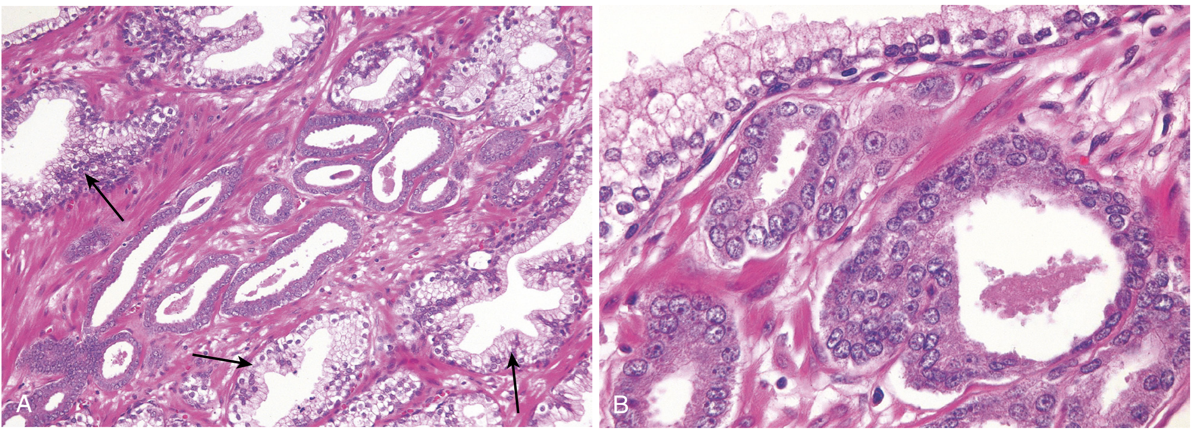
- Small, crowded glands without a basal cell layer
- Enlarged nuclei with prominent nucleoli (single, large, and central - a hallmark)
- Dark cytoplasm
- Perineural invasion (specific feature)
- AMACR (alpha-methylacyl-CoA racemase) - upregulated in cancer (sensitivity 82-100%)
- p63 / CK5/6 - basal cell markers absent in cancer (benign glands retain basal cells)
4. Gleason Grading & Grade Groups
| Grade Group | Gleason Score | Description | Prognosis |
|---|---|---|---|
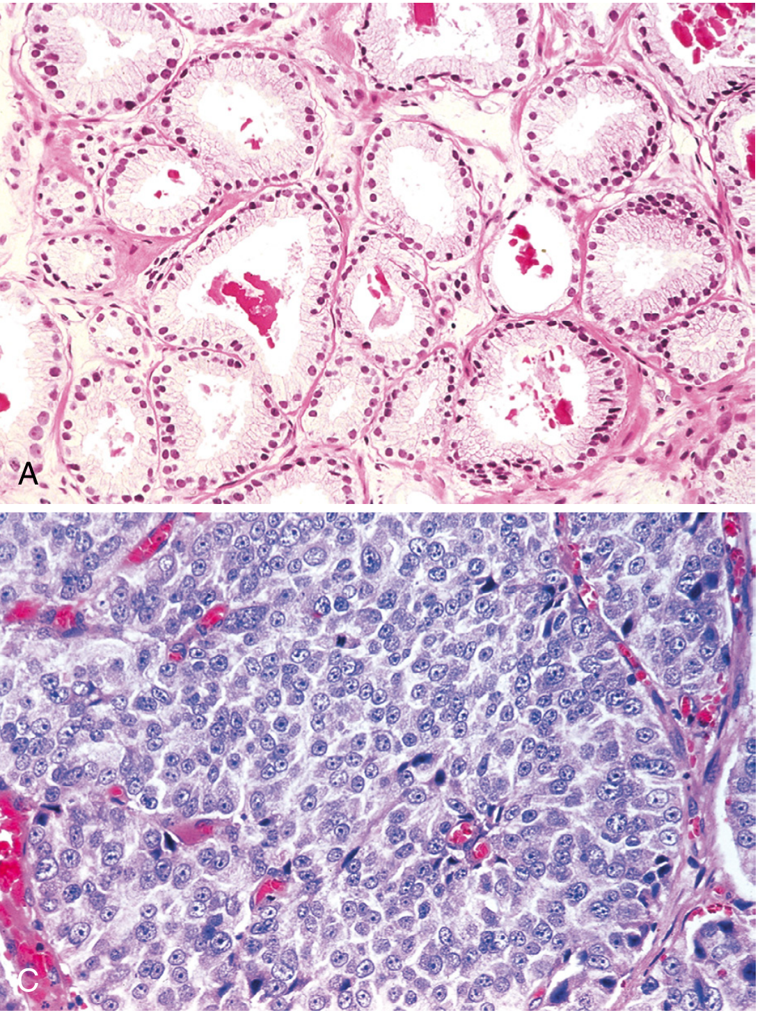
| GG 1 | ≤6 (3+3) | Well-formed, uniform individual glands | Excellent |
| GG 2 | 7 (3+4) | Predominantly well-formed glands + some poorly formed | Good |
| GG 3 | 7 (4+3) | Predominantly poorly formed glands + some well-formed | Intermediate |
| GG 4 | 8 (4+4 or 3+5 or 5+3) | Poorly formed / no glands or fused | Poor |
| GG 5 | 9-10 | No gland formation, sheets, single cells | Very poor |
"The Grade Group scheme has been investigated, and its correlation with prostate cancer-specific mortality has been validated." - Campbell-Walsh-Wein Urology
5. Clinical Features & Diagnosis
- Elevated or rising serum PSA
- Abnormal digital rectal exam (DRE) - hard, nodular, irregular, asymmetric
- Incidental finding on TURP specimen (T1a/T1b)
- Obstructive lower urinary tract symptoms
- Hematuria, hematospermia
- Bone pain from osteoblastic metastases (spine > pelvis > femur - a "blastic" pattern on plain X-ray and bone scan)
- Spinal cord compression (oncologic emergency)
- Elevated ALP, anemia (marrow involvement)
PSA (Prostate-Specific Antigen)
- Prostate cancer
- BPH
- Prostatitis
- Prostate manipulation/biopsy (wait 4-6 weeks after DRE to draw PSA)
- Ejaculation (wait 48 hours)
| Concept | Clinical Significance |
|---|---|
| Total PSA | >4 ng/mL commonly triggers biopsy |
| PSA density | PSA/gland volume; density >0.15 associated with cancer |
| Free PSA % | Lower free PSA % = higher cancer likelihood |
| PSA velocity | Rise >0.75 ng/mL/year suspicious |
| PSA doubling time (PSADT) | <3 months = high risk; >14 months = low risk |
"A single PSA measured at age 60 is associated (AUC of 0.90) with lifetime risk of death from prostate cancer." - Harrison's, p. 741
Diagnosis: Prostate Biopsy
- Transrectal ultrasound (TRUS)-guided biopsy or MRI-targeted biopsy (preferred when available)
- Standard: 12-core systematic biopsy of the peripheral zone
- MRI-TRUS fusion biopsy detects more clinically significant cancers and avoids overdiagnosis of insignificant disease
Advanced Imaging
| Modality | Role |
|---|---|
| Multiparametric MRI (mpMRI) | Best local staging - capsular extension, seminal vesicle invasion, NVB involvement |
| PSMA PET/CT | Gold standard for staging and biochemical recurrence; FDA-approved (¹⁸F-piflufolastat, ¹⁸F-DCFPyL) |
| Bone scan (⁹⁹ᵐTc-MDP) | Osteoblastic metastases; being replaced by PSMA PET |
| CT abdomen/pelvis | Lymph node assessment in intermediate/high-risk disease |
6. Staging (TNM)
| Stage | Description |
|---|---|
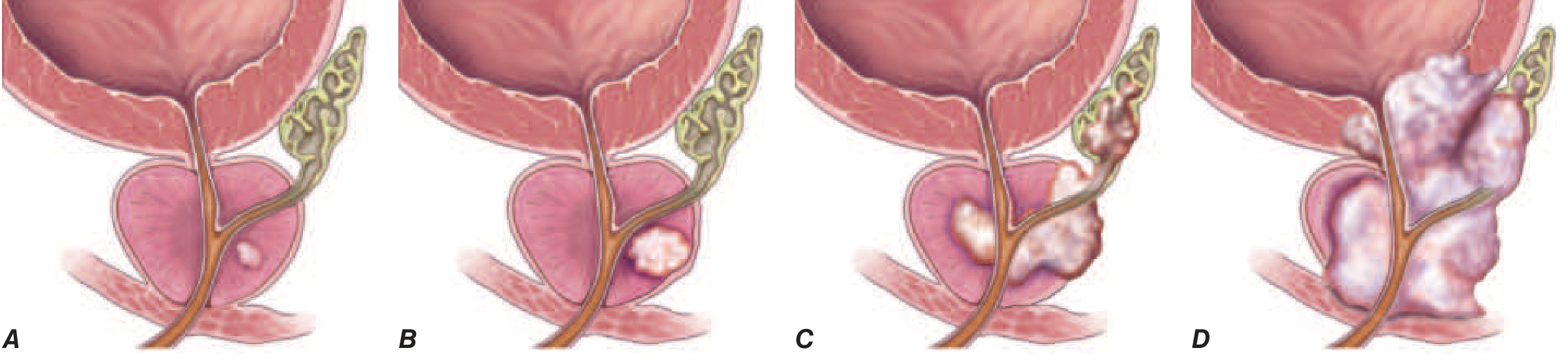
| T1a | Incidental finding, ≤5% of TURP chips |
| T1b | Incidental finding, >5% of TURP chips |
| T1c | Found on needle biopsy (elevated PSA) |
| T2a | Involves ≤50% of one lobe |
| T2b | >50% of one lobe, not both |
| T2c | Both lobes involved |
| T3a | Extracapsular extension |
| T3b | Seminal vesicle invasion |
| T4 | Invades adjacent structures (bladder, rectum, levator muscles) |
| N1 | Regional lymph node metastasis |
| M1 | Distant metastasis (M1a = non-regional LN, M1b = bone, M1c = other) |
| Risk | PSA | Grade Group | T Stage |
|---|---|---|---|
| Very Low | <10 | GG1 | T1c |
| Low | <10 | GG1 | T1-T2a |
| Intermediate | 10-20 OR GG2-3 OR T2b-T2c | - | - |
| High | >20 OR GG4-5 OR T3a | - | - |
| Very High | GG5, T3b-T4, or >4 biopsy cores GG4+ | - | - |
7. Treatment Overview
Localized Disease
"Patients with clinically localized disease are typically managed by radical prostatectomy, radiation therapy, or active surveillance. There is no clear evidence for the superiority of any one form of local therapy relative to another." - Harrison's, p. 742
| Approach | Indication |
|---|---|
| Active Surveillance (AS) | Very low/low risk; defers treatment to avoid overtreatment morbidity |
| Radical Prostatectomy (RP) | Localized disease, good performance status; curative intent |
| External Beam Radiation (EBRT) | Localized/locally advanced; alternative to surgery |
| Brachytherapy | Low-intermediate risk; radioactive seeds implanted |
| Combined EBRT + ADT | Intermediate/high-risk disease |
Metastatic / Advanced Disease
- Androgen Deprivation Therapy (ADT): GnRH agonists (leuprolide, goserelin) or antagonists (degarelix, relugolix) - reduces testosterone to castrate levels (<50 ng/dL)
- mCSPC (metastatic castration-sensitive): ADT + docetaxel, or ADT + abiraterone/enzalutamide - triplet therapy now standard in high-volume disease
- mCRPC (metastatic castration-resistant): Enzalutamide, abiraterone, cabazitaxel, PARP inhibitors (olaparib for BRCA mutations), ¹⁷⁷Lu-PSMA-617 (lutetium therapy)
- Bone-targeted therapy: Denosumab, zoledronic acid, Radium-223 (bone-specific)
PART 2: ROBOTIC PROSTATECTOMY (RARP/RALRP)
8. What is RALRP?
- 7 degrees of freedom (vs. 4 for laparoscopy)
- 10-12x 3D magnification
- Tremor filtration
- Articulating "Endowrist" instruments that mimic the human wrist
9. Indications & Contraindications
- Clinically localized prostate cancer (T1-T3a)
- Life expectancy >10 years
- Fit for general anesthesia and Trendelenburg position
- Particularly beneficial in patients with previous mesh hernia repair, obesity, or prior abdominal surgery (where open approach is difficult)
- Extensive prior pelvic surgery with dense adhesions
- Severe cardiopulmonary compromise (cannot tolerate steep Trendelenburg + pneumoperitoneum)
- Morbid obesity (though RALRP is often preferred over open in obese patients)
- Uncorrectable coagulopathy
10. Patient Positioning & OR Setup
"Fixed shoulder rests should be avoided because this can result in compression injury to the shoulders and brachial plexus when in the steep Trendelenburg position." - Campbell-Walsh-Wein Urology, p. 4677
- da Vinci S/Si/Xi system
- 0° and 30° robotic cameras
- Endowrist instruments: Maryland bipolar forceps, curved monopolar scissors, ProGrasp forceps, large needle drivers
- 12-mm and 8-mm trocars
- 18-Fr urethral catheter
11. Surgical Approach: Transperitoneal vs. Extraperitoneal
| Feature | Transperitoneal | Extraperitoneal |
|---|---|---|
| Working space | Larger | Smaller |
| Trendelenburg needed | Yes | May be less steep |
| Bowel involvement | More risk if adhesions | Peritoneum acts as barrier |
| Postoperative ileus | More common | Less common (diet quicker) |
| Operative time | Slightly longer | Slightly shorter |
| Previous mesh hernioplasty | Preferred | Difficult - space obliterated |
12. Key Surgical Steps (Transperitoneal Anterior RALRP)
- Port placement: 5-6 trocars placed in a fan arrangement (see trocar configuration figure in Campbell-Walsh Ch. 156). Camera port periumbilically.
- Pneumoperitoneum: CO₂ to 15 mmHg, steep Trendelenburg.
- Bowel mobilization: Sigmoid reflected, space of Retzius developed.
- Incision of the endopelvic fascia: Lateral to the prostate bilaterally.
- Dorsal venous complex (DVC) ligation: 0-polydioxanone suture around the DVC (Santorini's plexus) to control bleeding.
- Bladder neck dissection: Circumferential division of bladder neck, identification of ureteral orifices to avoid injury.
- Posterior dissection: Division of seminal vesicles and vas deferens; Denonvilliers' fascia incised.
- Neurovascular bundle (NVB) preservation (nerve-sparing technique - see below).
- Apical dissection: Division of the urethra at the apex with careful preservation of sphincter length.
- Specimen removal: Through a small extension of a port site (laparoscopic bag).
- Pelvic lymph node dissection (PLND): Performed in intermediate/high-risk cases; obturator fossa + external iliac nodes.
- Vesicourethral anastomosis (VUA): 3-0 Monocryl double-armed suture; a running (Van Velthoven) technique is standard. Watertight anastomosis over 18-Fr catheter.
13. Nerve-Sparing Technique - The Most Critical Step
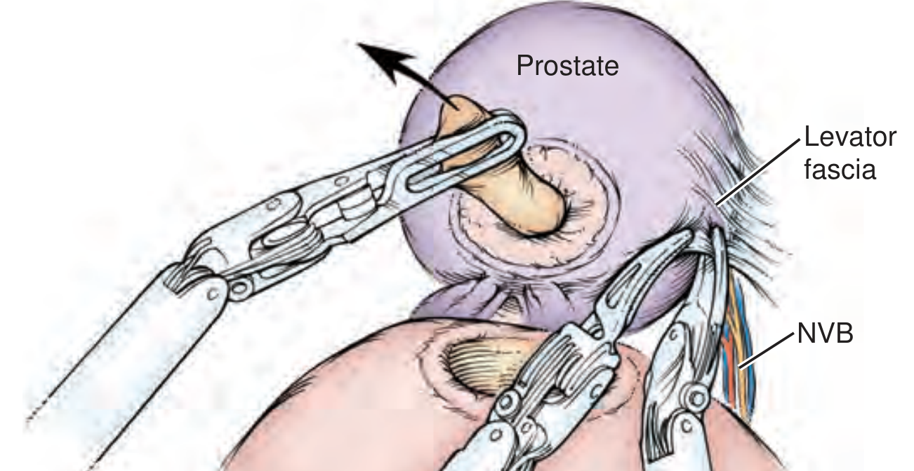
- Extrafascial: Between lateral prostatic fascia and levator fascia. Used for locally advanced disease (T3). Maximum cancer control, sacrifices NVB.
- Interfascial: Between levator fascia and prostatic fascia. Standard nerve-sparing for organ-confined disease.
- Intrafascial: Directly on prostatic capsule. Maximum nerve preservation for low-risk, smaller prostates.
"The superb visualization of the periprostatic tissues with minimally invasive surgery has led to a greater appreciation of the periprostatic fascial layers." - Campbell-Walsh-Wein Urology, p. 4678
- Cold scissors dissection preferred near NVB (avoid thermal energy)
- Antegrade vs. retrograde NVB release depending on anatomy
- Hemostasis with bipolar only where necessary
- "High anterior release" takes the nerve bundle off the anterior prostate surface - maximizes nerve tissue preservation
14. Outcomes of RALRP vs. Open Radical Prostatectomy
| Outcome | RALRP | Open RP |
|---|---|---|
| Blood loss | ~300 mL | ~1400 mL |
| Transfusion rate | 1-2% | 10-15% |
| Hospital stay | 1-2 days | 3-5 days |
| Return to activity | Faster | Slower |
| Positive surgical margins | Equivalent | Equivalent |
| Cancer control (PSA-free survival) | Equivalent | Equivalent |
| Continence recovery | Equivalent at 12 months | Equivalent at 12 months |
| Potency recovery | Equivalent (surgeon dependent) | Equivalent (surgeon dependent) |
"Outcomes are very dependent on the individual surgeon's ability and experience, and outcomes improve when surgery is performed by high-volume surgeons at high-volume centers." - Campbell-Walsh-Wein Urology, p. 4682
15. Complications of Robotic Prostatectomy
Intraoperative
| Complication | Notes |
|---|---|
| Bleeding | DVC injury; transfusion rare (1-2%) |
| Rectal injury | Rare but serious; recognized = primary repair + omental interposition; unrecognized = recto-vesical fistula, needs colostomy |
| Ureteral injury | At bladder neck (post-TURP, large median lobe); ureter passes close to vas; stent if at risk |
| Robot failure | Critical failure rate ~0.4%; conversion to laparoscopic or open |
Postoperative
| Complication | Notes |
|---|---|
| Urinary incontinence | Most common functional complication; 90% continent by 12 months; Kegel exercises help recovery |
| Erectile dysfunction | Related to NVB preservation; age and baseline function are key predictors |
| Anastomotic leak | Early urinary extravasation; catheter left for 7-14 days |
| DVT/PE | Risk from Trendelenburg + pneumoperitoneum; sequential compression stockings + early ambulation |
| Positive surgical margin (PSM) | ~10-20%; risk increases with extracapsular extension |
| Biochemical recurrence | PSA >0.2 ng/mL after RP; triggers salvage radiation ± ADT |
Positioning Complications (unique to steep Trendelenburg)
- Brachial plexus injury (arms not tucked properly)
- Radial/ulnar nerve compression
- Femoral neurapraxia (excessive hip flexion of table)
- Facial/laryngeal edema (prolonged Trendelenburg)
- Ocular complications (increased intraocular pressure)
- Rhabdomyolysis (prolonged compression of compartments)
16. Functional Recovery After RALRP
- Immediate post-catheter removal: variable incontinence
- 3 months: ~60-70% continent
- 12 months: ~85-95% continent (no pad or 1 security pad)
- Predictors of recovery: pre-op continence, sphincter length, preservation of urethra length at apex
- Bilateral nerve-sparing + young patient + good baseline function: ~60-80% potency at 12-18 months
- Unilateral nerve-sparing: ~40-50%
- Non-nerve-sparing: <10%
- PDE5 inhibitors (sildenafil, tadalafil) used early as "penile rehabilitation"
- PSA should be undetectable (<0.1 ng/mL) by 6 weeks post-RP
- Biochemical recurrence defined as PSA ≥0.2 ng/mL (confirmed on repeat)
- PSADT calculated - guides need for salvage radiation vs. observation
17. Special Procedure: Salvage RALRP
Summary Table: Prostate Cancer at a Glance
| Domain | Key Points |
|---|---|
| Most common histology | Adenocarcinoma (>95%) |
| Most common location | Peripheral zone (~70-80%) |
| Key marker | PSA (organ-specific, NOT cancer-specific) |
| Grading system | Gleason score / Grade Groups 1-5 |
| Pathological hallmark | Absent basal cell layer; prominent nucleoli; AMACR+ |
| Metastasis pattern | Osteoblastic bone metastases (spine, pelvis) |
| Local staging modality | mpMRI prostate |
| Systemic staging | PSMA PET/CT (gold standard) |
| Curative treatments | Surgery (RALRP), EBRT, Brachytherapy |
| Hormonal treatment | ADT (GnRH agonist/antagonist) |
| RALRP advantages | Less blood loss, faster recovery, equivalent oncological outcomes |
| Key RALRP step | NVB preservation (potency); watertight VUA (continence) |
| Post-RALRP surveillance | PSA should be undetectable by 6 weeks |
- Campbell-Walsh-Wein Urology (gold standard urology reference), Chapters 84, 85, 156
- Robbins & Cotran Pathologic Basis of Disease (2023), Chapter 21
- Harrison's Principles of Internal Medicine 22E (2025), Chapter 92
- Miller's Anesthesia 10E, Chapter 23 (RALRP anesthetic considerations)
- Wang et al., 2023 - RARP vs open RP meta-analysis (PMID 37721644)
- Nahas et al., 2024 - Randomized trial RARP vs open RP (PMID 38723593)
- Dinneen et al., 2025 - NeuroSAFE PROOF RCT, Lancet Oncology (PMID 40147459)
Make into a word document
ls /home/daytona/workspace/ 2>/dev/null && ls /home/daytona/skills/docx/
mkdir -p /home/daytona/workspace/prostate-cancer-doc && node -e "require('docx'); console.log('docx available')"
~/prostate-cancer-doc/build.js
const {
Document, Packer, Paragraph, TextRun, Table, TableRow, TableCell,
HeadingLevel, AlignmentType, BorderStyle, WidthType, ShadingType,
VerticalAlign, PageNumber, Header, Footer, TableOfContents,
UnderlineType, PageBreak
} = require('docx');
const fs = require('fs');
// ── helpers ──────────────────────────────────────────────────────────────────
function h1(text) {
return new Paragraph({ text, heading: HeadingLevel.HEADING_1, spacing: { before: 360, after: 120 } });
}
function h2(text) {
return new Paragraph({ text, heading: HeadingLevel.HEADING_2, spacing: { before: 280, after: 80 } });
}
function h3(text) {
return new Paragraph({ text, heading: HeadingLevel.HEADING_3, spacing: { before: 200, after: 60 } });
}
function para(text, opts = {}) {
return new Paragraph({
children: [new TextRun({ text, size: 22, ...opts })],
spacing: { before: 60, after: 60 },
});
}
function bold(text) {
return new Paragraph({
children: [new TextRun({ text, bold: true, size: 22 })],
spacing: { before: 60, after: 60 },
});
}
function bullet(text) {
return new Paragraph({
children: [new TextRun({ text, size: 22 })],
bullet: { level: 0 },
spacing: { before: 40, after: 40 },
});
}
function subbullet(text) {
return new Paragraph({
children: [new TextRun({ text, size: 22 })],
bullet: { level: 1 },
spacing: { before: 30, after: 30 },
});
}
function italic(text) {
return new Paragraph({
children: [new TextRun({ text, italics: true, size: 22, color: '555555' })],
spacing: { before: 40, after: 60 },
});
}
function divider() {
return new Paragraph({
children: [new TextRun({ text: '' })],
border: { bottom: { color: 'AAAAAA', space: 1, style: BorderStyle.SINGLE, size: 6 } },
spacing: { before: 120, after: 120 },
});
}
function pageBreak() {
return new Paragraph({ children: [new PageBreak()] });
}
function noteText(text) {
return new Paragraph({
children: [new TextRun({ text, italics: true, size: 20, color: '444488' })],
spacing: { before: 40, after: 60 },
indent: { left: 360 },
border: { left: { color: '4444AA', space: 10, style: BorderStyle.SINGLE, size: 12 } },
});
}
// ── table builder ─────────────────────────────────────────────────────────────
function makeTable(headers, rows, colWidths) {
const HEADER_COLOR = '1F4E79';
const ALT_COLOR = 'D6E4F0';
const headerRow = new TableRow({
tableHeader: true,
children: headers.map((h, i) =>
new TableCell({
children: [new Paragraph({
children: [new TextRun({ text: h, bold: true, color: 'FFFFFF', size: 20 })],
alignment: AlignmentType.CENTER,
})],
shading: { fill: HEADER_COLOR, type: ShadingType.CLEAR },
verticalAlign: VerticalAlign.CENTER,
width: colWidths ? { size: colWidths[i], type: WidthType.PERCENTAGE } : undefined,
})
),
});
const dataRows = rows.map((row, ri) =>
new TableRow({
children: row.map((cell, ci) =>
new TableCell({
children: [new Paragraph({
children: [new TextRun({ text: cell, size: 20, bold: ci === 0 && row.length > 1 })],
})],
shading: ri % 2 === 1 ? { fill: ALT_COLOR, type: ShadingType.CLEAR } : undefined,
width: colWidths ? { size: colWidths[ci], type: WidthType.PERCENTAGE } : undefined,
})
),
})
);
return new Table({
rows: [headerRow, ...dataRows],
width: { size: 100, type: WidthType.PERCENTAGE },
margins: { top: 80, bottom: 80, left: 120, right: 120 },
});
}
// ── DOCUMENT ─────────────────────────────────────────────────────────────────
const children = [];
// ── Title page ────────────────────────────────────────────────────────────────
children.push(
new Paragraph({
children: [new TextRun({ text: 'Prostate Cancer', bold: true, size: 64, color: '1F4E79' })],
alignment: AlignmentType.CENTER,
spacing: { before: 1440, after: 200 },
}),
new Paragraph({
children: [new TextRun({ text: '& Robotic Prostatectomy', bold: true, size: 48, color: '2E75B6' })],
alignment: AlignmentType.CENTER,
spacing: { before: 0, after: 400 },
}),
new Paragraph({
children: [new TextRun({ text: 'A Comprehensive Clinical Teaching Guide', size: 28, italics: true, color: '555555' })],
alignment: AlignmentType.CENTER,
spacing: { before: 0, after: 200 },
}),
new Paragraph({
children: [new TextRun({ text: 'Sources: Campbell-Walsh-Wein Urology | Robbins & Cotran Pathology | Harrison\'s Internal Medicine 22E', size: 20, color: '777777' })],
alignment: AlignmentType.CENTER,
spacing: { before: 0, after: 1440 },
}),
pageBreak()
);
// ── PART 1: Prostate Cancer ───────────────────────────────────────────────────
children.push(
new Paragraph({
children: [new TextRun({ text: 'PART 1: PROSTATE CANCER', bold: true, size: 36, color: '1F4E79' })],
alignment: AlignmentType.CENTER,
spacing: { before: 200, after: 300 },
})
);
// 1. Epidemiology
children.push(
h1('1. Epidemiology & Risk Factors'),
para('Prostate cancer is the most common non-cutaneous malignancy in men in the Western world and the second leading cause of cancer death in men.'),
para(''),
makeTable(
['Risk Factor', 'Details'],
[
['Age', 'Rare before 50; incidence rises steeply after 60'],
['Race', 'African-American men have the highest incidence and mortality worldwide'],
['Family History', '2–3× risk if first-degree relative affected; BRCA2 mutations confer high risk'],
['Diet', 'High fat, red meat associated with increased risk; lycopene/selenium may be protective'],
['Geography', 'High incidence in North America, Europe; low in Asia (rises with Westernisation)'],
],
[25, 75]
),
para(''),
noteText('Over 70–80% of newly diagnosed cancers are now clinically organ-confined, largely due to PSA screening. — Harrison\'s Principles of Internal Medicine 22E'),
divider()
);
// 2. Anatomy
children.push(
h1('2. Anatomy Review'),
para('The prostate is a walnut-sized gland at the bladder base surrounding the urethra. It has three zones relevant to cancer:'),
bullet('Peripheral zone (~70% of gland): Site of ~70–80% of prostate cancers — palpable on DRE'),
bullet('Transition zone (~25%): Site of BPH; ~20% of cancers'),
bullet('Central zone (~5%): Rarely affected by cancer'),
para(''),
para('Key adjacent structures during surgery: rectum (posterior), bladder neck (superior), external urethral sphincter (inferior), and the neurovascular bundles (NVBs) running posterolaterally — critical for erectile function preservation.'),
divider()
);
// 3. Pathology
children.push(
h1('3. Pathology'),
para('Over 95% of prostate cancers are adenocarcinomas arising from the secretory epithelial cells. Key pathological features:'),
para(''),
h2('Microscopic Features of Malignancy'),
bullet('Small, crowded glands without a basal cell layer'),
bullet('Enlarged nuclei with prominent nucleoli (single, large, central — a hallmark)'),
bullet('Dark cytoplasm'),
bullet('Perineural invasion (specific feature)'),
para(''),
h2('Useful Immunohistochemical Markers'),
makeTable(
['Marker', 'Role in Prostate Cancer'],
[
['AMACR (alpha-methylacyl-CoA racemase)', 'Upregulated in cancer; sensitivity 82–100%'],
['p63 / CK5/6', 'Basal cell markers — ABSENT in cancer (benign glands retain basal cells)'],
],
[40, 60]
),
para(''),
h2('High-Grade PIN (HGPIN)'),
para('Architecturally benign large acini with cytologically atypical cells. Found in ~80% of specimens harbouring carcinoma. It RETAINS a basal cell layer — unlike frank carcinoma.'),
divider()
);
// 4. Gleason Grading
children.push(
h1('4. Gleason Grading & Grade Groups'),
para('The Gleason system grades glandular patterns from 1 (most differentiated) to 5 (least differentiated). The two most prevalent patterns are summed to give a Gleason Score (2–10). The modern system uses Grade Groups (GG):'),
para(''),
makeTable(
['Grade Group', 'Gleason Score', 'Description', 'Prognosis'],
[
['GG 1', '≤6 (3+3)', 'Well-formed, uniform individual glands', 'Excellent'],
['GG 2', '7 (3+4)', 'Predominantly well-formed + some poorly formed', 'Good'],
['GG 3', '7 (4+3)', 'Predominantly poorly formed + some well-formed', 'Intermediate'],
['GG 4', '8 (4+4/3+5/5+3)', 'Poorly formed / no glands or fused', 'Poor'],
['GG 5', '9–10', 'No gland formation, sheets, single cells', 'Very poor'],
],
[15, 20, 45, 20]
),
para(''),
noteText('"The Grade Group scheme has been investigated, and its correlation with prostate cancer-specific mortality has been validated." — Campbell-Walsh-Wein Urology'),
divider()
);
// 5. Clinical Features & Diagnosis
children.push(
h1('5. Clinical Features & Diagnosis'),
para('Localized disease is typically asymptomatic. It is usually found via:'),
bullet('Elevated or rising serum PSA'),
bullet('Abnormal digital rectal exam (DRE) — hard, nodular, irregular, asymmetric'),
bullet('Incidental finding on TURP specimen (T1a/T1b)'),
para(''),
h2('Advanced Disease Presentation'),
bullet('Obstructive lower urinary tract symptoms'),
bullet('Hematuria, hematospermia'),
bullet('Bone pain from osteoblastic metastases (spine > pelvis > femur)'),
bullet('Spinal cord compression (oncologic emergency)'),
bullet('Elevated ALP, anemia (marrow involvement)'),
para(''),
h2('PSA (Prostate-Specific Antigen)'),
para('PSA is a kallikrein-related serine protease (KLK3) produced by prostatic epithelium — organ-specific but NOT cancer-specific. Elevated by: prostate cancer, BPH, prostatitis, prostate manipulation, ejaculation.'),
para(''),
makeTable(
['PSA Concept', 'Clinical Significance'],
[
['Total PSA', '>4 ng/mL commonly triggers biopsy'],
['PSA Density', 'PSA / gland volume; density >0.15 associated with cancer'],
['Free PSA %', 'Lower free PSA % = higher cancer likelihood'],
['PSA Velocity', 'Rise >0.75 ng/mL/year is suspicious'],
['PSA Doubling Time (PSADT)', '<3 months = high risk; >14 months = low risk'],
],
[35, 65]
),
para(''),
h2('Imaging Modalities'),
makeTable(
['Modality', 'Role'],
[
['Multiparametric MRI (mpMRI)', 'Best local staging — capsular extension, seminal vesicle invasion, NVB involvement'],
['PSMA PET/CT', 'Gold standard for staging and biochemical recurrence; FDA-approved'],
['Bone Scan (99mTc-MDP)', 'Osteoblastic metastases; being replaced by PSMA PET'],
['CT Abdomen/Pelvis', 'Lymph node assessment in intermediate/high-risk disease'],
],
[30, 70]
),
divider()
);
// 6. Staging
children.push(
h1('6. Staging (TNM)'),
makeTable(
['Stage', 'Description'],
[
['T1a', 'Incidental finding, ≤5% of TURP chips'],
['T1b', 'Incidental finding, >5% of TURP chips'],
['T1c', 'Found on needle biopsy (elevated PSA)'],
['T2a', 'Involves ≤50% of one lobe'],
['T2b', '>50% of one lobe, not both'],
['T2c', 'Both lobes involved'],
['T3a', 'Extracapsular extension'],
['T3b', 'Seminal vesicle invasion'],
['T4', 'Invades adjacent structures (bladder, rectum, levator muscles)'],
['N1', 'Regional lymph node metastasis'],
['M1a/b/c', 'Distant metastasis (non-regional LN / bone / other)'],
],
[15, 85]
),
para(''),
h2('Risk Stratification (NCCN)'),
makeTable(
['Risk Group', 'PSA', 'Grade Group', 'T Stage'],
[
['Very Low', '<10', 'GG1', 'T1c'],
['Low', '<10', 'GG1', 'T1–T2a'],
['Intermediate', '10–20 OR GG2–3', 'GG2–3', 'T2b–T2c'],
['High', '>20 OR GG4–5', 'GG4–5', 'T3a'],
['Very High', 'Any', 'GG5 or >4 cores GG4+', 'T3b–T4'],
],
[20, 25, 30, 25]
),
divider()
);
// 7. Treatment
children.push(
h1('7. Treatment Overview'),
h2('Localized Disease'),
makeTable(
['Approach', 'Indication'],
[
['Active Surveillance (AS)', 'Very low/low risk; defers treatment to avoid overtreatment morbidity'],
['Radical Prostatectomy (RP)', 'Localized disease, good performance status; curative intent'],
['External Beam Radiation (EBRT)', 'Localized/locally advanced; alternative to surgery'],
['Brachytherapy', 'Low–intermediate risk; radioactive seeds implanted'],
['Combined EBRT + ADT', 'Intermediate/high-risk disease'],
],
[30, 70]
),
para(''),
h2('Metastatic / Advanced Disease'),
bullet('Androgen Deprivation Therapy (ADT): GnRH agonists (leuprolide, goserelin) or antagonists (degarelix, relugolix)'),
bullet('mCSPC: ADT + docetaxel, or ADT + abiraterone/enzalutamide — triplet therapy now standard in high-volume disease'),
bullet('mCRPC: Enzalutamide, abiraterone, cabazitaxel, PARP inhibitors (olaparib for BRCA mutations), 177Lu-PSMA-617'),
bullet('Bone-targeted therapy: Denosumab, zoledronic acid, Radium-223'),
divider()
);
// ── PART 2: Robotic Prostatectomy ────────────────────────────────────────────
children.push(
pageBreak(),
new Paragraph({
children: [new TextRun({ text: 'PART 2: ROBOTIC PROSTATECTOMY', bold: true, size: 36, color: '1F4E79' })],
alignment: AlignmentType.CENTER,
spacing: { before: 200, after: 300 },
})
);
// 8. What is RALRP
children.push(
h1('8. What is RALRP?'),
para('Robot-Assisted Laparoscopic Radical Prostatectomy (RALRP) uses the da Vinci Surgical System to perform minimally invasive removal of the prostate with curative intent. Introduced into urology in 2002, it has rapidly become the dominant surgical approach — today >85% of radical prostatectomies in the USA are performed robotically.'),
para(''),
para('The da Vinci system provides:'),
bullet('7 degrees of freedom (vs. 4 for laparoscopy)'),
bullet('10–12× 3D magnification'),
bullet('Tremor filtration'),
bullet('Articulating "Endowrist" instruments that mimic the human wrist'),
divider()
);
// 9. Indications & Contraindications
children.push(
h1('9. Indications & Contraindications'),
h2('Indications'),
bullet('Clinically localized prostate cancer (T1–T3a)'),
bullet('Life expectancy >10 years'),
bullet('Fit for general anaesthesia and Trendelenburg position'),
bullet('Particularly beneficial in patients with previous mesh hernia repair, obesity, or prior abdominal surgery'),
para(''),
h2('Relative Contraindications'),
bullet('Extensive prior pelvic surgery with dense adhesions'),
bullet('Severe cardiopulmonary compromise (cannot tolerate steep Trendelenburg + pneumoperitoneum)'),
bullet('Morbid obesity (RALRP is often still preferred over open in these patients)'),
bullet('Uncorrectable coagulopathy'),
divider()
);
// 10. Patient Positioning & OR Setup
children.push(
h1('10. Patient Positioning & OR Setup'),
para('Positioning: Steep Trendelenburg (~22–25°) with arms tucked at sides, padded with "egg crate" foam. Sequential compression stockings. Feet may be in stirrups or spread on foot rests.'),
para(''),
noteText('"Fixed shoulder rests should be avoided because this can result in compression injury to the shoulders and brachial plexus when in the steep Trendelenburg position." — Campbell-Walsh-Wein Urology, p. 4677'),
para(''),
h2('Key Equipment'),
bullet('da Vinci S/Si/Xi system with 0° and 30° robotic cameras'),
bullet('Endowrist instruments: Maryland bipolar forceps, curved monopolar scissors, ProGrasp forceps, large needle drivers'),
bullet('12-mm and 8-mm trocars (5–6 total)'),
bullet('18-Fr urethral catheter'),
bullet('0-polydioxanone suture for DVC ligation; 3-0 Monocryl for anastomosis'),
divider()
);
// 11. Transperitoneal vs Extraperitoneal
children.push(
h1('11. Surgical Approach: Transperitoneal vs. Extraperitoneal'),
makeTable(
['Feature', 'Transperitoneal', 'Extraperitoneal'],
[
['Working space', 'Larger', 'Smaller'],
['Trendelenburg needed', 'Yes, steep', 'May be less steep'],
['Bowel involvement', 'More risk if adhesions', 'Peritoneum acts as barrier'],
['Postoperative ileus', 'More common', 'Less common (diet quicker)'],
['Operative time', 'Slightly longer', 'Slightly shorter'],
['Previous mesh hernioplasty', 'Preferred approach', 'Difficult — space obliterated'],
],
[34, 33, 33]
),
divider()
);
// 12. Surgical Steps
children.push(
h1('12. Key Surgical Steps (Transperitoneal Anterior RALRP)'),
para(''),
new Paragraph({
children: [new TextRun({ text: '1. Port Placement', bold: true, size: 22 }), new TextRun({ text: ' — 5–6 trocars placed in a fan arrangement. Camera port periumbilically.', size: 22 })],
spacing: { before: 60, after: 40 },
}),
new Paragraph({
children: [new TextRun({ text: '2. Pneumoperitoneum', bold: true, size: 22 }), new TextRun({ text: ' — CO₂ to 15 mmHg, steep Trendelenburg.', size: 22 })],
spacing: { before: 40, after: 40 },
}),
new Paragraph({
children: [new TextRun({ text: '3. Bowel Mobilization', bold: true, size: 22 }), new TextRun({ text: ' — Sigmoid reflected, space of Retzius developed.', size: 22 })],
spacing: { before: 40, after: 40 },
}),
new Paragraph({
children: [new TextRun({ text: '4. Endopelvic Fascia Incision', bold: true, size: 22 }), new TextRun({ text: ' — Lateral to prostate bilaterally.', size: 22 })],
spacing: { before: 40, after: 40 },
}),
new Paragraph({
children: [new TextRun({ text: '5. Dorsal Venous Complex (DVC) Ligation', bold: true, size: 22 }), new TextRun({ text: ' — 0-polydioxanone suture around Santorini\'s plexus to control bleeding.', size: 22 })],
spacing: { before: 40, after: 40 },
}),
new Paragraph({
children: [new TextRun({ text: '6. Bladder Neck Dissection', bold: true, size: 22 }), new TextRun({ text: ' — Circumferential division; identify ureteral orifices to avoid injury.', size: 22 })],
spacing: { before: 40, after: 40 },
}),
new Paragraph({
children: [new TextRun({ text: '7. Posterior Dissection', bold: true, size: 22 }), new TextRun({ text: ' — Division of seminal vesicles and vas deferens; Denonvilliers\' fascia incised.', size: 22 })],
spacing: { before: 40, after: 40 },
}),
new Paragraph({
children: [new TextRun({ text: '8. NVB Preservation', bold: true, size: 22 }), new TextRun({ text: ' — (See Section 13 — the most critical step).', size: 22 })],
spacing: { before: 40, after: 40 },
}),
new Paragraph({
children: [new TextRun({ text: '9. Apical Dissection', bold: true, size: 22 }), new TextRun({ text: ' — Division of urethra at apex; preserve maximum sphincter length for continence.', size: 22 })],
spacing: { before: 40, after: 40 },
}),
new Paragraph({
children: [new TextRun({ text: '10. Specimen Removal', bold: true, size: 22 }), new TextRun({ text: ' — Through a small extension of a port site in a laparoscopic bag.', size: 22 })],
spacing: { before: 40, after: 40 },
}),
new Paragraph({
children: [new TextRun({ text: '11. Pelvic Lymph Node Dissection (PLND)', bold: true, size: 22 }), new TextRun({ text: ' — Obturator fossa + external iliac nodes; performed in intermediate/high-risk cases.', size: 22 })],
spacing: { before: 40, after: 40 },
}),
new Paragraph({
children: [new TextRun({ text: '12. Vesicourethral Anastomosis (VUA)', bold: true, size: 22 }), new TextRun({ text: ' — 3-0 Monocryl double-armed suture; Van Velthoven running technique; watertight over 18-Fr catheter.', size: 22 })],
spacing: { before: 40, after: 60 },
}),
divider()
);
// 13. Nerve Sparing
children.push(
h1('13. Nerve-Sparing Technique — The Most Critical Step'),
para('The neurovascular bundles (NVBs) run posterolaterally along the prostate within the peri-prostatic fascia and are responsible for penile erections. Their preservation determines post-operative erectile function.'),
para(''),
h2('Fascial Dissection Planes (least to most nerve-sparing)'),
makeTable(
['Plane', 'Location', 'Indication'],
[
['Extrafascial', 'Between lateral prostatic fascia and levator fascia', 'Locally advanced (T3); maximum cancer control, sacrifices NVB'],
['Interfascial', 'Between levator fascia and prostatic fascia', 'Standard nerve-sparing for organ-confined disease'],
['Intrafascial', 'Directly on prostatic capsule', 'Maximum nerve preservation for low-risk, smaller prostates'],
],
[18, 50, 32]
),
para(''),
h2('Principles of Nerve Sparing'),
bullet('Cold scissors dissection preferred near NVB — avoid thermal energy'),
bullet('Antegrade vs. retrograde NVB release depending on anatomy'),
bullet('Haemostasis with bipolar only where strictly necessary'),
bullet('"High anterior release" takes nerve bundle off the anterior prostate surface — maximises nerve tissue preserved'),
para(''),
noteText('"The superb visualization of the periprostatic tissues with minimally invasive surgery has led to a greater appreciation of the periprostatic fascial layers." — Campbell-Walsh-Wein Urology, p. 4678'),
divider()
);
// 14. Outcomes
children.push(
h1('14. Outcomes: RALRP vs. Open Radical Prostatectomy'),
makeTable(
['Outcome', 'RALRP', 'Open RP'],
[
['Blood loss', '~300 mL', '~1,400 mL'],
['Transfusion rate', '1–2%', '10–15%'],
['Hospital stay', '1–2 days', '3–5 days'],
['Return to activity', 'Faster', 'Slower'],
['Positive surgical margins', 'Equivalent', 'Equivalent'],
['Cancer control (PSA-free survival)', 'Equivalent', 'Equivalent'],
['Continence recovery at 12 months', 'Equivalent', 'Equivalent'],
['Potency recovery', 'Equivalent (surgeon dependent)', 'Equivalent (surgeon dependent)'],
],
[40, 30, 30]
),
para(''),
noteText('"Outcomes are very dependent on the individual surgeon\'s ability and experience, and outcomes improve when surgery is performed by high-volume surgeons at high-volume centers." — Campbell-Walsh-Wein Urology, p. 4682'),
para(''),
h2('Recent Evidence'),
bullet('Wang et al. 2023 (PMID 37721644) — Systematic review & meta-analysis confirming RALRP achieves equivalent oncological outcomes with significantly less blood loss and shorter hospital stay vs. open RP.'),
bullet('Nahas et al. 2024 (PMID 38723593) — Randomized clinical trial confirming equivalent oncological and functional outcomes; RALRP favoured for blood loss and length of stay.'),
bullet('Dinneen et al. 2025 — NeuroSAFE PROOF Phase 3 RCT (Lancet Oncology, PMID 40147459): NeuroSAFE-guided RARP (intraoperative frozen section of NVB interface) significantly improved erectile function and urinary continence recovery vs. standard RARP.'),
divider()
);
// 15. Complications
children.push(
h1('15. Complications of Robotic Prostatectomy'),
h2('Intraoperative Complications'),
makeTable(
['Complication', 'Notes'],
[
['Bleeding (DVC)', 'Transfusion rare (1–2%); CO2 pneumoperitoneum compresses venous bleeding'],
['Rectal injury', 'Rare but serious; recognized = primary repair + omental interposition; unrecognized = recto-vesical fistula (requires colostomy)'],
['Ureteral injury', 'At bladder neck (post-TURP, large median lobe); stent if at risk'],
['Robot failure', 'Critical failure ~0.4%; conversion to laparoscopic or open'],
],
[25, 75]
),
para(''),
h2('Postoperative Complications'),
makeTable(
['Complication', 'Notes'],
[
['Urinary incontinence', 'Most common functional issue; 90% continent by 12 months; Kegel exercises help'],
['Erectile dysfunction', 'Related to NVB preservation; age and baseline function are key predictors'],
['Anastomotic leak', 'Early urinary extravasation; catheter left 7–14 days'],
['DVT / PE', 'Risk from Trendelenburg + pneumoperitoneum; compression stockings + early ambulation'],
['Positive surgical margin', '~10–20%; risk increases with extracapsular extension'],
['Biochemical recurrence', 'PSA >0.2 ng/mL after RP; triggers salvage radiation ± ADT'],
],
[30, 70]
),
para(''),
h2('Positioning Complications (Unique to Steep Trendelenburg)'),
bullet('Brachial plexus injury (arms not tucked properly)'),
bullet('Radial / ulnar nerve compression'),
bullet('Femoral neurapraxia (excessive hip flexion of table)'),
bullet('Facial / laryngeal oedema (prolonged Trendelenburg)'),
bullet('Raised intraocular pressure / ocular complications'),
bullet('Rhabdomyolysis (prolonged compression of compartments)'),
divider()
);
// 16. Functional Recovery
children.push(
h1('16. Functional Recovery After RALRP'),
h2('Urinary Continence'),
bullet('Immediate post-catheter removal: variable incontinence'),
bullet('3 months: ~60–70% continent'),
bullet('12 months: ~85–95% continent (no pad or 1 security pad)'),
bullet('Predictors of recovery: pre-op continence status, sphincter length, preservation of urethra length at apex'),
para(''),
h2('Erectile Function'),
bullet('Bilateral nerve-sparing + young patient + good baseline function: ~60–80% potency at 12–18 months'),
bullet('Unilateral nerve-sparing: ~40–50%'),
bullet('Non-nerve-sparing: <10%'),
bullet('PDE5 inhibitors (sildenafil, tadalafil) used early as "penile rehabilitation"'),
para(''),
h2('PSA Monitoring (Oncological Surveillance)'),
bullet('PSA should be undetectable (<0.1 ng/mL) by 6 weeks post-RP'),
bullet('Biochemical recurrence: PSA ≥0.2 ng/mL confirmed on repeat testing'),
bullet('PSA doubling time (PSADT) calculated — guides need for salvage radiation vs. observation'),
divider()
);
// 17. Summary Table
children.push(
pageBreak(),
h1('17. Summary: Prostate Cancer at a Glance'),
makeTable(
['Domain', 'Key Points'],
[
['Most common histology', 'Adenocarcinoma (>95%)'],
['Most common location', 'Peripheral zone (~70–80%)'],
['Key marker', 'PSA (organ-specific, NOT cancer-specific)'],
['Grading system', 'Gleason score / Grade Groups 1–5'],
['Pathological hallmark', 'Absent basal cell layer; prominent nucleoli; AMACR+'],
['Metastasis pattern', 'Osteoblastic bone metastases (spine, pelvis)'],
['Local staging modality', 'mpMRI prostate'],
['Systemic staging', 'PSMA PET/CT (gold standard)'],
['Curative treatments', 'Surgery (RALRP), EBRT, Brachytherapy'],
['Hormonal treatment', 'ADT (GnRH agonist/antagonist)'],
['RALRP advantages', 'Less blood loss, faster recovery, equivalent oncological outcomes'],
['Key RALRP steps', 'NVB preservation (potency); watertight VUA (continence)'],
['Post-RALRP surveillance', 'PSA should be undetectable by 6 weeks'],
],
[35, 65]
),
divider()
);
// Sources
children.push(
h2('Sources & References'),
bullet('Campbell-Walsh-Wein Urology (Chapters 84, 85, 156) — Gold standard urology reference'),
bullet('Robbins & Cotran Pathologic Basis of Disease (2023), Chapter 21'),
bullet("Harrison's Principles of Internal Medicine 22E (2025), Chapter 92"),
bullet('Miller\'s Anesthesia 10E, Chapter 23 (RALRP anaesthetic considerations)'),
bullet('Wang et al. 2023 — Robot-assisted versus open radical prostatectomy: systematic review and meta-analysis of prospective studies. J Robot Surg. PMID 37721644'),
bullet('Nahas et al. 2024 — Perioperative, Oncological, and Functional Outcomes Between RALRP and Open RRP: A Randomized Clinical Trial. J Urol. PMID 38723593'),
bullet('Dinneen et al. 2025 — NeuroSAFE PROOF: multicentre, patient-blinded, randomised, controlled phase 3 trial. Lancet Oncol. PMID 40147459'),
);
// ── Build document ────────────────────────────────────────────────────────────
const doc = new Document({
creator: 'Orris Medical AI',
title: 'Prostate Cancer & Robotic Prostatectomy',
description: 'Comprehensive clinical teaching guide',
styles: {
default: {
document: {
run: { font: 'Calibri', size: 22 },
},
heading1: {
run: { font: 'Calibri', bold: true, size: 32, color: '1F4E79' },
paragraph: { spacing: { before: 360, after: 120 } },
},
heading2: {
run: { font: 'Calibri', bold: true, size: 26, color: '2E75B6' },
paragraph: { spacing: { before: 240, after: 80 } },
},
heading3: {
run: { font: 'Calibri', bold: true, size: 24, color: '2E75B6' },
paragraph: { spacing: { before: 160, after: 60 } },
},
},
},
sections: [{
properties: {
page: {
margin: { top: 1080, bottom: 1080, left: 1080, right: 1080 },
},
},
headers: {
default: new Header({
children: [new Paragraph({
children: [
new TextRun({ text: 'Prostate Cancer & Robotic Prostatectomy | Clinical Teaching Guide', size: 18, color: '888888' }),
],
alignment: AlignmentType.RIGHT,
border: { bottom: { color: 'CCCCCC', style: BorderStyle.SINGLE, size: 6, space: 4 } },
})],
}),
},
footers: {
default: new Footer({
children: [new Paragraph({
children: [
new TextRun({ text: 'Source: Campbell-Walsh-Wein Urology | Robbins & Cotran Pathology | Harrison\'s 22E Page ', size: 18, color: '888888' }),
new TextRun({ children: [PageNumber.CURRENT], size: 18, color: '888888' }),
],
alignment: AlignmentType.CENTER,
border: { top: { color: 'CCCCCC', style: BorderStyle.SINGLE, size: 6, space: 4 } },
})],
}),
},
children,
}],
});
Packer.toBuffer(doc).then(buffer => {
fs.writeFileSync('/home/daytona/workspace/prostate-cancer-doc/Prostate_Cancer_Robotic_Prostatectomy.docx', buffer);
console.log('Done');
});
cd /home/daytona/workspace/prostate-cancer-doc && node build.js 2>&1
python /home/daytona/skills/docx/scripts/office/validate.py /home/daytona/workspace/prostate-cancer-doc/Prostate_Cancer_Robotic_Prostatectomy.docx 2>&1
python3 -c " import zipfile, os path = '/home/daytona/workspace/prostate-cancer-doc/Prostate_Cancer_Robotic_Prostatectomy.docx' size = os.path.getsize(path) print(f'File size: {size} bytes') with zipfile.ZipFile(path) as z: names = z.namelist() print('Contents:', names[:8]) # Check word/document.xml exists if 'word/document.xml' in names: print('document.xml: OK') xml = z.read('word/document.xml').decode('utf-8', errors='replace') print(f'document.xml length: {len(xml)} chars') "
Prostate Cancer Robotic Prostatectomy
Word Document · DOCX
- Title page with styled heading
- Header and footer on every page (title + page numbers)
- 17 fully formatted sections covering:
- Epidemiology & risk factors
- Anatomy, pathology, Gleason grading
- Clinical features, PSA, imaging, staging (TNM + NCCN risk groups)
- Treatment (localized through metastatic)
- RALRP: indications, positioning, OR setup, surgical steps (numbered)
- Nerve-sparing technique and fascial planes
- Outcomes (RALRP vs. open RP comparison table)
- Complications (intraoperative, postoperative, positioning)
- Functional recovery (continence + erectile function)
- Summary table + full references with PMIDs
- Color-coded tables (navy headers, alternating row shading)
- Blockquote callouts for key citations from Campbell-Walsh, Harrison's, and Robbins