
Does a 3 lead ECG monitor or 5 lead ECG monitor will show ST if the lead position is wrong How can we place it What are the basic physics about it Explain me in details about the ecg lead position and ecg leads Specially 3 and 5 lead used in Anaesthesia Alsoa little about 12 Lead ecg basics
Reading File
Finding Sources
Finding Sources
Finding Sources
Searching PubMed
electrocardiography AND electrode placement
Searching the Web
AHA practice standards ECG monitoring 3 lead 5 lead ST segment electrode placement
Searching Images
diagram 5 lead ECG electrode placement anaesthesia monitoring
Finding Sources
Finding Sources
Finding Sources
Reading Web Page
https://www.ahajournals.org/doi/10.1161/cir.0000000000000527
Reading File
Finding Sources
Finding Sources
query_images_db
Searching the Web
5 lead ECG electrode placement RA LA RL LL V1 V5 operating room monitoring site official clinical guideline
Reading File
Reading File
Reading File
Short answer
Yes. Wrong electrode position can change the displayed ST segment on both 3-lead and 5-lead monitors. It can cause a false ST alarm, hide real ischemia, or make comparison with a previous ECG invalid.
- A 3-lead monitor can display an ST trend in a limb lead such as II, but it has limited sensitivity for myocardial ischemia because it has no true chest/precordial lead such as V5.
- A 5-lead monitor can display a selected chest lead, commonly V5, and is much better for perioperative ST monitoring.
- In anaesthesia, the usual useful pair is lead II for rhythm/P waves and V5 for ST-segment ischemia monitoring. Miller's Anesthesia specifically recommends continuous II and V5 in settings at risk of rhythm disturbance and ST change (p. 4710).
An ST change on a bedside monitor is a warning, not a diagnosis of infarction. Check electrodes, patient position, signal quality, filters, blood pressure, oxygenation, haemoglobin, rhythm, and obtain a properly placed diagnostic 12-lead ECG if clinically indicated.
1. First distinction: electrode versus lead
These words are often used interchangeably, but they are different.
- Electrode: the sticky pad attached to the skin.
- Lead: an electrical view of cardiac electrical activity, calculated from one or more electrodes.
A 12-lead ECG uses only 10 electrodes:
- 4 limb electrodes: RA, LA, RL, LL
- 6 chest electrodes: V1-V6
Those 10 electrodes generate 12 electrical views, not 12 adhesive pads.
2. Basic ECG physics
The heart depolarizes and repolarizes through changing electrical currents. At any instant, the combined cardiac electrical activity behaves approximately like a moving electrical vector or arrow.
Each ECG lead measures the part, or projection, of that moving vector along its own viewing direction.
The basic rule
- Electrical activity moving toward the positive electrode produces an upward deflection.
- Electrical activity moving away from the positive electrode produces a downward deflection.
- Electrical activity moving roughly perpendicular to the lead produces a small or biphasic signal.
This is why the same heartbeat can look different in lead II, aVR, V1, and V5.
Why moving an electrode changes the ECG
Moving an electrode changes the line of sight of that lead. Therefore it can change:
- P-wave visibility and polarity
- QRS size and axis
- T-wave appearance
- Baseline
- Apparent ST elevation or depression
This is not merely a technical nuisance. It changes the electrical measurement itself.
The right-leg electrode is generally a reference or ground electrode. Moving it usually does not alter the calculated ECG lead, whereas moving RA, LA, LL, or the chest electrode can. Miller's Anesthesia explains that the monitoring electrodes should remain outside the cardiac borders; placing a limb electrode too near the heart distorts the displayed tracing (pp. 4712-13).
3. What is the ST segment?
The ST segment is the relatively flat portion between:
- the end of the QRS complex, called the J point, and
- the start of the T wave.
It represents the period when ventricular myocardium is broadly depolarized.
At standard ECG calibration:
- 1 small square vertically = 1 mm = 0.1 mV
- 1 small square horizontally = 0.04 seconds, at 25 mm/s
- 1 large square = 0.20 seconds
Most monitors measure ST deviation at about J + 60 ms or J + 80 ms, compared with an isoelectric reference point, usually the PR segment. Miller's Anesthesia, p. 4719.
A monitor can make an incorrect ST measurement if it misidentifies:
- the J point
- the PR baseline
- the QRS end
- the T wave
- a paced beat or bundle branch block waveform
4. Why false ST alarms happen in anaesthesia
A new monitor ST deviation can be real ischemia, but often it is artifact or geometry.
Common non-ischemic causes
- Electrode migration or wrong placement
- Change in patient position: supine to lateral, Trendelenburg, sitting, prone
- Change in heart position within the chest
- Poor contact: sweat, hair, oily skin, dried gel, loose pad
- Respiratory baseline wander
- Electrocautery or electrical interference
- Monitor filtering
- Gain or calibration setting changes
- Bundle branch block, ventricular pacing, ventricular hypertrophy, tachycardia
- Monitor algorithm error
Bedside monitors use filters to remove low-frequency baseline wander and high-frequency electrical noise. Those filters may also distort or exaggerate ST deviations. Miller's Anesthesia, p. 4705.
A helpful clue: positional ST changes often occur with a simultaneous change in QRS amplitude or shape. That suggests a changed lead vector rather than true ischemia, but it does not safely exclude ischemia. Miller's Anesthesia, p. 4720.
5. Three-lead ECG monitoring
Electrodes
A conventional 3-electrode monitoring system uses:
- RA: right arm
- LA: left arm
- LL: left leg
For anaesthesia monitoring, these are usually placed on the torso, not literally on the arms and legs.
Practical 3-lead placement
| Electrode | Practical torso site |
|---|---|
| RA | Right upper chest, just below right clavicle |
| LA | Left upper chest, just below left clavicle |
| LL | Left lower torso, below cardiac border, near left iliac/hip region |
Do not place LL over the precordium or close to the heart.
What can a 3-lead monitor show?
It can derive the three standard limb leads:
[
\text{Lead I} = LA - RA
]
[
\text{Lead II} = LL - RA
]
[
\text{Lead III} = LL - LA
]
This yields the familiar Einthoven relationship:
[
\text{Lead II} = \text{Lead I} + \text{Lead III}
]
Best use of 3-lead monitoring
- Continuous heart-rate monitoring
- Rhythm monitoring
- Detection of significant bradycardia, tachycardia, pauses, ventricular arrhythmia
- Synchronization for cardioversion
- Transport and procedural monitoring
Lead II is often chosen because it commonly shows an upright P wave and a clear QRS complex, making atrial activity and AV relationships easier to see.
Can 3-lead ECG detect ST changes?
It can show ST deviation in the selected limb lead, but it is not a reliable system for ischemia surveillance.
Why:
- It has no V3, V4, or V5 chest view.
- Perioperative subendocardial ischemia often manifests as ST depression in anterolateral precordial leads.
- The single most useful perioperative ischemia-monitoring lead is traditionally V5, which a standard 3-lead configuration cannot produce.
Three-lead systems remain useful for rhythm and transport monitoring but are inferior to 5-lead monitoring for perioperative ST surveillance. Miller's Anesthesia, pp. 4717-18.
6. Five-lead ECG monitoring in anaesthesia
A standard 5-electrode monitoring system uses:
- RA: right arm
- LA: left arm
- RL: right leg/reference
- LL: left leg
- C or V: precordial/chest electrode
The system can derive:
- I, II, III
- aVR, aVL, aVF
- one selected chest lead: V1, V2, V3, V4, V5, or V6
Usually the monitor displays two channels, commonly II and V5. Miller's Anesthesia, p. 4712.
Practical 5-lead placement for anaesthesia
| Electrode | Typical torso position | Purpose |
|---|---|---|
| RA | Just below right clavicle, outside cardiac border | Limb lead calculation |
| LA | Just below left clavicle, outside cardiac border | Limb lead calculation |
| RL | Right lower abdomen/hip, outside cardiac border | Reference/ground |
| LL | Left lower abdomen/hip, outside cardiac border | Limb lead calculation |
| C/V | Selected true chest-lead location | V1-V6 view |
Important practical point
For monitoring, placing RA/LA high on the torso and RL/LL low on the torso is called a Mason-Likar-type torso placement. It reduces movement artifact, but the limb-lead waveform is not identical to a diagnostic ECG recorded from wrists and ankles.
Therefore:
- Use torso electrodes for routine OR monitoring.
- Do not call it a standard diagnostic 12-lead ECG.
- If diagnostic interpretation is required, obtain a proper 12-lead ECG with standard placement and document any nonstandard electrode location.
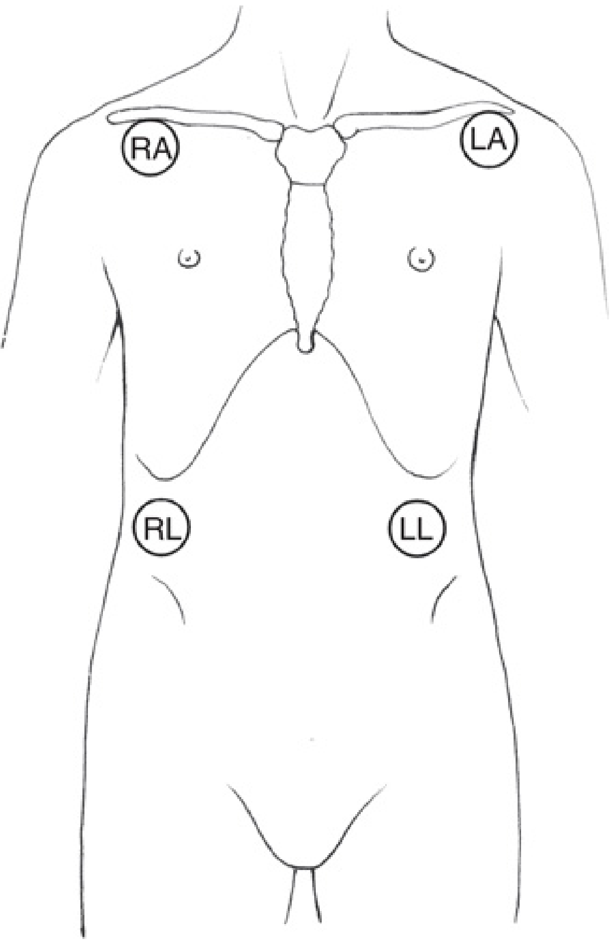
Monitoring limb-electrode locations shown in Miller's Anesthesia, p. 4713.
7. Which 5-lead chest position should be selected?
V5: routine choice for ischemia monitoring
V5 location:
- Fifth intercostal space
- Left anterior axillary line
This is the standard high-yield choice for perioperative ischemia monitoring.
Why V5?
- It looks at the left lateral/anterior-lateral LV region.
- It has historically been the most sensitive single lead for intraoperative ischemia detection.
- It is often accessible even when the sternum is in the operative field.
Miller's Anesthesia states that V5 is most commonly selected for patients at risk of myocardial ischemia and that V3, V4, or V5 should be used for sensitive ischemia detection (pp. 4705, 4714).
Lead II: routine choice for rhythm
Use lead II alongside V5 for:
- P-wave detection
- Sinus versus non-sinus rhythm
- AV conduction assessment
- Bradyarrhythmia/tachyarrhythmia recognition
V1: useful for arrhythmia and conduction questions
V1 location:
- Fourth intercostal space
- Right sternal border
It may help with:
- P-wave assessment
- Wide-complex rhythm interpretation
- Bundle branch block patterns
- Some atrial arrhythmia analysis
But V1 is generally less sensitive than V3-V5 for perioperative ischemia monitoring.
V4 and V3
- V4: fifth intercostal space, left mid-clavicular line
- V3: midway between V2 and V4
These may be chosen if surgical access makes V5 impractical or in selected high-risk patients. Do not place the C/V electrode merely “somewhere on the left chest.” That loses the meaning of the lead.
8. How to find V5 correctly
- Palpate the sternal angle of Louis at the manubriosternal junction.
- The rib level there is the second rib.
- Move down to identify the second intercostal space.
- Count down to the fifth intercostal space.
- Find the left anterior axillary line.
- Place the C/V electrode there for V5.
The correct related positions are:
| Lead | Location |
|---|---|
| V1 | 4th intercostal space, right sternal border |
| V2 | 4th intercostal space, left sternal border |
| V3 | Midway between V2 and V4 |
| V4 | 5th intercostal space, left mid-clavicular line |
| V5 | Same horizontal level as V4, left anterior axillary line |
| V6 | Same horizontal level as V4/V5, left mid-axillary line |
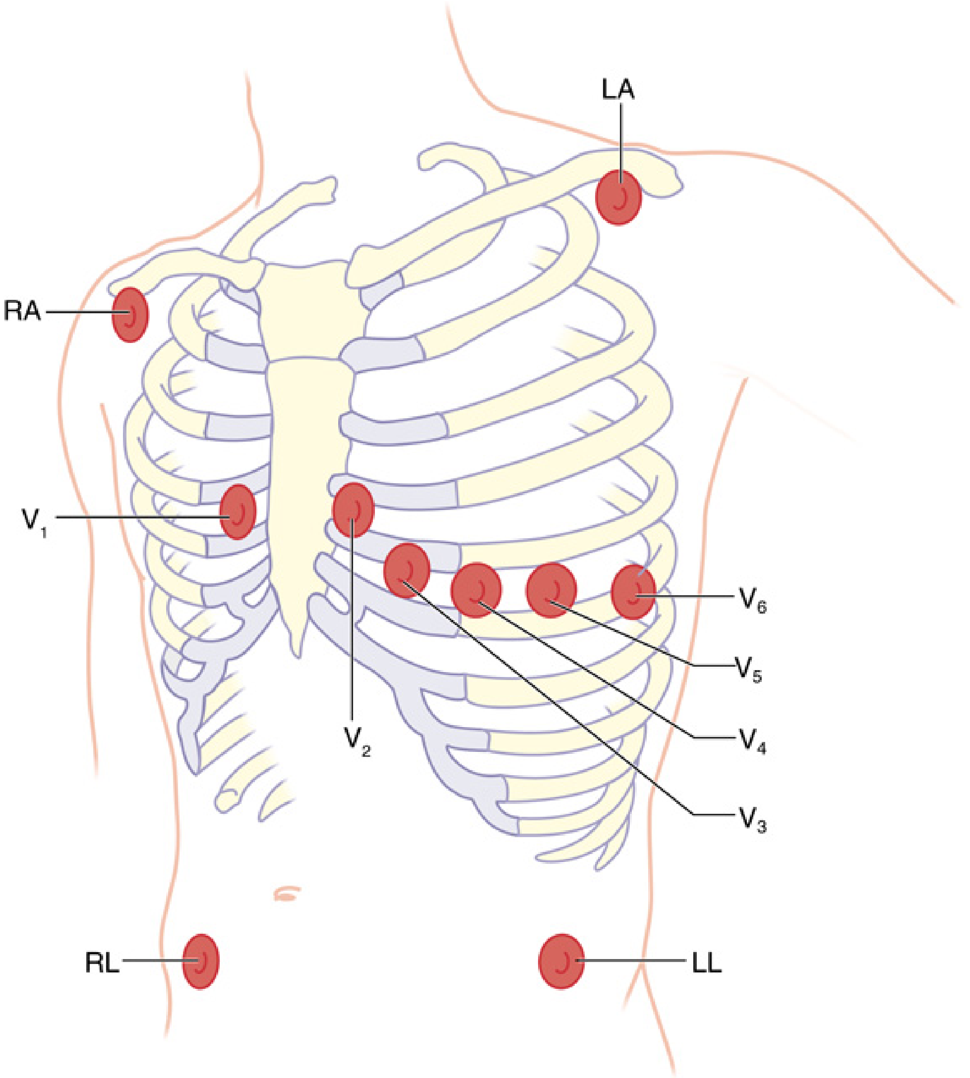
V4, V5, and V6 must be at the same horizontal level. Miller's description places V4 at the fifth interspace mid-clavicular line, V5 directly lateral at the anterior axillary line, and V6 at the mid-axillary line (Miller's Anesthesia, pp. 4714-15).
9. What happens if the 5-lead electrodes are wrongly placed?
If the C/V electrode is too high or too low
The monitor may still label the tracing “V5,” but it is not actually V5.
Consequences:
- Different R-wave amplitude
- Different T-wave shape
- Changed ST baseline
- False ST depression/elevation
- Reduced ability to detect ischemia
A V5 electrode placed too high can behave more like V4 or even V3. If placed too lateral, it can become V6-like. It may also be affected by arm position, breast tissue, surgical drapes, or lateral positioning.
If LL is placed over the left chest
This changes lead II and III because LL is part of those calculations. Lead I is relatively unaffected because it uses only RA and LA. This is a classic example of why limb electrodes must stay outside the cardiac border. Miller's Anesthesia, p. 4715.
If RA and LA are reversed
Lead I may invert, and the apparent frontal-axis pattern changes. This can mimic pathology or make a normal tracing look abnormal.
If RA and LL are reversed
The distortion is usually more dramatic and may mimic an abnormal axis, inferior ischemia, or unusual rhythm morphology.
If pads are loose or skin contact is poor
Expect:
- Baseline wander
- Large slow oscillation with ventilation
- Intermittent noise
- Sudden false ST alarms
- “Leads off” alarms
- QRS detection failure and false heart-rate values
10. How to place monitoring ECG electrodes well
Skin preparation
- Select intact skin away from incisions, defibrillator pads, central-line dressings, surgical prep, and bony prominences.
- Remove excessive hair if needed.
- Clean and dry the skin.
- Gently abrade only if local policy permits and signal quality is poor.
- Use fresh electrodes.
- Press each pad firmly around its edges.
- Secure cable loops so traction does not pull the electrode off during positioning.
Positioning principles
- Keep limb electrodes outside the cardiac silhouette.
- Keep right and left upper electrodes roughly symmetrical.
- Keep lower electrodes below the heart, near the iliac/hip region.
- Use the same sites throughout the case if serial ST comparison matters.
- Record/document nonstandard placement when unavoidable.
- Recheck electrodes after moving the patient, applying warming devices, placing drapes, or changing to lateral/prone/Trendelenburg positions.
Color-code warning
Do not rely only on a memorized color scheme. US AHA and IEC/international color systems differ. Follow the printed RA, LA, RL, LL, C/V labels on the monitor cable.
In a common AHA color system:
- RA white
- LA black
- RL green
- LL red
- C/V brown
But some equipment uses IEC colors, which are different. Miller's Anesthesia, p. 4712.
11. Anaesthesia practical setup
Routine healthy patient
- 3-lead or 5-lead depending on local policy and case.
- Select lead II if rhythm observation is the main issue.
Higher cardiac-risk patient or major surgery
Use 5-lead monitoring, ideally:
- Display II + V5
- Turn on ST trending if available
- Ensure appropriate monitor filter/diagnostic mode according to the device and local protocol
- Establish a stable baseline after induction and positioning
- Correlate ST changes with haemodynamics and clinical context
For anterolateral ischemia detection, V3, V4, or V5 is recommended; perioperative ischemia is commonly manifest as ST depression in these leads. Miller's Anesthesia, p. 4705.
If ST depression/elevation appears during surgery
A safe sequence:
- Check whether this is new and persistent.
- Look for QRS-shape or amplitude change suggesting electrode/patient-position effect.
- Confirm electrode adhesion and correct positions.
- Check monitor gain, ST reference points, and filter setting.
- Assess blood pressure, heart rate, oxygenation, ventilation, temperature, haemoglobin/bleeding, and surgical stimulus.
- Treat important supply-demand abnormalities: hypotension, tachycardia, hypoxaemia, anaemia, severe hypertension.
- Obtain a 12-lead ECG when feasible and involve the appropriate senior/cardiology team based on the clinical situation.
Do not dismiss persistent ST deviation simply because the patient is anaesthetized and cannot report chest pain.
12. Basic 12-lead ECG overview
A diagnostic 12-lead ECG gives 12 views of the heart.
Limb leads: frontal-plane views
| Lead | Formula / viewpoint |
|---|---|
| I | LA - RA |
| II | LL - RA |
| III | LL - LA |
| aVR | looks from right shoulder |
| aVL | looks from left shoulder |
| aVF | looks upward from the feet |
These assess the heart in the frontal plane.
Precordial leads: horizontal-plane views
| Leads | Main territory viewed |
|---|---|
| V1-V2 | Septal/right-sided region |
| V3-V4 | Anterior region |
| V5-V6 | Lateral region |
These assess the heart in the horizontal plane.
A 12-lead ECG is a snapshot, while a 3-lead or 5-lead monitor is continuous surveillance. They are complementary, not interchangeable.
For suspected acute ischemia
A 12-lead ECG is needed because:
- ST change must be assessed across contiguous leads.
- Localization matters.
- Reciprocal changes may be present.
- A single monitor lead cannot adequately characterize ST-elevation MI or rule it out.
Key takeaways
- Wrong electrode position can create or hide apparent ST changes.
- 3-lead ECG is mainly for rhythm. It can show ST deviation but is not preferred for ischemia surveillance.
- 5-lead ECG is preferred in anaesthesia when ischemia monitoring matters.
- Use II for rhythm and V5 for ST monitoring in many high-risk cases.
- Place V5 at the fifth intercostal space, left anterior axillary line.
- Keep limb electrodes on the torso but outside the cardiac borders.
- A monitor reading “V5” is meaningful only if the chest electrode is actually in the V5 position.
- Confirm any concerning monitor ST change with clinical assessment and, when feasible, a correctly placed diagnostic 12-lead ECG.
A recent systematic-review search did not identify a review that changes these established placement principles. The detailed perioperative recommendations above are from Miller's Anesthesia, 10th ed., pp. 4705 and 4712-18. The AHA monitoring standards also emphasize accurate electrode placement and appropriate lead selection for continuous ECG monitoring.
This is a shared conversation. Sign in to Orris to start your own chat.