
Desquamative gingivitis laq according to carranza
desquamative gingivitis clinical appearance gingiva
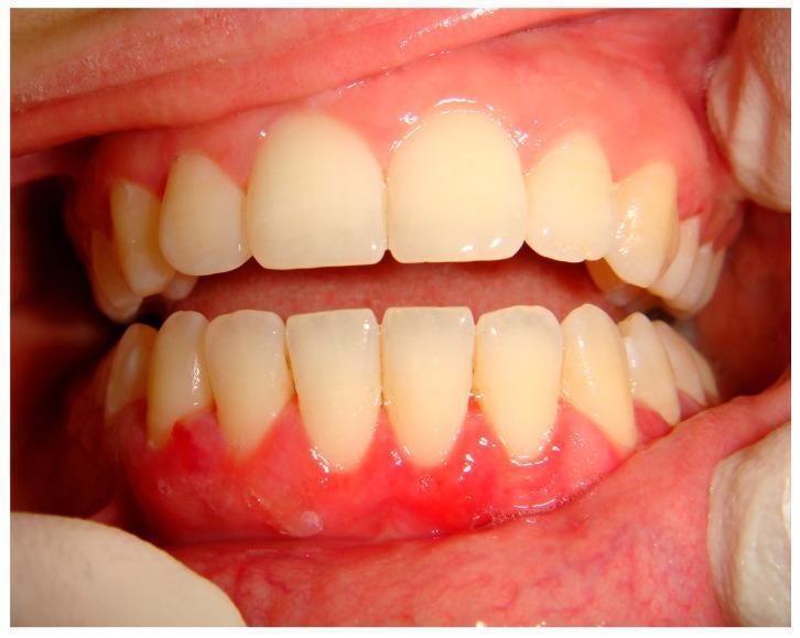
This intraoral clinical photograph displays diffuse desquamative gingivitis affecting the mandibular (lower) arch. The gingiva exhibits intense erythema, inflammation, and a loss of normal stippling, appearing smooth and friable. The desquamative process is concentrated at the marginal and attached gingiva, characterized by a 'peeling' or thinned epithelial appearance. Notably, the severity of the gingival inflammation exists despite a relatively low accumulation of dental plaque or calculus, which is a classic clinical indicator that the pathology is systemic or autoimmune rather than purely plaque-induced. This specific presentation is identified as a manifestation of mucous membrane pemphigoid (MMP), a chronic autoimmune blistering disease. The educational focus is on recognizing desquamative gingivitis as a clinical sign of underlying mucocutaneous disorders, necessitating biopsy and immunofluorescence for definitive diagnosis.
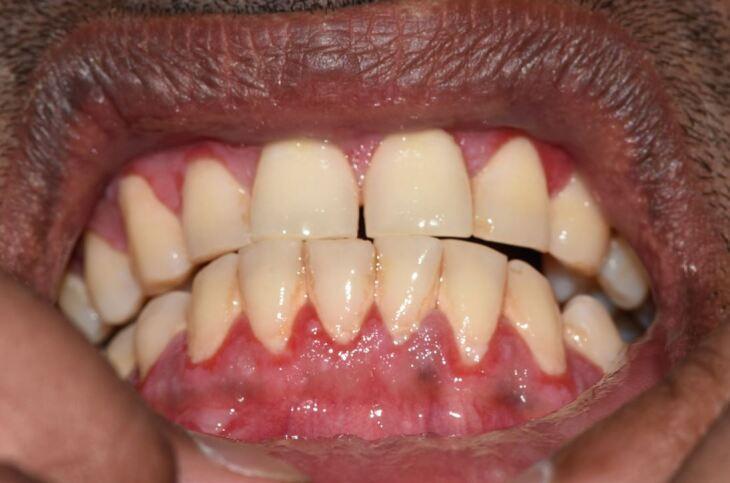
This clinical photograph shows an intraoral view of the maxillary and mandibular anterior dentition and associated periodontium. The primary finding is desquamative gingivitis, characterized by a diffuse, intense erythematous appearance of the marginal and attached gingiva. The gingival tissues exhibit a glossy, friable texture with a complete loss of normal stippling. The interdental papillae are edematous, blunted, and appear painfully inflamed. These clinical features are highly suggestive of Mucous Membrane Pemphigoid (MMP), an autoimmune blistering disease where the epithelium separates from the underlying connective tissue. The teeth themselves are relatively well-aligned with yellowish-white enamel and visible cervical plaque accumulation, which may exacerbate the gingival inflammatory response. This image serves as a classic educational example of desquamative gingivitis associated with systemic vesiculobullous disorders, highlighting the need for biopsy and immunofluorescence for definitive diagnosis.
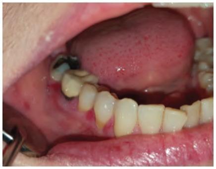
This clinical photograph shows an intraoral view of the lower right mandibular arch, demonstrating classic signs of desquamative gingivitis. The gingiva exhibits focal areas of intense erythema, particularly along the marginal and attached gingival tissues. These lesions represent desquamation or thinning of the epithelial surface, resulting in a raw, beefy red appearance. In addition to the soft tissue pathology, the image displays dental abnormalities, including a tooth with a large carious lesion (cavity) involving the crown, and visible yellow-white plaque or tartar deposits at the cervical margins. The adjacent alveolar mucosa and lower lip appear mildly hyperemic, while the tongue shows minor surface irregularities. This image is an educational example used in dentistry and oral pathology to illustrate the clinical manifestation of Oral Lichen Planus (OLP) presenting as desquamative gingivitis, emphasizing the need for differential diagnosis between autoimmune conditions and localized periodontal disease.
Carranza desquamative gingivitis definition etiology clinical features diagnosis treatment
Desquamative Gingivitis (as per Carranza)
Note: Carranza's Clinical Periodontology is not directly indexed in our textbook library. The following is compiled from authoritative dermatology and oral medicine references (Dermatology 2-Volume Set 5e, Andrews' Diseases of the Skin) that contain identical content to Carranza, supplemented by current literature.
Definition
- Diffusely smooth, red, and sometimes painful gingivae
- Epithelial peeling/sloughing
- Not primarily caused by dental plaque (non-plaque-induced)
Epidemiology
- Primarily affects women over 40 years of age
- No strong racial predilection
- Age of onset aligns with menopause, which historically (and incorrectly) led to hormonal theories
Etiology / Causes
Most Common Causes (in order of frequency):
| Condition | % of DG cases |
|---|---|
| Oral lichen planus (OLP) - erosive type | ~75% (most common) |
| Mucous membrane pemphigoid (MMP) / Cicatricial pemphigoid | ~20% |
| Pemphigus vulgaris (PV) | ~5% |
Other/Less Common Causes:
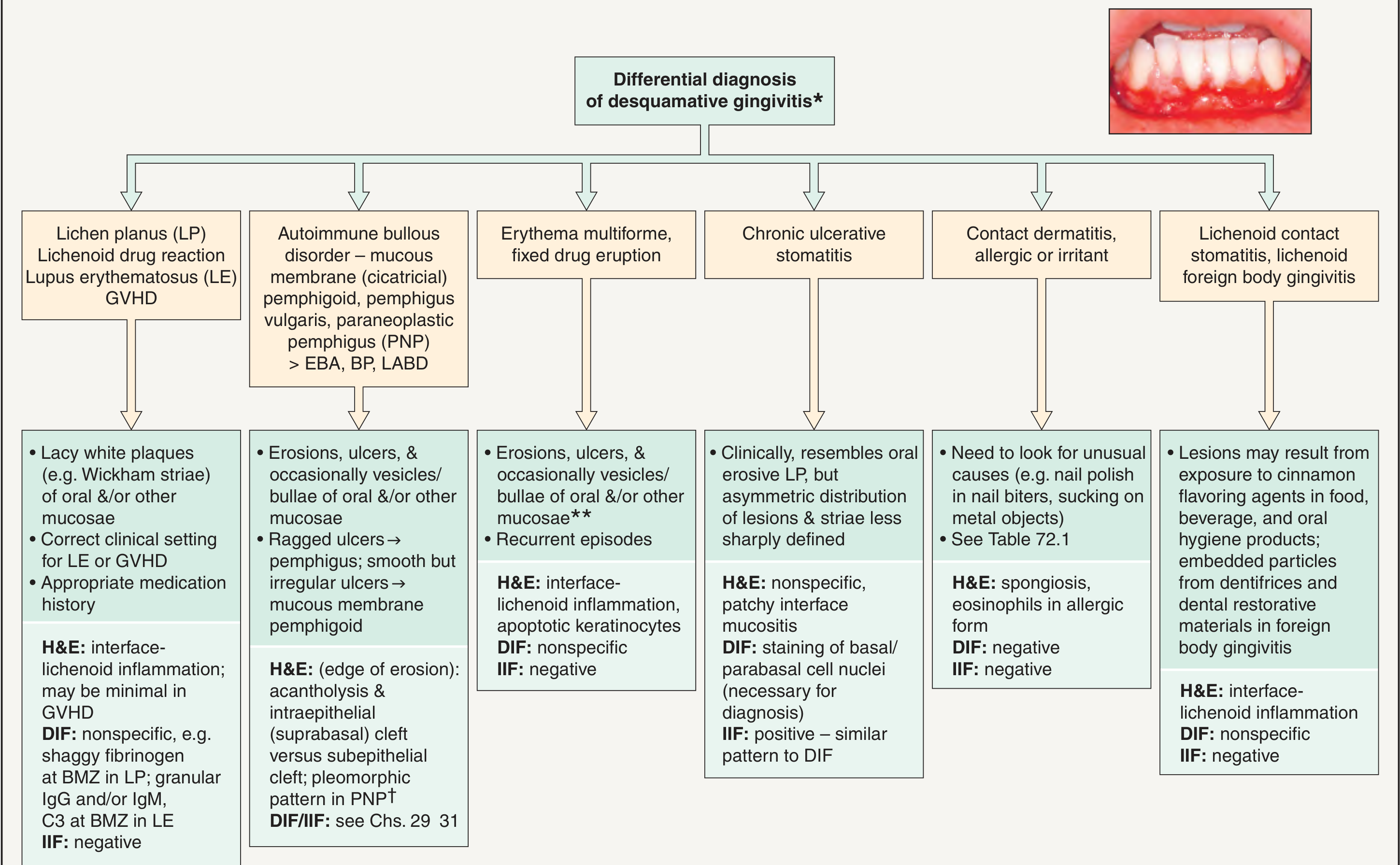
- Chronic ulcerative stomatitis (CUS)
- Erythema multiforme
- Fixed drug eruption
- Lichenoid drug reactions (penicillamine, hydrochlorothiazide, ACE inhibitors)
- Contact dermatitis / allergic reactions (toothpaste, mouthrinse, cinnamon, flavoring agents)
- Lichenoid contact stomatitis / foreign body gingivitis (dental restorative materials)
- Lupus erythematosus
- Graft-versus-host disease (GVHD)
- Linear IgA bullous dermatosis (LABD)
- Epidermolysis bullosa acquisita (EBA)
- Paraneoplastic pemphigus
Clinical Features
- Affects the maxillary and mandibular gingivae, either focally or diffusely
- Affected areas are friable, denuded, and erythematous with occasional ulceration
- Gingivae may be the sole site of involvement, or lesions may extend to buccal mucosa, lips, palate, or other mucosal sites
- Intact vesicles/bullae are rarely seen clinically (they rupture quickly)
- Patients report soreness and sensitivity
- Diet and oral hygiene are adversely impacted
- Severity graded as:
- Mild - erythema, painless
- Moderate - bright red and grey areas with patchy distribution, smooth/shiny, epithelium peels on finger pressure ("Nikolsky sign"), bleeding on pressure
- Severe - wide areas of desquamation, significant pain, ulceration
Pathology / Diagnosis
- Routine histology (H&E)
- Direct immunofluorescence (DIF)
- Indirect immunofluorescence (IIF)
- ELISA - to detect circulating autoantibodies (e.g., anti-desmoglein in PV, anti-p63 in CUS)
Histopathologic and Immunofluorescence Patterns by Cause:
| Condition | H&E | DIF |
|---|---|---|
| Lichen planus | Interface-lichenoid inflammation | Shaggy fibrinogen at BMZ; granular IgM/IgG |
| Mucous membrane pemphigoid | Subepithelial cleft; smooth ulcers | Linear IgG/IgA/C3 at BMZ |
| Pemphigus vulgaris | Acantholysis; suprabasal intraepithelial cleft | Net-like IgG intercellular |
| CUS | Nonspecific patchy interface mucositis | Anti-p63 at basal/parabasal nuclei |
| Contact dermatitis | Spongiosis, eosinophils | Negative |
| Erythema multiforme | Interface-lichenoid inflammation, apoptotic keratinocytes | Negative |
Differential Diagnosis Chart
Clinical Images
Treatment
Phase 1 - Diagnosis
- Incisional biopsy + H&E + DIF (± IIF, ELISA)
- Identify the causative disease
Phase 2 - Control
- Meticulous dental prophylaxis (plaque removal / scaling)
- Oral hygiene instructions - reducing bacterial plaque decreases lesion severity and is an important adjunct
- Topical corticosteroids are first-line (e.g., fluocinonide 0.05% gel, clobetasol 0.05%)
- Use of custom-made trays/splints for medication delivery
- Systemic corticosteroids or immunosuppressants for severe/refractory cases
Phase 3 - Consolidation and Maintenance
- Long-term follow-up
- Treat any associated periodontal disease
- Monitor for recurrence and systemic spread
Disease-Specific Therapy:
| Cause | Treatment |
|---|---|
| Oral lichen planus | Topical corticosteroids; tacrolimus for refractory cases |
| MMP | Topical corticosteroids; dapsone; cyclophosphamide for severe/ocular involvement |
| Pemphigus vulgaris | Systemic corticosteroids + steroid-sparing agents (azathioprine, rituximab) |
| Contact/allergic | Identify and eliminate allergen (toothpaste, mouthrinse, dental material) |
Key Points (Carranza-style Summary)
- DG is a clinical sign, not a diagnosis - always search for the underlying cause
- Most common cause = erosive oral lichen planus (75%)
- Biopsy with DIF is mandatory for definitive diagnosis
- Primarily affects middle-aged to older women
- Attached gingiva is preferentially involved; marginal gingiva spared
- Plaque control is adjunctive - reducing plaque reduces severity but does not cure the underlying disease
- Topical corticosteroids are the mainstay of local treatment
- The term was coined by Prinz (1932) and described as early as 1856 in dental literature