POST GRADUATE SCHOLAR THESIS LECTURE REVIEW OF 1. PHYSIOLOGY OF BLOOD PRESSURE 2. PATHOPHYSIOLOGY OF HYPERTENSION

Reading File
Finding Sources
Finding Sources
Searching PubMed

"hypertension" AND "pathophysiology"

Finding Sources
Reading File
Reading File
Finding Sources
Reading File
Finding Sources
Reading File
Reading File
Reading File
Finding Sources
Reading File
Finding Sources
Reading File
Searching the Web

physiology blood pressure regulation cardiac output peripheral resistance baroreceptors 2024 2025

Loading Image
Loading Image
Loading Image
Loading Image
I now have all the material needed for a comprehensive postgraduate-level thesis lecture review. Here is the full document:

POST GRADUATE SCHOLAR THESIS LECTURE REVIEW

PHYSIOLOGY OF BLOOD PRESSURE AND PATHOPHYSIOLOGY OF HYPERTENSION

Source: Fuster and Hurst's The Heart, 15th Edition | Histology: A Text and Atlas with Correlated Cell and Molecular Biology | Comprehensive Clinical Nephrology, 7th Edition

PART I: PHYSIOLOGY OF BLOOD PRESSURE


1.1 The Fundamental Equation of Blood Pressure

Blood pressure (BP) regulation is understood through the following governing relationship:
BP = Cardiac Output (CO) × Total Peripheral Resistance (TPR)
This equation frames the two major determinants of arterial pressure - the pump (heart) and the resistance (vasculature). However, long-term BP control and the pathogenesis of hypertension require a third dimension: renal regulation of body fluid volume and time-dependent interactions among multiple control systems.

1.2 Cardiac Output and Its Determinants

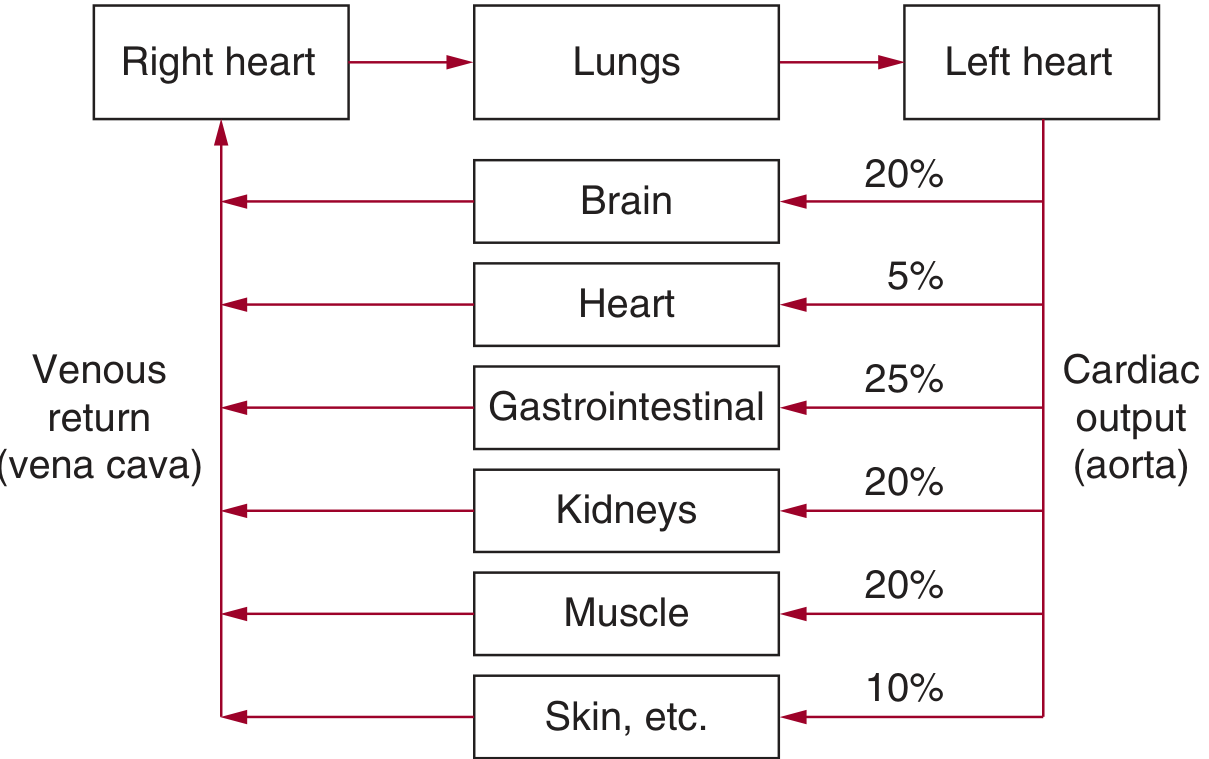
Cardiac output is the sum of blood flows to all tissues and organs in the body. Normal distribution of CO at rest:
Cardiac output distribution to organs
Figure 1. Relationship between cardiac output, peripheral blood flow, and venous return. CO is distributed as: Brain 20%, Kidneys 20%, Muscle 20%, GI tract 25%, Heart 5%, Skin/other 10%.
Key physiological points:
  • CO is determined not only by cardiac pumping ability, but critically by the peripheral circulation and tissue metabolic demands
  • Amputation of a limb reduces venous return and CO without significantly altering BP - demonstrating that CO adapts to peripheral needs
  • During exercise, pregnancy, or growth, increased CO may occur without BP change, because TPR falls proportionally
  • In severe heart failure, cardiac pumping becomes limiting, but under normal conditions CO is governed by metabolic demands of tissues
Determinants of CO:
  • Stroke Volume (SV) = preload (ventricular filling), afterload (wall stress during ejection), contractility (inotropy)
  • Heart Rate (HR) - regulated by the sinoatrial node under autonomic and humoral control
  • CO = SV × HR (normal resting value: ~5 L/min)

1.3 Total Peripheral Resistance

TPR is the aggregate resistance offered by the entire systemic vasculature. From Poiseuille's law:
Resistance = (8 × viscosity × length) / (π × radius⁴)
The radius (r) dominates because resistance varies with r⁴. Therefore, even small changes in arteriolar caliber have enormous effects on resistance and perfusion pressure.
Arterioles are the principal site of TPR regulation - they are the "resistance vessels" whose tone is modulated by:
  • Neurogenic factors (sympathetic vasoconstrictor tone)
  • Hormonal factors (angiotensin II, endothelin, vasopressin)
  • Metabolic autacoids (adenosine, CO₂, H⁺, lactate)
  • Endothelium-derived signals (nitric oxide, prostacyclin, endothelin-1)

1.4 Short-Term Blood Pressure Regulation

1.4.1 The Baroreflex (Baroreceptor Reflex)

The arterial baroreceptors - high-pressure mechanoreceptors located in the carotid sinus and aortic arch - respond to vessel wall stretch caused by elevated arterial pressure:
  1. Increased BP → increased wall stretch → increased baroreceptor firing frequency
  2. Afferent signals travel via CN IX (carotid sinus nerve/Hering's nerve) and CN X (aortic depressor nerve) to the Nucleus Tractus Solitarius (NTS) in the medulla oblongata
  3. NTS modulates activity of sympathetic and parasympathetic (vagal) neurons in the cardiovascular center
  4. Net result: decreased sympathetic output + increased vagal tone → decreased HR, decreased contractility, vasodilation → BP falls back toward normal
This reflex provides rapid, beat-to-beat BP stabilization and can alter BP within seconds.
  • Maximum carotid sinus sensitivity occurs near normal mean arterial pressure (MAP), so tiny deviations from the "set point" trigger powerful corrective responses
  • The baroreflex is reset upward in chronic hypertension (see Part II)

1.4.2 Chemoreceptor Reflexes

  • Peripheral chemoreceptors (carotid and aortic bodies) respond to hypoxia, hypercapnia, and acidosis - stimulating the vasomotor center to increase sympathetic tone
  • Central chemoreceptors in the medulla respond primarily to CO₂/pH changes in cerebrospinal fluid

1.4.3 CNS Ischemic Response

When cerebral perfusion pressure falls critically (MAP < 50 mmHg), the vasomotor center itself becomes ischemic and fires maximally, producing the Cushing reflex - a massive surge in sympathetic output raising BP as an emergency response.

1.5 Long-Term Blood Pressure Regulation: Tissue Blood Flow Autoregulation

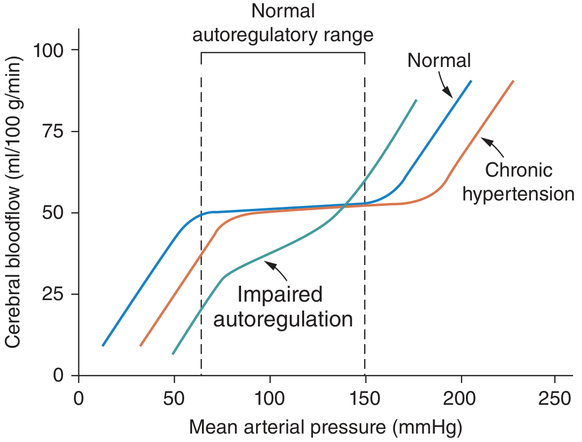
Autoregulation is the intrinsic ability of each tissue to maintain constant blood flow over a wide range of perfusion pressures (approximately 75-150 mmHg MAP).
Autoregulation of cerebral blood flow
Figure 2. Autoregulation curves: In normal individuals (blue), cerebral blood flow remains constant across a wide MAP range. In chronic hypertension (red), the autoregulatory curve shifts rightward - patients are protected from high pressures but vulnerable to hypoperfusion at "normal" MAPs. In impaired autoregulation (green), flow is pressure-dependent.
Mechanisms:
  • Acute (seconds to minutes): Myogenic response (Bayliss effect) - vessel wall contracts in response to stretch. Metabolic vasodilation (local O₂ depletion, CO₂/H⁺ accumulation) restores flow after vasoconstriction
  • Chronic (days to weeks): Structural vascular remodeling - wall thickening, capillary rarefaction (reduced capillary density), angiogenesis with tissue growth
Clinical implication for hypertension: Elevated TPR in chronic hypertension is often a consequence (autoregulatory response) rather than a primary cause, as vessels constrict to protect tissues from chronically elevated perfusion pressure.

1.6 The Renal-Body Fluid System: The Ultimate Long-Term BP Controller

The kidneys exert the dominant influence on long-term BP through pressure natriuresis - the relationship between arterial pressure and urinary sodium excretion:
  • As BP rises → kidneys excrete more sodium and water (natriuresis and diuresis)
  • This reduces blood volume → reduces venous return → reduces CO → BP falls toward normal
  • Conversely, a fall in BP → sodium and water retention → volume expansion → restored BP
This system is the only one capable of indefinitely maintaining sodium balance at a precise blood pressure level. Any sustained alteration in BP is therefore a reflection of a reset or impaired pressure-natriuresis relationship.

1.7 The Renin-Angiotensin-Aldosterone System (RAAS)

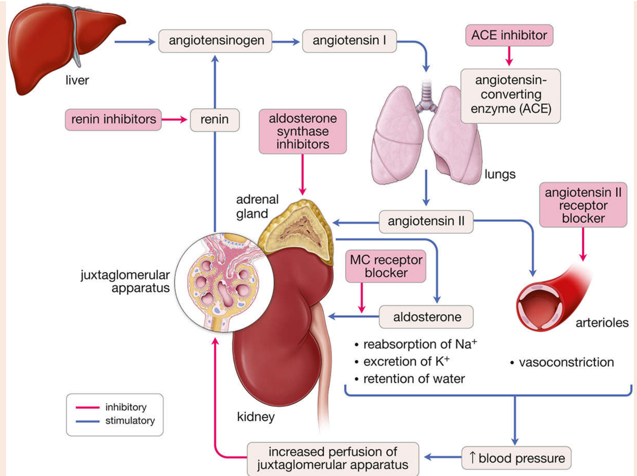
The RAAS is the central hormonal axis integrating renal, cardiovascular, and adrenal function in BP regulation:
RAAS cascade and pharmacological sites of intervention
Figure 3. The RAAS cascade: Liver produces angiotensinogen → converted by renin (from juxtaglomerular cells) to Angiotensin I → converted by ACE in lung endothelium to Angiotensin II → acts on arterioles (vasoconstriction) and adrenal cortex (aldosterone release). Aldosterone acts on the collecting duct to reabsorb Na⁺, excrete K⁺, and retain water. Pharmacological targets are shown (ACE inhibitors, ARBs, renin inhibitors, mineralocorticoid receptor blockers).
Steps in RAAS activation:
StepSiteEvent
1LiverSynthesis of angiotensinogen (alpha-2 globulin)
2JGA of kidneyRenin released in response to: ↓ renal perfusion pressure, ↓ Na⁺ delivery to macula densa, ↑ sympathetic (β₁) stimulation
3BloodstreamRenin cleaves angiotensinogen → Angiotensin I (decapeptide)
4Pulmonary capillary endotheliumACE cleaves 2 amino acids → Angiotensin II (octapeptide - most potent vasoconstrictor)
5Systemic arteriolesAng II → vasoconstriction (AT₁ receptors) → ↑ TPR
6Adrenal zona glomerulosaAng II → aldosterone release → Na⁺/water retention → ↑ blood volume
7KidneyAng II → direct tubular Na⁺ reabsorption, reset pressure-natriuresis curve upward
Additional Ang II effects:
  • Stimulates ADH (vasopressin) release from posterior pituitary
  • Stimulates thirst via hypothalamic centers
  • Stimulates sympathetic ganglia and adrenal medulla (positive feedback)
  • Promotes vascular smooth muscle hypertrophy (trophic/remodeling effect)

1.8 The Sympathetic Nervous System in BP Regulation

The sympathetic nervous system (SNS) can:
  • Raise BP within seconds by causing vasoconstriction, increased heart rate, and enhanced cardiac contractility
  • Sudden SNS inhibition can reduce BP to as low as half of normal within 1 minute
The SNS plays a major long-term role through renal sympathetic innervation - extensive fibers reach renal blood vessels, the juxtaglomerular apparatus, and renal tubules:
  • Stimulate renin secretion
  • Promote sodium reabsorption in proximal tubule, loop of Henle, and distal segments
  • Impair renal pressure-natriuresis (shift the curve upward)

1.9 Other Vasoactive Systems

SystemBP EffectMechanism
Nitric Oxide (NO)VasodilatoryEndothelium-derived, activates guanylate cyclase in VSMCs → ↓ Ca²⁺ → relaxation
Endothelin-1 (ET-1)VasoconstrictiveMost potent endogenous vasoconstrictor; acts via ETA receptors
Atrial Natriuretic Peptide (ANP)Vasodilatory/natriureticReleased by atrial cardiomyocytes in response to stretch; increases GFR, inhibits tubular Na⁺ reabsorption, suppresses RAAS
Prostacyclin (PGI₂)VasodilatoryEndothelium-derived; opposes thromboxane A₂
Vasopressin (ADH)VasoconstrictiveV₁ receptors on vessels; also promotes water reabsorption via V₂ receptors
BradykininVasodilatoryStimulates NO and PGI₂ release; degraded by ACE (hence ACE inhibitors also preserve bradykinin)

PART II: PATHOPHYSIOLOGY OF HYPERTENSION


2.1 Definition and Classification

Hypertension is defined as a sustained elevation in arterial blood pressure above accepted thresholds. Based on current JNC/ACC/AHA guidelines:
CategorySystolic (mmHg)Diastolic (mmHg)
Normal< 120< 80
Elevated120-129< 80
Stage 1 HTN130-13980-89
Stage 2 HTN≥ 140≥ 90
Hypertensive crisis> 180> 120
Classification by cause:
  • Primary (Essential) Hypertension: >90% of all cases; no single identifiable cause
  • Secondary Hypertension: <10% of cases; identifiable underlying disorder

2.2 Blood Flow Regulation in Hypertension

One characteristic of most patients with chronic hypertension is elevated TPR. However, tissue blood flows remain approximately normal - indicating that increased resistance is an autoregulatory adaptation, not a primary cause:
  • The autoregulatory curve shifts rightward in chronic hypertension (Figure 2 above)
  • This protects tissues from overperfusion at high BP, but renders patients vulnerable to ischemia at "normal" MAPs (e.g., rapid antihypertensive treatment can precipitate cerebral ischemia)
  • Hemodynamic pattern in non-obese primary hypertension: normal CO, normal tissue blood flow, elevated TPR

2.3 The Central Role of the Kidneys: Pressure Natriuresis and Hypertension

The kidney is the final arbiter of long-term BP. For hypertension to be sustained, the pressure-natriuresis relationship must be impaired or reset. This can occur through:
  1. Reduced nephron number (congenital or acquired) - decreased filtration surface means natriuresis requires higher BP; slope of pressure-natriuresis curve decreases (salt-sensitive pattern)
  2. Decreased glomerular filtration coefficient (Kf) - similar effect; ↑ glomerular pressure, risk of glomerular injury
  3. Increased tubular sodium reabsorption - e.g., excess aldosterone, increased sympathetic tone, primary ENaC activation (Liddle syndrome)
  4. Increased preglomerular resistance - renal artery stenosis or aortic coarctation causes a "parallel shift" in the pressure-natriuresis curve (salt-insensitive pattern); renin and Ang II transiently elevated but normalize as a new equilibrium is established at a higher BP
Salt-Sensitive vs. Salt-Insensitive Hypertension:
FeatureSalt-SensitiveSalt-Insensitive
Cause↓ nephron mass, ↓ Kf, ↑ tubular reabsorption↑ preglomerular resistance
Pressure-natriuresis curveDecreased slopeParallel rightward shift
Glomerular injuryYes (↑ glomerular pressure)No (glomeruli protected by upstream resistance)
Plasma renin activityLow-normalElevated initially, then normal
(Source: Fuster and Hurst's The Heart, 15th Edition, Table MLE5-3)

2.4 The Sympathetic Nervous System in Hypertension

Excessive SNS activation is a major contributor to hypertension through:
  1. Renal nerve activation: Even mild increases in renal sympathetic activity stimulate renin secretion and Na⁺ reabsorption in the proximal tubule, loop of Henle, and distal segments - impairing pressure-natriuresis
  2. Vascular effects: Sympathetic vasoconstriction increases TPR
  3. Cardiac effects: Increased HR and contractility raise CO
Evidence from Renal Denervation (RDN):
  • RDN reduces BP in experimental hypertension models (spontaneously hypertensive rats)
  • Human trials (SYMPLICITY series): catheter-based radiofrequency RDN lowered office BP for up to 36 months in obese patients with resistant hypertension
  • Current limitation: catheter-based RDN achieves only <50% nerve ablation; branches of main renal artery are inaccessible; renal nerves may regrow
Mechanisms of SNS activation in primary hypertension:
  • Hypothalamic CRH: Obesity and stress chronically elevate corticotropin-releasing hormone, activating the hypothalamic-pituitary-adrenal axis and the SNS
  • Leptin: Adipose tissue-derived hormone; stimulates the SNS (hypothalamic pathways) - links obesity to hypertension
  • Melanocortin (MC4R) pathway: Leptin and central nervous system melanocortin receptor activation raise sympathetic outflow and BP

2.5 The RAAS in Hypertension

While 24-hour urine renin levels in essential hypertension are often "normal," this should be interpreted in context: any measurable renin activity in the setting of volume expansion is inappropriately elevated. The "lesion" in chronic essential hypertension is now believed to be excessive production of Angiotensin II, particularly in pulmonary endothelium.
Key pathological mechanisms:
  • Ang II impairs the pressure-natriuresis relationship - shifts it upward, requiring higher BP to achieve the same sodium excretion
  • Ang II promotes vascular hypertrophy and remodeling - structural narrowing of arterioles (functional narrowing → anatomical narrowing)
  • Ang II promotes oxidative stress via NADPH oxidase activation → superoxide generation → scavenges NO → endothelial dysfunction
  • Chronic Ang II excess promotes glomerulosclerosis and nephron loss - a vicious cycle maintaining hypertension

2.6 Nitric Oxide Deficiency and Endothelial Dysfunction

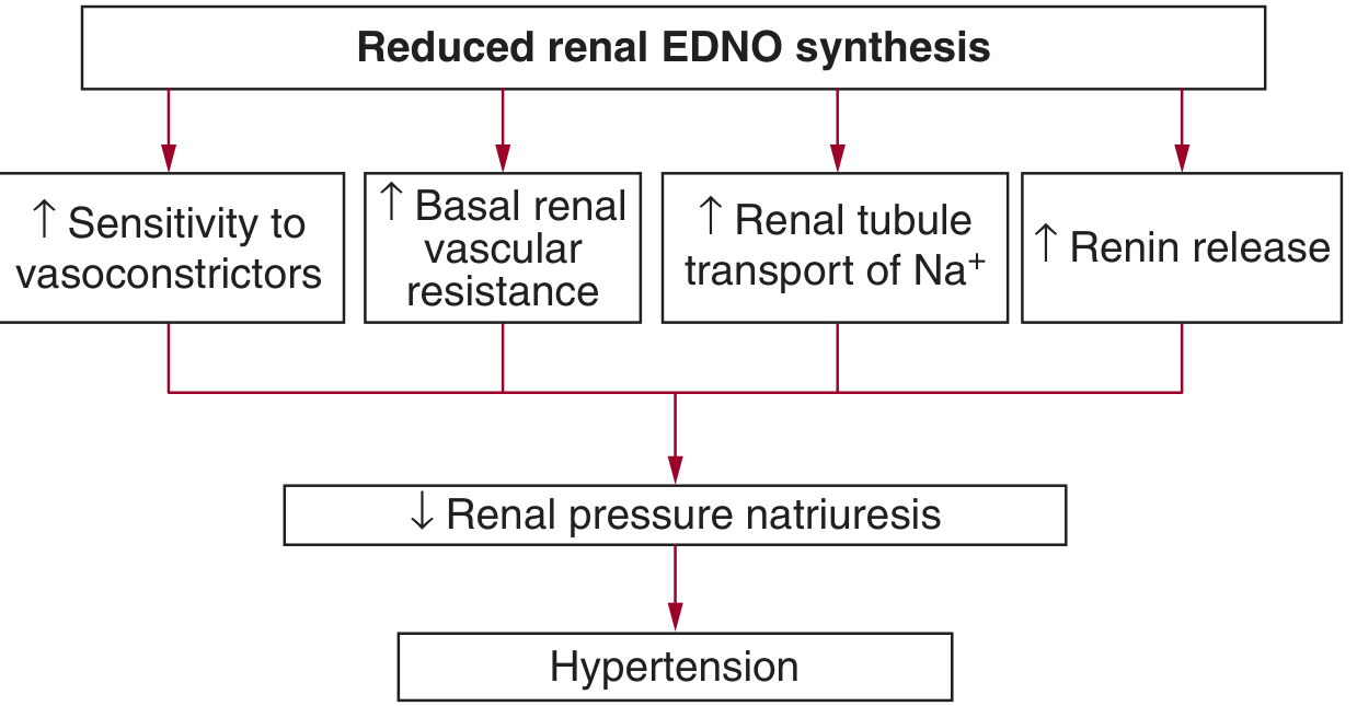
Reduced endothelial-derived nitric oxide (EDNO) synthesis is a key mechanism in hypertension:
Reduced renal EDNO and hypertension
Figure 4. Consequences of reduced renal endothelial NO synthesis: increased sensitivity to vasoconstrictors (Ang II, norepinephrine), increased basal renal vascular resistance, increased renal tubular Na⁺ transport, and increased renin release. All four mechanisms converge to reduce renal pressure natriuresis and ultimately produce hypertension.
Mechanisms of reduced NO in hypertension:
  • Oxidative stress: superoxide (O₂⁻) reacts with NO to form peroxynitrite (ONOO⁻), reducing NO bioavailability
  • eNOS uncoupling: insufficient tetrahydrobiopterin (BH4) as cofactor causes eNOS to produce superoxide instead of NO
  • Reduced L-arginine substrate availability
  • Hemodynamic shear stress reduction (physical inactivity)

2.7 Oxidative Stress in Hypertension

Considerable evidence supports a role for reactive oxygen species (ROS) in experimental hypertension:
  • Dahl Salt-sensitive (DS) rats: increased vascular and renal superoxide production, increased H₂O₂, decreased superoxide dismutase expression
  • Stroke-prone spontaneously hypertensive rats: elevated superoxide, decreased total antioxidant capacity
  • Chronic Tempol (SOD mimetic) administration significantly decreases BP and attenuates renal damage in animal models
  • Ang II hypertension is partly mediated by NADPH oxidase-derived superoxide (apocynin reduces BP)
However, clinical translation has been disappointing: Large-scale clinical trials of antioxidant therapy (Vitamins C and E) have failed to confirm BP-lowering effects in humans. Assessment of ROS in human tissues is technically challenging due to low levels and short half-lives.

2.8 Atrial Natriuretic Peptide (ANP): A Counterregulatory System

ANP is a 28-amino-acid peptide released from atrial cardiomyocytes in response to stretch/volume expansion:
  • Enhances GFR (but not prerequisite for natriuresis)
  • Directly inhibits renal tubular Na⁺ reabsorption
  • Alters medullary blood flow and peritubular capillary physical factors
  • Suppresses antinatriuretic hormones (Ang II, aldosterone)
In primary hypertension, plasma ANP is elevated in volume-expanded states. Exogenous ANP infusions at physiological concentrations can lower BP. Deficient ANP signaling - from reduced release or receptor downregulation - may contribute to impaired natriuresis in salt-sensitive hypertension.

2.9 Pathophysiology of Primary (Essential) Hypertension

Essential hypertension accounts for >90% of all cases and is multifactorial:

2.9.1 Known Contributing Causes

Overweight/Obesity - The Dominant Risk Factor (65-75% of risk for primary hypertension):
Obesity raises BP through multiple mechanisms:
  1. Increased renal tubular sodium reabsorption - adipose tissue compresses the kidneys (renal adipose capsule), directly increasing sodium retention ("reno-pressor effect" of obesity)
  2. SNS activation - leptin from adipose tissue activates hypothalamic SNS outflow
  3. RAAS activation - adipose tissue locally produces angiotensinogen and angiotensin; insulin resistance raises Ang II levels
  4. Insulin resistance and hyperinsulinemia - insulin directly stimulates renal sodium reabsorption and SNS activity
  5. Inflammation - adipose tissue macrophages and cytokines (TNF-α, IL-6) impair NO bioavailability and promote vascular inflammation
Hemodynamic pattern in obesity-hypertension differs from non-obese hypertension:
  • Elevated CO (to meet metabolic demands of excess adipose tissue)
  • Relatively normal TPR initially (may rise later due to autoregulation)
  • Expanded blood volume
Other modifiable causes:
  • Excess dietary sodium chloride
  • Excess alcohol intake
  • Sedentary lifestyle
  • Low dietary potassium

2.9.2 Genetic Determinants

Although primary hypertension has no single gene mutation, BP has substantial heritability (~30-50%). Genome-wide association studies have identified >1000 genetic loci associated with BP. Monogenic forms (rare but mechanistically instructive):
Genetic SyndromeMechanismInheritanceTreatment
Liddle SyndromeGain-of-function ENaC mutation → ↑ Na⁺ reabsorptionADAmiloride, triamterene (not aldosterone antagonists)
FH-I / GRAChimeric gene → ACTH-driven aldosterone synthaseADGlucocorticoids (suppress ACTH)
FH-IIAldosterone excess, normal ACTHADMineralocorticoid receptor antagonist
Gordon Syndrome (PHA Type II)SPAK/WNK kinase gain → ↑ NCC activityADThiazide diuretics
AME (Apparent Mineralocorticoid Excess)11β-HSD2 deficiency → cortisol activates MRARDexamethasone, MR antagonist, amiloride
Brachydactyly + HypertensionChromosome 12p11 vascular anomalyADMilrinone
(Source: Fuster and Hurst's The Heart, Table 5-5)

2.10 Pathophysiology of Secondary Hypertension

2.10.1 Renovascular Hypertension (RAS / Goldblatt Hypertension)

  • Mechanism: Renal artery stenosis (atherosclerosis or fibromuscular dysplasia) → reduced renal perfusion pressure → JGA activation → ↑ renin → ↑ Ang II → ↑ aldosterone
  • In two-kidney one-clip (2K1C) model: the clipped kidney drives RAAS; contralateral kidney has natriuresis; renin-mediated, salt-insensitive hypertension
  • In one-kidney one-clip (1K1C) model: volume-dependent hypertension; renin normalizes once volume is expanded
  • Preglomerular resistance is protective for glomeruli in the stenosed kidney but the contralateral kidney faces elevated pressure → glomerular injury risk

2.10.2 Primary Hyperaldosteronism (Conn Syndrome)

  • Excessive aldosterone from adrenal adenoma or bilateral hyperplasia
  • Mechanism: continuous Na⁺ retention, K⁺ wasting, hypokalemia, suppressed renin
  • The aldosterone-to-renin ratio (ARR) > 30 with aldosterone > 15 ng/dL is diagnostic screening test
  • Most common surgically correctable form of hypertension

2.10.3 Pheochromocytoma

  • Rare (0.05% of hypertensive patients) but potentially fatal if unrecognized
  • Neuroectodermal chromaffin cell tumor; 85% adrenal medulla, 15-30% extraadrenal
  • Mechanism: excessive catecholamine (epinephrine/norepinephrine) release
  • Norepinephrine predominant: α-adrenergic vasoconstriction → sustained diastolic hypertension
  • Epinephrine predominant: β-adrenergic cardiac stimulation → mainly systolic hypertension with tachycardia, sweating, tremors, flushing; 5% remain normotensive
  • Hypertension may be sustained or paroxysmal depending on secretory pattern

2.10.4 Cushing Syndrome

  • Glucocorticoid excess (endogenous or exogenous): cortisol activates mineralocorticoid receptors (bypassing 11β-HSD2 saturation) → Na⁺ retention
  • Also increases angiotensinogen synthesis → ↑ RAAS
  • Clinical features: central obesity, striae, buffalo hump, moon face, hyperglycemia

2.10.5 Preeclampsia

  • New-onset hypertension (SBP ≥140 or DBP ≥90 mmHg) after 20 weeks of gestation, with proteinuria and multisystem involvement
  • Mechanism: abnormal cytotrophoblast invasion of spiral arterioles → placental ischemia → maternal vascular endothelial dysfunction → ↑ ET-1, thromboxane, superoxide, ↑ vascular sensitivity to Ang II, ↓ NO and prostacyclin
  • Key pathway: anti-angiogenic sFlt-1 (soluble VEGF receptor) is released by the ischemic placenta → scavenges VEGF and PlGF → endothelial injury → hypertension, proteinuria, multi-organ damage
  • Hypertension remits after delivery - placenta is the central driver

2.10.6 Coarctation of the Aorta

  • Structural narrowing, usually at the level of the ductus arteriosus
  • Mechanism: increased preglomerular resistance to kidneys (distal to coarctation) activates RAAS - salt-insensitive hypertension pattern
  • Upper extremity hypertension with lower extremity hypotension/absent pulses is the classic presentation

2.11 Target Organ Damage in Hypertension

Sustained hypertension injures multiple organs:
OrganPathological ChangeClinical Consequence
HeartLV hypertrophy (concentric) → diastolic dysfunction → systolic failure; coronary artery diseaseHeart failure, arrhythmias, sudden cardiac death
BrainLacunar infarcts (small vessel disease), hemorrhagic stroke, hypertensive encephalopathyStroke, vascular dementia, hypertensive emergency
KidneysBenign nephrosclerosis (hyalinosis of afferent arterioles, glomerulosclerosis) → nephron loss → CKDProgressive CKD, ESRD - creates a vicious cycle
RetinaArteriovenous nipping, copper/silver wiring (grade 1-2), cotton wool spots (grade 3), papilledema (grade 4)Visual impairment, optic neuropathy
Large vesselsAccelerated atherosclerosis, aortic dissection, aneurysm formationAortic dissection, peripheral vascular disease

2.12 The Vicious Cycle of Hypertension and Renal Damage

A clinically critical concept:
Hypertension → Glomerular hypertension/hyperfiltration
             → Glomerular injury → Nephron loss
             → Reduced filtration surface
             → Impaired pressure-natriuresis
             → Further sodium retention
             → Worsening hypertension (self-perpetuating cycle)
This is why effective antihypertensive therapy (especially RAAS blockade) is nephroprotective beyond BP reduction alone - by reducing glomerular hypertension directly.

2.13 Summary: Integrated Model of Hypertension Pathophysiology

The following unified model integrates all mechanisms:
PRIMARY DRIVER(S)
      ↓
[Renal impairment of pressure-natriuresis] ← Central axis
      ↑                    ↑                    ↑
SNS excess          RAAS activation       ↓ NO / oxidative stress
(obesity, CNS)    (RAS, Conn, Ang II)    (endothelial dysfunction)
      ↑                    ↑
Genetic factors     Environmental factors
(monogenic or       (obesity, Na⁺ excess,
 polygenic)          sedentary, alcohol)
      ↓
Sustained ↑ BP → Target organ damage
The kidneys must be impaired (structurally or functionally) for hypertension to persist long-term. All pressor mechanisms ultimately act by shifting the renal pressure-natriuresis relationship to the right - requiring a higher BP to achieve sodium balance.

SUMMARY TABLE: PHYSIOLOGY vs. PATHOPHYSIOLOGY

ParameterNormal PhysiologyHypertension
BPCO × TPR; tightly regulatedSustained elevation; pressure-natriuresis reset
Cardiac Output~5 L/min; adapts to metabolic needNormal in non-obese; ↑ in obesity-HTN
TPRAutoregulated per tissue need↑ in most patients; often compensatory
RAASActivated by ↓ perfusion/volumeChronically upregulated (even if "normal" levels are inappropriate)
SNSRapid BP controllerChronically elevated in many forms; ↑ renal Na⁺ retention
NOVasodilatory, natriureticReduced; scavenged by superoxide
KidneysPressure-natriuresis is the long-term controllerImpaired; shifted curve; glomerular injury → nephron loss
BaroreceptorsSet point ~120/80Reset upward; less responsive at normal BP
ANPReleased with volume expansion; promotes natriuresisElevated but may have blunted renal response

REFERENCES

  • Fuster and Hurst's The Heart, 15th Edition, Chapter 5: Pathophysiology of Hypertension, pp. 228-250 - primary source for this review
  • Histology: A Text and Atlas with Correlated Cell and Molecular Biology, Clinical Correlation: RAAS and Hypertension, pp. 1888-1890
  • Comprehensive Clinical Nephrology, 7th Edition, Hypertension and RAAS Blockade
  • Deepshikha et al., Curr Hypertens Rev, 2025 - Recent review: Hypertension Pathophysiology, Risk Factors, and Modern Management (PMID: 40051353)
  • Gallo & Savoia, Int J Mol Sci, 2024 - Hypertension and Heart Failure: From Pathophysiology to Treatment (PMID: 38928371)

Prepared for Post Graduate Scholar Thesis Lecture | July 2026 | Orris Medical Education

Hypertension each symptoms behind is each symptom pathophysiology for literature review

Finding Sources
Finding Sources
Searching PubMed

"hypertension" AND "symptoms" AND "pathophysiology"

Finding Sources
Finding Sources
Finding Sources
Finding Sources
Finding Sources
Finding Sources
Finding Sources
Finding Sources
Searching the Web

hypertension symptoms pathophysiology mechanism headache epistaxis palpitations edema nocturia 2024

I now have comprehensive data from the medical library (Textbook of Family Medicine, Fuster & Hurst, Braunwald's Heart Disease) plus current literature. Let me compile the complete symptom-by-symptom pathophysiology review.

LITERATURE REVIEW: HYPERTENSION - SYMPTOM-BY-SYMPTOM PATHOPHYSIOLOGY

For Post Graduate Scholar Thesis | Fuster & Hurst's The Heart, 15th Ed. | Braunwald's Heart Disease | Textbook of Family Medicine 9th Ed. | Current Literature

IMPORTANT PREFACE: THE PARADOX OF THE "SILENT KILLER"

Hypertension is classically called the "silent killer" because the majority of patients with even severe hypertension remain asymptomatic for years to decades. The pressure itself does not directly stimulate pain or discomfort - symptoms arise only when BP is high enough or sustained long enough to:
  1. Damage blood vessel walls (endothelial dysfunction, vascular remodeling)
  2. Injure target organs (heart, brain, kidneys, eyes, great vessels)
  3. Exceed physiological compensatory mechanisms (autoregulation breakthrough)
A landmark 2024 population-based study from Pakistan (PMID: 39184750) confirmed that most "hypertensive symptoms" (headache, vertigo, edema, fatigue) were not statistically different between normotensive and hypertensive groups - only palpitations, vision problems, and sleep apnea showed significant association with hypertension (p < 0.05). This underscores that symptoms attributed to BP elevation are often non-specific.

SYMPTOM 1: HEADACHE

Clinical Pattern

  • Classically described as occipital, throbbing, worse in the morning, improving as the day progresses
  • Most commonly associated with severe hypertension (BP > 180/110 mmHg) and hypertensive crises
  • Episodic headache occurs in 80% of pheochromocytoma cases (sudden episodic BP surges)

Pathophysiology

Mechanism 1 - Cerebral Vascular Distension: In severe acute hypertension, when BP exceeds the upper limit of cerebral autoregulation (~150-180 mmHg MAP), the cerebral vasculature can no longer vasoconstrict adequately to protect the brain. Forced vasodilation of intracranial arteries occurs - stretch and distension of pain-sensitive vessel walls and meningeal structures (innervated by trigeminal C-fibers) generates pain signals. This is the "breakthrough autoregulation" mechanism.
Mechanism 2 - Increased Intracranial Pressure: In hypertensive encephalopathy and grade 4 hypertensive retinopathy, papilledema indicates raised intracranial pressure (ICP) secondary to cerebral edema. Elevated ICP stretches pain-sensitive meninges and dural venous sinuses → holocranial or occipital headache.
Mechanism 3 - Morning Predominance: Morning headaches correspond to the circadian BP surge on awakening (cortisol, catecholamine release, orthostatic activation of RAAS). Recumbent position during sleep also slightly increases ICP compared to upright posture. Both factors converge to make morning the peak risk period.
Mechanism 4 - Neurohumoral: Research from Gus et al., PMC1781314 proposes that vasopressin and endorphin (released during chronic BP elevation) have analgesic properties - paradoxically, the antinociceptive tone from chronically elevated BP may actually reduce headache perception in some patients with sustained hypertension, explaining why headache is not always proportional to BP level.
Key Point: Headache alone is an unreliable indicator of BP level. Most morning headaches in hypertensive patients may reflect obstructive sleep apnea (itself associated with hypertension) rather than elevated BP directly.

SYMPTOM 2: EPISTAXIS (NOSEBLEED)

Clinical Pattern

  • Posterior epistaxis (from Woodruff's plexus at the posterior nasal septum, sphenopalatine artery)
  • Associated with acute hypertensive crises rather than chronic stable hypertension
  • Prevalence in hypertensive patients: ~4% vs 1% in normotensives (2024 study)

Pathophysiology

Mechanism 1 - Increased Transmural Pressure: Chronically elevated systemic BP raises the hydrostatic pressure within the Kiesselbach's plexus (Little's area, anterior septum) and the sphenopalatine artery branches (posterior). This increased transmural pressure across fragile submucosal capillaries and arterioles - combined with any minor mechanical trauma (nose-blowing, dry air, digital trauma) - ruptures the vessel wall.
Mechanism 2 - Vascular Structural Changes: Chronic hypertension causes arteriolar remodeling - medial hypertrophy, reduced lumen-to-wall ratio, and loss of vascular compliance. These structurally abnormal vessels are paradoxically more fragile (reduced elastic reserve) and rupture more easily under pressure spikes.
Mechanism 3 - Catecholamine Surge (in Hypertensive Crisis): Sudden BP surges from catecholamine storms (pheochromocytoma, extreme stress) rapidly distend nasal submucosal vessels that lack time for autoregulatory vasoconstriction, causing acute rupture.
Mechanism 4 - Impaired Platelet Function: Severe hypertension-associated endothelial dysfunction reduces prostacyclin (PGI₂) and NO production, impairing normal hemostatic mechanisms - but this is a minor contributor compared to the mechanical pressure effect.

SYMPTOM 3: DIZZINESS AND VERTIGO

Clinical Pattern

  • Non-specific; among the most common complaints in hypertensive patients (62% in the 2024 Pakistan study)
  • BUT statistically not significantly different from normotensive population - suggesting it may reflect anxiety, medication side effects, or coexisting cerebrovascular disease

Pathophysiology

Mechanism 1 - Cerebral Hypoperfusion (Paradoxical): In patients with chronic hypertension where autoregulation is right-shifted, the lower limit of autoregulation has moved upward (e.g., from ~60 mmHg to ~100 mmHg MAP). When BP is rapidly lowered to "normal" levels (MAP 80-90 mmHg) - whether by antihypertensive drugs or spontaneous variation - cerebral blood flow may fall below the autoregulatory floor, causing orthostatic hypoperfusion → dizziness/presyncope.
Mechanism 2 - Small Vessel Disease of the Vestibular System: Chronic hypertension causes lacunar infarcts in the small perforating vessels of the brainstem and cerebellum (basilar artery territory). Microangiopathy of the labyrinthine artery (which has no collateral supply) can impair cochlear and vestibular function → vertigo, tinnitus, hearing loss.
Mechanism 3 - Posterior Fossa Ischemia: White matter hyperintensities (leukoaraiosis) from hypertensive small vessel disease in the periventricular and posterior fossa regions impair balance and spatial processing → chronic dizziness and gait instability.
Mechanism 4 - Medication-Induced: Antihypertensive drugs (particularly alpha-blockers, calcium channel blockers, and diuretics) cause postural hypotension - orthostatic dizziness within 1-3 minutes of standing due to inadequate baroreceptor-mediated compensatory vasoconstriction. This is the most common mechanism in treated hypertensives.

SYMPTOM 4: PALPITATIONS

Clinical Pattern

  • One of the few symptoms significantly associated with hypertension (40.1% hypertensives vs 22.7% normotensives; p < 0.001 in the 2024 study)
  • Characterized as awareness of rapid, forceful, or irregular heartbeat

Pathophysiology

Mechanism 1 - Left Ventricular Hypertrophy (LVH): Chronic pressure overload from hypertension → concentric LV hypertrophy (increased wall thickness, preserved or reduced cavity volume). The hypertrophied ventricle:
  • Has increased contractility initially → more forceful ejection → patient perceives heartbeat forcefully
  • Develops diastolic dysfunction (stiff ventricle, impaired relaxation) → elevated LV filling pressure → left atrial enlargement → atrial irritability
  • LV hypertrophy itself is arrhythmogenic (electrical remodeling, fibrosis, re-entry pathways)
Mechanism 2 - Sympathetic Nervous System Activation: Chronically elevated SNS tone in hypertension (especially obesity-related and neurogenic hypertension) directly stimulates β₁ adrenergic receptors on the SA node → sinus tachycardia, awareness of rapid heartbeat.
Mechanism 3 - Atrial Fibrillation: Hypertension is the most common risk factor for atrial fibrillation globally. The mechanism is:
  1. LV diastolic dysfunction → ↑ LV end-diastolic pressure → ↑ left atrial pressure
  2. Atrial wall stretch → atrial fibrosis (driven by Ang II, TGF-β, aldosterone)
  3. Atrial remodeling → re-entry circuits → paroxysmal or persistent AF → irregular palpitations
  4. Hypertension is responsible for 14-20% of AF attributable risk in population studies
Mechanism 4 - Catecholamine Surges (Secondary HTN): In pheochromocytoma, episodic epinephrine release causes β₁ stimulation → paroxysmal tachycardia with palpitations, pallor, diaphoresis, and severe headache (the "hypertensive paroxysm triad").

SYMPTOM 5: DYSPNEA (BREATHLESSNESS)

Clinical Pattern

  • Ranges from exertional dyspnea (early) to orthopnea and paroxysmal nocturnal dyspnea (advanced)
  • Reflects the cardiac consequences of chronic hypertension

Pathophysiology

Mechanism 1 - Diastolic Heart Failure (HFpEF): This is the most common mechanism:
  1. Pressure overload → concentric LVH → ↓ LV compliance (stiff ventricle)
  2. Impaired relaxation (lusitropic failure) → elevated LV filling pressures
  3. ↑ Left atrial pressure → ↑ pulmonary venous pressure → pulmonary congestion
  4. Pulmonary interstitial/alveolar edema → activated J-receptors (juxtacapillary receptors) → stimulate vagal afferents → dyspnea sensation
  5. Reduced lung compliance from edema → increased work of breathing → exertional dyspnea
Mechanism 2 - Systolic Heart Failure (HFrEF - Late Stage): With progressive hypertensive heart disease:
  • Sustained pressure overload → myocyte apoptosis, replacement fibrosis
  • Transition from compensated hypertrophy to dilated, failing ventricle
  • ↓ Ejection fraction → ↓ forward output → systemic congestion + pulmonary congestion
  • Frank pulmonary edema in hypertensive emergency → acute severe dyspnea, pink frothy sputum
Mechanism 3 - Pulmonary Hypertension (Secondary): Chronic left-sided filling pressure elevation → pulmonary venous hypertension → reactive pulmonary arterial hypertension → right ventricular strain → dyspnea, fatigue, peripheral edema (right heart failure).
Mechanism 4 - Sleep-Disordered Breathing: Hypertension has a strong bidirectional relationship with obstructive sleep apnea (OSA):
  • OSA → intermittent hypoxia → SNS activation → RAAS → hypertension
  • OSA itself causes nocturnal dyspnea, non-restorative sleep, daytime fatigue
  • 40% of hypertensive patients had sleep apnea vs 17.5% normotensives (2024 study, p < 0.001)

SYMPTOM 6: VISUAL DISTURBANCES

Clinical Pattern

  • Blurred vision, visual field defects, sudden visual loss
  • Statistically significantly associated with hypertension (58.7% vs 39.8%; p < 0.05)

Pathophysiology

The retinal vasculature provides a direct window into systemic vascular changes of hypertension, as it is the only part of the circulation directly visible without surgery.
Keith-Wagener-Barker / Scheie Classification of Hypertensive Retinopathy:
GradeVascular ChangeMechanismVisual Symptom
Grade 1Generalized arteriolar narrowing (silver/copper wiring); broadened light reflexArteriolar smooth muscle hypertrophy + intimal fibrosis → reduced lumen → increased light reflection from thickened wallsUsually asymptomatic
Grade 2Focal arteriolar narrowing; arteriovenous (AV) nipping/nickingSclerosed arteriole compresses underlying vein at AV crossing (shared adventitial sheath) → venous compression, impaired venous drainageUsually asymptomatic; risk of branch retinal vein occlusion
Grade 3Flame hemorrhages (from arteriolar rupture), cotton-wool spots (nerve fiber layer infarcts from arteriolar occlusion), hard exudates (lipid leakage from damaged vessels)Arteriolar necrosis → fibrinoid necrosis → rupture (hemorrhages); pre-capillary arteriolar occlusion → ischemic axoplasmic stasis in nerve fibers → cotton-wool spotsBlurred vision, scotomata
Grade 4All Grade 3 changes + papilledema (disc swelling)Raised ICP from hypertensive encephalopathy; or local axoplasmic stasis at optic nerve head due to disrupted axonal transportVisual field constriction, possible sudden severe visual loss
(Source: Textbook of Family Medicine, 9th Ed., p. 3459)
Mechanism of Cotton-Wool Spots: Hypertensive arteriolar spasm/occlusion in pre-capillary arterioles → acute ischemia of retinal nerve fiber layer → blockage of axoplasmic transport → accumulation of organelles and axoplasmic material in swollen nerve fibers → white fluffy patches on fundoscopy.
Central/Branch Retinal Artery Occlusion: Hypertension is the leading risk factor. Atherosclerotic plaque in the central retinal artery or embolus from a hypertension-damaged carotid artery occludes retinal circulation → sudden painless monocular visual loss ("curtain coming down"); irreversible if not treated within 90 minutes.

SYMPTOM 7: CHEST PAIN AND ANGINA

Pathophysiology

Mechanism 1 - Supply-Demand Mismatch (Hypertensive LVH): LVH from hypertension increases myocardial oxygen demand (more muscle mass to perfuse, increased wall stress during systole). Simultaneously:
  • Coronary perfusion is impaired: subendocardial compression during systole is worsened by elevated LVEDP
  • Coronary vascular reserve is reduced: hypertensive arteriolar remodeling in intramyocardial vessels → impaired hyperemic response → microvascular angina (cardiac syndrome X)
  • Net: supply-demand mismatch → exertional angina without epicardial coronary stenosis
Mechanism 2 - Epicardial Coronary Artery Disease: Hypertension is a major risk factor for atherosclerosis:
  • Endothelial shear stress from turbulent flow → endothelial dysfunction → LDL oxidation, monocyte adhesion → foam cell formation → plaque
  • Ang II promotes VSMC proliferation and plaque instability
  • Plaque rupture → acute coronary syndrome → acute chest pain
Mechanism 3 - Aortic Dissection: Hypertension is the most common predisposing cause of aortic dissection:
  • Chronic pressure + pulse wave trauma → cystic medial necrosis (fragmentation of elastic lamellae, smooth muscle loss) in aortic wall
  • Intimal tear → blood enters media → dissecting hematoma propagates along aorta
  • Sudden severe "tearing" or "ripping" chest pain radiating to the back, maximal at onset - distinguishes from MI where pain builds gradually

SYMPTOM 8: EDEMA (PERIPHERAL/ANKLE SWELLING)

Pathophysiology

Mechanism 1 - Sodium and Water Retention: RAAS activation in hypertension → aldosterone → ENaC stimulation in collecting duct → Na⁺ reabsorption → water follows → expanded extracellular fluid volume → increased capillary hydrostatic pressure → transudation into interstitium → pitting edema.
Mechanism 2 - Hypertensive Heart Failure: Right-sided heart failure (from hypertension-induced left heart failure or pulmonary hypertension) → elevated central venous pressure → elevated systemic capillary hydrostatic pressure → bilateral dependent pitting edema, hepatomegaly, ascites (cardiac cirrhosis late stage).
Mechanism 3 - Calcium Channel Blocker Side Effect: A critical distinction: Dihydropyridine CCBs (amlodipine, nifedipine) cause dose-dependent, non-pitting dependent edema through preferential dilation of pre-capillary arterioles without corresponding venous dilation → increased capillary hydrostatic pressure → interstitial fluid accumulation. This is not a sign of heart failure - it does not respond to diuretics but resolves with CCB dose reduction or switching to ACE inhibitor/ARB combination (which also dilates the post-capillary venule, equalizing the pressure gradient).
Mechanism 4 - Hypertensive Nephropathy: Progressive hypertensive glomerulosclerosis → loss of albumin (proteinuria) → reduced plasma oncotic pressure → oncotic-pressure driven edema (nephrotic-type) + fluid overload from impaired renal sodium excretion.

SYMPTOM 9: NOCTURIA (EXCESSIVE NOCTURNAL URINATION)

Pathophysiology

Mechanism 1 - Loss of Nocturnal BP Dipping: Normal individuals show a 10-20% fall in BP during sleep ("dipper" pattern), mediated by reduced SNS activity and RAAS. In hypertensives - especially salt-sensitive, elderly, and those with CKD - this dipping is blunted or absent ("non-dippers" or "reverse-dippers").
During the day, peripheral edema accumulates (Na⁺ retained by upright posture + gravity). On lying down at night, fluid redistributes from the periphery back to the central circulation → increased cardiac filling → ANP release → natriuresis/diuresis → nocturia (the kidneys excrete the fluid loaded during daytime recumbency).
Mechanism 2 - Impaired Pressure-Natriuresis Curve: In hypertension, sodium excretion during the day is reduced (the pressure-natriuresis curve is right-shifted), creating a sodium/fluid debt. At night, even modest increases in renal perfusion (from supine position) may tip the kidneys into catching up on sodium excretion → compensatory nocturnal natriuresis and diuresis.
Mechanism 3 - Renal Tubular Impairment: Hypertensive nephrosclerosis → tubular dysfunction → reduced concentrating ability → polyuria and nocturia even without volume excess.

SYMPTOM 10: FATIGUE AND COGNITIVE IMPAIRMENT

Pathophysiology

Mechanism 1 - Reduced Cerebral Perfusion: Chronic hypertension → cerebral small vessel disease → white matter hyperintensities (leukoaraiosis) → slowed neural conduction, reduced processing speed → cognitive fatigue, reduced executive function, memory impairment (vascular cognitive impairment/dementia).
Mechanism 2 - Sleep Disruption: Hypertension-associated OSA → fragmented sleep → non-restorative sleep → chronic fatigue, which is then misattributed to the BP itself.
Mechanism 3 - Cardiac Output Reduction: In advanced hypertensive heart disease with diastolic or systolic dysfunction, reduced CO at peak exertion limits skeletal muscle oxygen delivery → early fatigue on exertion, reduced exercise tolerance.
Mechanism 4 - Medication Side Effects: Beta-blockers → reduced HR and CO → exertional fatigue; central-acting agents (methyldopa, clonidine) → sedation and fatigue; diuretics → hypokalemia → muscle weakness and fatigue.

SYMPTOM 11: HEMATURIA AND PROTEINURIA (RENAL SYMPTOMS)

Pathophysiology

Hypertensive Nephrosclerosis - The Cascade:
Chronic ↑ BP
    ↓
Afferent arteriolar hyalinosis (smooth muscle replaced by amorphous pink material)
    ↓
Reduced glomerular perfusion → Glomerular ischemia (benign nephrosclerosis)
              OR
Elevated glomerular capillary pressure if preglomerular resistance inadequate
    ↓
Glomerular basement membrane disruption → Proteinuria (microalbuminuria initially)
Capillary tuft rupture → Microscopic/macroscopic hematuria
    ↓
Tubular atrophy, interstitial fibrosis → Progressive CKD
    ↓
Reduced nephron mass → Further impaired pressure-natriuresis → Worsening hypertension
(vicious cycle)
Microalbuminuria (urine albumin-to-creatinine ratio > 30 mg/g) is an early marker of both hypertensive renal damage and generalized endothelial dysfunction, indicating elevated cardiovascular risk beyond the BP level alone.

SYMPTOMS SPECIFIC TO HYPERTENSIVE EMERGENCIES

When BP rises acutely and severely (typically > 180/120 mmHg with end-organ damage), a cascade of acute symptoms emerges:
SymptomMechanismClinical Syndrome
Severe headache + confusion + seizuresBreakthrough cerebral autoregulation → forced dilation → cerebral edema → raised ICPHypertensive Encephalopathy
Sudden focal neurological deficitLacunar infarct or hemorrhagic stroke from vessel ruptureHypertensive stroke
Acute dyspnea + pink frothy sputumAcute LV failure → pulmonary edemaHypertensive acute pulmonary edema
Chest pain radiating to backAortic wall intimal tearAcute aortic dissection
Visual loss + papilledemaOptic nerve head axoplasmic flow obstruction from ICP; retinal arteriolar necrosisMalignant hypertension
Oliguria + hematuriaFibrinoid necrosis of renal arterioles → acute kidney injuryMalignant hypertensive nephropathy

SUMMARY: SYMPTOM-PATHOPHYSIOLOGY MATRIX

SymptomPrimary MechanismOrganBP Threshold
HeadacheCerebrovascular dilation / raised ICP / autoregulation breakthroughBrainUsually > 180/110
EpistaxisTransmural pressure excess + arteriolar fragilityNasal vesselsAcute crises
DizzinessCerebral hypoperfusion (right-shifted autoregulation); small vessel diseaseBrain/vestibularVariable
PalpitationsLVH + SNS activation + AF (Ang II-mediated atrial fibrosis)HeartSustained HTN
DyspneaDiastolic dysfunction → pulmonary congestionHeart/LungsSustained HTN
Visual changesRetinal arteriolar changes (grades 1-4); CRAORetinaGrades 3-4 → severe HTN
Chest painMicrovascular angina (LVH); CAD; aortic dissectionHeart/AortaSustained + severe
EdemaNa⁺ retention (RAAS) + right heart failure + hypertensive nephropathyKidneys/HeartSustained HTN
NocturiaNon-dipping pattern + nocturnal natriuresis + tubular dysfunctionKidneysSustained HTN
FatigueCerebral SVD; sleep apnea; reduced CO; medicationsMulti-systemSustained HTN
Hematuria/ProteinuriaGlomerular capillary injury / arteriolar hyalinosisKidneysSustained HTN

REFERENCES

  • Fuster and Hurst's The Heart, 15th Edition - Blood flow regulation and hypertension pathophysiology
  • Textbook of Family Medicine, 9th Edition - Hypertensive retinopathy grading (Scheie's classification), pp. 3439-3459
  • Braunwald's Heart Disease, 12th Edition - LVH, diastolic dysfunction, hypertensive heart disease
  • Kidwai et al., Cureus, 2024 - High Blood Pressure-Associated Symptoms: Insights from a Population-Based Study in Pakistan (PMID: 39184750)
  • Gus et al., J Human Hypertension, PMC1781314 - Systemic Hypertension, Headache, and Ocular Hemodynamics: A New Hypothesis
  • Wang et al., J Cardiovasc Dev Dis, 2025 - Diagnosis and Management of Hypertensive Heart Disease incorporating 2023 ESH / 2024 ESC Guidelines

Prepared for Post Graduate Scholar Thesis Literature Review | July 2026 | Orris Medical Education

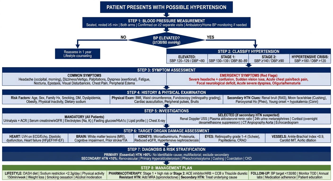
Make a clinical presentation flowchart for hypertensive symptoms and diagnosis

Generating Image

A comprehensive clinical presentation flowchart for hypertension symptoms and diagnosis, medical poster style suitable for postgraduate scholars. The flowchart begins at the top with a box titled "PATIENT PRESENTS WITH POSSIBLE HYPERTENSION" with an arrow pointing down. Below that, a wide box labeled "STEP 1: BLOOD PRESSURE MEASUREMENT" containing: "Seated, rested ≥5 min | Both arms | Confirmed on ≥2 separate visits | Ambulatory/Home BP monitoring if needed" Arrow down to a DIAMOND decision box: "BP ELEVATED? (≥130/80 mmHg)" Two branches - YES (right arrow) and NO (left arrow to "Reassess in 1 year / Lifestyle counseling" box) YES branch flows down to a box "STEP 2: CLASSIFY HYPERTENSION" with 4 sub-boxes side by side: - "ELEVATED: SBP 120–129 / DBP <80" - "STAGE 1: SBP 130–139 / DBP 80–89" - "STAGE 2: SBP ≥140 / DBP ≥90" - "HYPERTENSIVE CRISIS: SBP >180 / DBP >120" Below that, "STEP 3: SYMPTOM ASSESSMENT" with two columns: Left column - "COMMON SYMPTOMS" listing: Headache (occipital, morning), Dizziness/Vertigo, Palpitations, Dyspnea (exertional), Fatigue, Nocturia, Epistaxis, Visual Disturbances, Chest Pain, Peripheral Edema Right column - "EMERGENCY SYMPTOMS (Red Flags)" in red text: Severe headache + confusion, Sudden vision loss, Acute chest pain/back pain, Focal neurological deficit, Acute severe dyspnea, Oliguria/hematuria Arrow down to "STEP 4: HISTORY & PHYSICAL EXAMINATION" with 3 sub-boxes: - "Risk Factors: Age, Sex, Family Hx, Smoking, DM, Dyslipidemia, Obesity, Physical inactivity, Dietary sodium" - "Physical Exam: BMI, Waist circumference, Fundoscopy (retinopathy grading), Cardiac auscultation, Peripheral pulses, Bruits" - "Secondary HTN Clues: Renal bruit (RAS), Moon face/striae (Cushing), Paroxysmal Hx (Pheo), Young onset + hypokalemia (Conn)" Arrow down to "STEP 5: INVESTIGATIONS" with two rows: Row 1 - "MANDATORY (All Patients)": Urinalysis + ACR | Serum creatinine/eGFR | Electrolytes (Na, K) | Fasting glucose/HbA1c | Lipid profile | ECG | Chest X-ray Row 2 - "SELECTED (if secondary HTN suspected)": Renal Doppler USS | Plasma aldosterone:renin ratio | 24h urine metanephrines | Cortisol (overnight dexamethasone suppression) | CT Angiography Aorta | Echocardiogram Arrow down to "STEP 6: TARGET ORGAN DAMAGE ASSESSMENT" with 5 organ boxes in a row: - HEART: LVH on ECG/Echo, Diastolic dysfunction, Heart failure (HFpEF/HFrEF) - BRAIN: White matter lesions (MRI), Cognitive impairment, Prior stroke/TIA - KIDNEYS: Microalbuminuria, Proteinuria, Reduced eGFR, Hypertensive nephrosclerosis - EYES: Retinopathy grade 1–4 (Scheie), Papilledema, CRAO - VESSELS: Ankle-Brachial Index <0.9, Carotid IMT, Aortic dilation Arrow down to "STEP 7: DIAGNOSIS & RISK STRATIFICATION" diamond: - "PRIMARY (Essential) HTN >90%: No identifiable cause; multifactorial; exclude secondary" - "SECONDARY HTN <10%: Renovascular | Primary Hyperaldosteronism | Pheochromocytoma | Cushing | Coarctation | CKD" Final box at bottom: "STEP 8: MANAGEMENT PLAN" with 3 columns: - "LIFESTYLE: DASH diet | Sodium restriction <2.3g/day | Physical activity 150min/week | Weight loss | Smoking cessation | Alcohol moderation" - "PHARMACOTHERAPY: Stage 1 + high risk or Stage 2: ACE inhibitor/ARB + CCB ± Thiazide diuretic | Resistant HTN: Add MRA (spironolactone) | Secondary HTN: Treat underlying cause" - "FOLLOW-UP: BP target <130/80 | Monitor TOD | Annual labs | Medication adherence | Patient education" Use a clean white background, navy blue headers, red for emergency boxes, green for management, organized professional medical diagram layout with clear arrows and clean typography.

Here is the complete clinical presentation flowchart:
Hypertension Clinical Presentation & Diagnosis Flowchart

The flowchart walks through 8 sequential clinical steps:
StepContent
Step 1BP Measurement - correct technique, both arms, confirmation on ≥2 visits
Step 2BP Classification - Elevated / Stage 1 / Stage 2 / Hypertensive Crisis
Step 3Symptom Assessment - common symptoms vs. red flag emergency symptoms (severe headache+confusion, sudden vision loss, tearing chest pain, focal neurology)
Step 4History & Physical Examination - risk factors, BMI/fundoscopy, clues to secondary HTN
Step 5Investigations - mandatory (ECG, urine ACR, eGFR, electrolytes, lipids, glucose) vs. selected (aldosterone:renin ratio, 24h metanephrines, renal Doppler)
Step 6Target Organ Damage Assessment - Heart / Brain / Kidneys / Eyes / Vessels
Step 7Diagnosis - Primary HTN (>90%) vs. Secondary HTN (<10%) with specific causes
Step 8Management - Lifestyle + Pharmacotherapy (ACEi/ARB + CCB ± thiazide) + Follow-up targets
You can right-click the image above to save or download it for your thesis presentation. If you need this as a downloadable PPTX slide or PDF poster, let me know!
This is a shared conversation. Sign in to Orris to start your own chat.