
from dm vasudevan a textbook of biochemistry for Medical Students 11th edition write a 5 mark short note with proper content including fowchart, diagram, tables and clinical application for biochemistry theory university exam on Hyperuricemia
purine catabolism uric acid synthesis pathway diagram
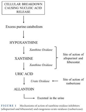
Summary : This flowchart illustrates the biochemical pathway of purine catabolism leading to uric acid production and its further breakdown, highlighting the sites of action for xanthine oxidase inhibitors (allopurinol, febuxostat) and urate oxidase (rasburicase). flowchart: # Nodes : • CELLULAR BREAKDOWN CAUSING NUCLEIC ACID RELEASE (rectangle) • Excess purine catabolism (rectangle) • HYPOXANTHINE (rectangle) • XANTHINE (rectangle) • URIC ACID (rectangle) • ALLANTOIN (rectangle) • Excreted in the urine (rectangle) • Site of action of allopurinol and febuxostat (annotation, right of XANTHINE) • Site of action of rasburicase (annotation, right of URIC ACID) # Connectors : • Downward arrows connect each node in sequence: CELLULAR BREAKDOWN → Excess purine catabolism → HYPOXANTHINE → XANTHINE → URIC ACID → ALLANTOIN → Excreted in the urine. • "Xanthine Oxidase" is labeled on the arrows from HYPOXANTHINE to XANTHINE and from XANTHINE to URIC ACID. • "Urate Oxidase" is labeled on the arrow from URIC ACID to ALLANTOIN. • Brackets and arrows indicate the sites of drug action: allopurinol/febuxostat act at the conversion of hypoxanthine/xanthine to uric acid; rasburicase acts at the conversion of uric acid to allantoin. # Layout : • Linear vertical arrangement, with annotations branching to the right at relevant steps. # Analysis : • The flowchart clarifies the sequential enzymatic steps in purine degradation, pinpointing where pharmacological agents intervene to reduce uric acid levels. Xanthine oxidase inhibitors (allopurinol, febuxostat) block the formation of uric acid, while rasburicase facilitates the conversion of uric acid to the more soluble allantoin, which is then excreted in urine.
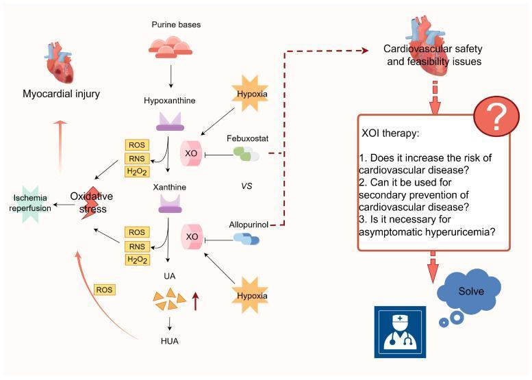
This pathophysiology diagram illustrates the metabolic pathway of purine degradation and its implications for cardiovascular health. The central pathway shows the sequential conversion of purine bases to hypoxanthine, then to xanthine, and finally to uric acid (UA) by the enzyme xanthine oxidase (XO). Excessive UA production leads to hyperuricemia (HUA). The diagram highlights that the XO-catalyzed steps concurrently generate reactive oxygen species (ROS), reactive nitrogen species (RNS), and hydrogen peroxide (H2O2), contributing to oxidative stress and subsequent myocardial injury, particularly during ischemia-reperfusion events. Hypoxia is shown to stimulate XO activity and UA levels. On the right, the clinical relevance is depicted through the use of xanthine oxidase inhibitors (XOIs), specifically Febuxostat and Allopurinol. The diagram raises critical clinical questions regarding XOI therapy, including its potential cardiovascular risks, its utility in secondary cardiovascular prevention, and its necessity in treating asymptomatic hyperuricemia. This flowchart integrates biochemistry with clinical cardiology to explore the therapeutic balance and safety profile of urate-lowering therapies in cardiovascular disease management.
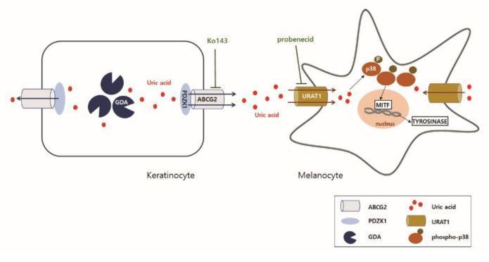
This pathophysiology diagram illustrates the cellular signaling pathway between a keratinocyte and a melanocyte involving Guanine Deaminase (GDA)-induced melanogenesis. In the keratinocyte (left), GDA facilitates the production of uric acid. Uric acid is subsequently exported from the cell via the ABCG2 transporter, which is stabilized by the scaffold protein PDZK1. This export process is inhibited by Ko143. Once in the extracellular space, uric acid is imported into the adjacent melanocyte (right) through the URAT1 transporter, a process inhibited by probenecid. Inside the melanocyte, uric acid triggers the phosphorylation and activation of p38 mitogen-activated protein kinase (MAPK). Phospho-p38 signaling leads to increased expression of Microphthalmia-associated Transcription Factor (MITF) within the nucleus and Tyrosinase in the cytoplasm, ultimately driving melanin synthesis. This schematic highlights the paracrine role of keratinocyte-derived uric acid in promoting skin hyperpigmentation associated with conditions like seborrheic keratosis and photoaging.
gout tophus monosodium urate crystals joint aspiration polarized light
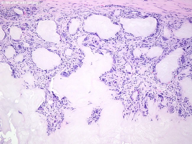
Light microscopy of a soft-tissue biopsy surrounding a gouty tophus shows extensive chalky, eosinophilic to basophilic amorphous deposits with adjacent granulomatous inflammation. Monosodium urate crystals are deposited within the periarticular soft tissue and synovium, appearing as refractile needle-shaped material that often dissolves during routine processing, leaving voids. The tophus is surrounded by a dense inflammatory capsule composed of macrophages, histiocytes, and numerous foreign body giant cells actively attempting to phagocytose crystalline material. Fibroblasts and scant lymphocytes form a fibrous stroma, with variable vascular proliferation. The gross histology may mimic a tumor or infection, but localization near joints and the characteristic granulomatous reaction supports chronic tophaceous gout. Polarized light examination would demonstrate negative birefringent crystals consistent with monosodium urate. Clinically, this pattern confirms chronic hyperuricemia with urate overproduction or reduced excretion and correlates with long-standing gout. The diagnostic significance lies in distinguishing inflammatory tophaceous gout from rheumatoid nodules, infectious arthritis, or soft-tissue neoplasms. Potential clinical use cases include confirming gout in patients with hyperuricemia, evaluating painful soft-tissue swellings around joints, and guiding urate-lowering therapy or surgical planning. This image emphasizes the pathognomonic granulomatous response to urate crystals and its impact on joint function. Correlation with serum uric acid improves diagnostic accuracy.
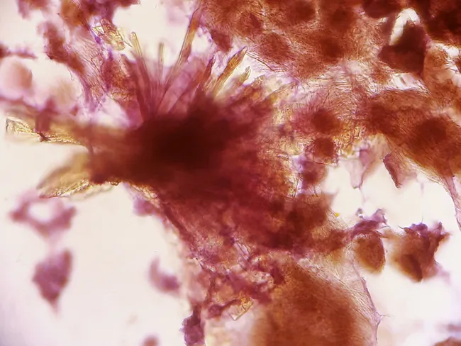
This fine-needle aspiration cytology image demonstrates a gouty tophus in soft tissue of the distal extremity (ankle/toes) from a 55-year-old female. FNA performed with a 23-gauge needle yielded material that, on light microscopy, comprises amorphous granular debris with dense aggregates of slender, needle-shaped crystals possessing pointed tips. The crystals are characteristic of monosodium urate and the appearance aligns with gouty tophus formation. The surrounding background may include inflammatory cells, macrophages and necrotic debris; the crystals may appear birefringent under polarized light (negatively birefringent under compensated polarized microscopy). These findings confirm the cytologic diagnosis of gout and help distinguish a potentially infiltrative soft-tissue lesion from infectious or neoplastic processes. Clinically, this supports uric acid–lowering therapy and appropriate anti-inflammatory management, with surgical excision considered for symptomatic tophi. In differential considerations, calcium pyrophosphate deposition (pseudogout) can mimic crystals but typically shows rhomboid crystals; infectious etiologies (tenosynovitis, septic arthritis) and soft tissue tumors must be excluded. This image is valuable for pathology education, differential diagnosis training, and clinical decision-making in patients with chronic gout presenting with foot and ankle nodules. This case exemplifies cytology's role in confirming crystal-induced pathology and guiding targeted therapy, reducing unnecessary procedures and improving diagnostic confidence in soft-tissue gout diagnosis.
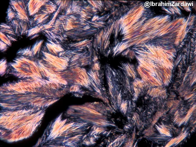
Polarized light microscopy image of monosodium urate crystals in synovial fluid from a joint, illustrating classic gout crystallography. Under cross-polarized illumination, slender, needle-shaped crystals appear colorless individually but generate bright interference colors when overlapped. These crystals are elongated with pointed termini and align to yield yellow-blue birefringence, creating a spectrum. The image showcases monosodium urate deposition, a hallmark of gout, with characteristic needle morphology and negative birefringence. The sample is not stained; polarization highlights crystalline structure and inter-crystal interference. Diagnostic significance includes definitive confirmation of gout and differentiation from calcium pyrophosphate crystals (pseudogout), which are rhomboid and weakly birefringent. Clinical relevance encompasses acute gout assessment, urate-lowering therapy decisions, and teaching crystal pathology in rheumatology education. Techniques: high magnification polarized light imaging with standard filters; interference colors reflect crystal overlap rather than stain. Credit: image courtesy of Dr. Ibrahim Zardawi. In PLM, monosodium urate crystals exhibit strong negative birefringence; their yellow-to-blue color shift depends on crystal orientation relative to the slow axis. By contrast, calcium pyrophosphate crystals in pseudogout are rhomboid and show weak positive birefringence, aiding differential diagnosis. This image is valuable for education, research on crystal pathophysiology, and confirming joint effusion analysis in rheumatologic clinics. Cited for teaching laboratories.
HYPERURICEMIA
Short Note - 5 Marks | Biochemistry University Exam
DEFINITION
Normal serum urate: 3.5-7.0 mg/dL (males); 2.5-6.0 mg/dL (females)
BIOCHEMICAL BASIS - PURINE CATABOLISM PATHWAY
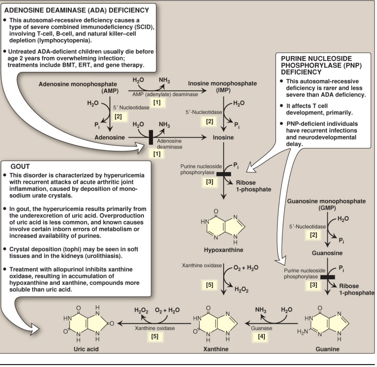
- AMP → Adenosine → Inosine → Hypoxanthine (via AMP deaminase, 5'-nucleotidase, adenosine deaminase, purine nucleoside phosphorylase)
- GMP → Guanosine → Guanine → Xanthine (via 5'-nucleotidase, purine nucleoside phosphorylase, guanase)
- Hypoxanthine + Xanthine → Uric Acid (catalyzed by Xanthine Oxidase - a molybdenum-containing enzyme)
FLOWCHART - CAUSES AND CONSEQUENCES OF HYPERURICEMIA
         PURINES (Dietary + Endogenous synthesis)
                        │
                        ▼
            Purine Catabolism Pathway
                        │
               Xanthine Oxidase
                        │
                        ▼
                   URIC ACID
                  ┌────┴────┐
         OVERPRODUCTION   UNDEREXCRETION
         (< 10% cases)    (> 90% cases)
                  └────┬────┘
                        ▼
              HYPERURICEMIA (>6.8 mg/dL)
                        │
            ┌───────────┼──────────────┐
            ▼           ▼              ▼
     Acute Gout    Chronic Gout    Uric Acid
    (Arthritis)   (Tophi/Joints)  Nephrolithiasis
                                  /Nephropathy
CAUSES OF HYPERURICEMIA
TABLE 1 - Classification of Hyperuricemia
| Type | Mechanism | Examples |
|---|---|---|
| PRIMARY - Overproduction | Increased purine synthesis de novo | PRPP synthetase superactivity (X-linked); Lesch-Nyhan syndrome (HGPRT deficiency) |
| PRIMARY - Underexcretion | Idiopathic renal urate retention | Unknown inherent excretory defects |
| SECONDARY - Overproduction | Increased cell turnover → more nucleic acid catabolism | Myeloproliferative disorders, chemotherapy, hemolytic anemia, psoriasis |
| SECONDARY - Underexcretion | Impaired renal urate excretion | Chronic renal failure, lactic acidosis, lead nephropathy (saturnine gout), thiazide diuretics, cyclosporine |
| Metabolic disease | Increased PRPP availability | Von Gierke disease (G-6-phosphatase deficiency), hereditary fructose intolerance |
PATHOGENESIS OF GOUT (The Key Consequence)
TABLE 2 - Clinical Stages of Gout
| Stage | Features |
|---|---|
| Asymptomatic hyperuricemia | Elevated serum urate; no symptoms; may have occult urate deposits |
| Acute gouty arthritis | Sudden severe joint pain, swelling, erythema; 1st MTP (podagra) most common; self-limiting (days-weeks) |
| Intercritical gout | Symptom-free intervals between acute attacks |
| Chronic tophaceous gout | Tophi in soft tissue (ears, tendons, joints); joint destruction; usually >10 years of disease |
BIOCHEMICAL ENZYME DEFECTS (Exam Favorites)
TABLE 3 - Genetic Causes of Hyperuricemia
| Disorder | Deficient Enzyme | Mechanism | Inheritance |
|---|---|---|---|
| Lesch-Nyhan Syndrome | HGPRT (Hypoxanthine-Guanine Phosphoribosyl Transferase) | Defective salvage → ↑ PRPP → ↑ de novo purine synthesis → ↑ uric acid | X-linked recessive |
| PRPP Synthetase Superactivity | PRPP synthetase (overactive) | ↑ PRPP production → ↑ purine synthesis → ↑ uric acid | X-linked |
| Von Gierke Disease | Glucose-6-phosphatase | ↑ G-6-P → ↑ pentose phosphate pathway → ↑ PRPP → ↑ uric acid | Autosomal recessive |
Key point: Lesch-Nyhan presents with hyperuricemia + self-mutilation + intellectual disability + choreoathetosis (HGPRT deficiency leads to no feedback inhibition of purine synthesis).
TREATMENT FLOWCHART

        HYPERURICEMIA MANAGEMENT
                │
    ┌───────────┼──────────────────┐
    ▼           ▼                  ▼
Acute Gout  Urate Lowering    Dietary/Lifestyle
Treatment   Therapy (ULT)
    │           │                  │
NSAIDs,   Xanthine Oxidase    Low purine diet
Colchicine,  Inhibitors:      Adequate hydration
Steroids   - Allopurinol*    Avoid alcohol
           - Febuxostat      Avoid thiazides
               OR
         Uricosurics:
         - Probenecid
         - Benzbromarone
               OR
         Uricases:
         - Rasburicase
         - Pegloticase
         (refractory gout)
CLINICAL APPLICATIONS
| Clinical Scenario | Biochemical Basis | Significance |
|---|---|---|
| Chemotherapy-induced hyperuricemia / Tumor Lysis Syndrome | Massive cell death → release of nucleic acids → purine catabolism → uric acid surge | Rasburicase (recombinant urate oxidase) given prophylactically to convert uric acid → allantoin (more soluble) |
| Lesch-Nyhan Syndrome | HGPRT deficiency → loss of salvage pathway → excess PRPP → excess uric acid | Allopurinol controls uricemia but does NOT correct neurological features (self-mutilation, spasticity, intellectual disability) |
| Saturnine Gout | Lead nephropathy reduces renal tubular excretion of urate | Seen in lead poisoning; treat underlying lead toxicity |
| Lactic acidosis / Starvation | Lactate/ketones compete with urate for tubular secretion | Acute gout flare can occur in fasting or diabetic ketoacidosis |
| Renal Uric Acid Stones (Urolithiasis) | Uric acid relatively insoluble at acidic urine pH | Urine alkalinization + allopurinol; abundant fluid intake |
| Febuxostat vs Allopurinol | Both are XO inhibitors, but febuxostat is non-purine and selective; useful in allopurinol-hypersensitive patients | Febuxostat preferred in renal impairment |
| Asymptomatic Hyperuricemia | Urate > 6.8 mg/dL but no gout symptoms | May indicate hypertension, metabolic syndrome; not routinely treated with drugs unless sustained and very high |
DIAGNOSTIC CRITERIA (Exam Point)
- Gold standard: Synovial fluid aspiration + polarized light microscopy showing needle-shaped, negatively birefringent MSU crystals
- Serum uric acid: >7 mg/dL males; >6 mg/dL females = hyperuricemia
- 24-hour urinary uric acid: >800 mg/day = overproduction; <800 mg/day with hyperuricemia = underexcretion
- Imaging: X-ray shows "rat-bite erosions" with overhanging edges in chronic tophaceous gout
QUICK REVISION SUMMARY BOX
| Feature | Detail |
|---|---|
| Definition | Serum uric acid >6.8 mg/dL |
| End product | Uric acid (humans lack uricase) |
| Key enzyme | Xanthine Oxidase (Mo-containing) |
| >90% cases due to | Underexcretion |
| Genetic causes | Lesch-Nyhan (HGPRT↓), PRPP synthetase superactivity |
| Key drug | Allopurinol (xanthine oxidase inhibitor - suicide inactivation) |
| Gold standard diagnosis | Polarized light microscopy - needle-shaped negative birefringent crystals |
| Complication | Gout, nephrolithiasis, nephropathy, tophi |