
Granulomatous amoebic encephalitis
"granulomatous amoebic encephalitis" Acanthamoeba Balamuthia
Acanthamoeba granulomatous amoebic encephalitis histology brain
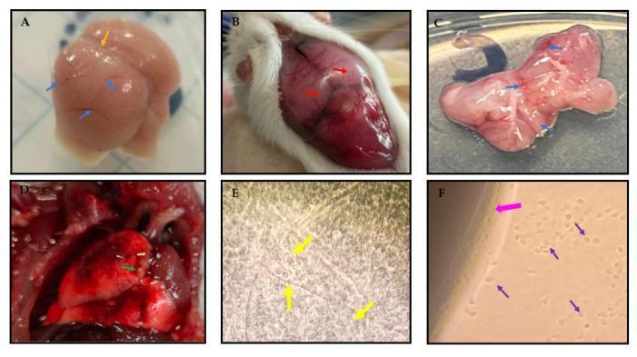
This composite educational graphic illustrates the macroscopic and microscopic pathology of Granulomatous Amoebic Encephalitis (GAE) and systemic dissemination caused by Acanthamoeba culbertsoni in a murine model. Panels A through C display the progression of neuroanatomical damage: Panel A (4 days post-infection) shows early inflammation with loss of central sulcus integrity and incipient hemorrhage. Panel B shows hyperemic meninges and severe congestion. Panel C demonstrates advanced encephalomalacia, characterized by total loss of brain tissue integrity, extensive bleeding, and edema. Panel D depicts secondary pulmonary involvement, showing a hyperemic lung with peripheral hemorrhage and granular lesions suggestive of granulomatous inflammation. Panels E and F provide microscopic confirmation: Panel E (40x optical microscopy) reveals rounded amoebic trophozoites embedded within brain tissue imprints. Panel F (10x inverted microscopy) shows trophozoites migrating from brain tissue explants onto Non-Nutrient Agar (NNA). This series serves as a clinical reference for understanding the fulminant pathophysiology of opportunistic free-living amoebic infections, highlighting tissue necrosis, vascular congestion, and morphological identification of the pathogen.
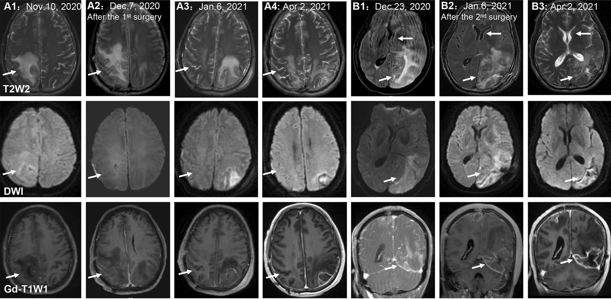
This diagnostic imaging composite displays a series of brain MRI scans illustrating the longitudinal progression of granulomatous amoebic encephalitis (GAE) caused by Balamuthia mandrillaris. The image is organized into two panels (A and B) across three imaging modalities: T2-weighted (T2W2), Diffusion-Weighted Imaging (DWI), and Gadolinium-enhanced T1-weighted (Gd-T1WI). Panel A (A1–A4) follows a right parietal lobe lesion from initial presentation through post-surgical follow-up. The lesion shows high T2 signal intensity and restricted diffusion on DWI, with peripheral enhancement on Gd-T1WI. Over time (Nov 2020 to Apr 2021), the lesion and associated perilesional edema significantly decrease in size. Panel B (B1–B3) tracks a subsequent left occipital lobe lesion. Key features include high T2 and DWI signal intensity, marked contrast enhancement, and evidence of a left cerebral subfalcine herniation (indicated by white arrows). Follow-up scans after a second surgery and combined antimicrobial therapy (sulfamethoxazole/trimethoprim, azithromycin, flucytosine, and fluconazole) show gradual stabilization of the lesion and slight improvement of the midline shift. This series demonstrates the characteristic multifocal, infiltrating, and necrotic nature of GAE and its response to intensive medical and surgical management.
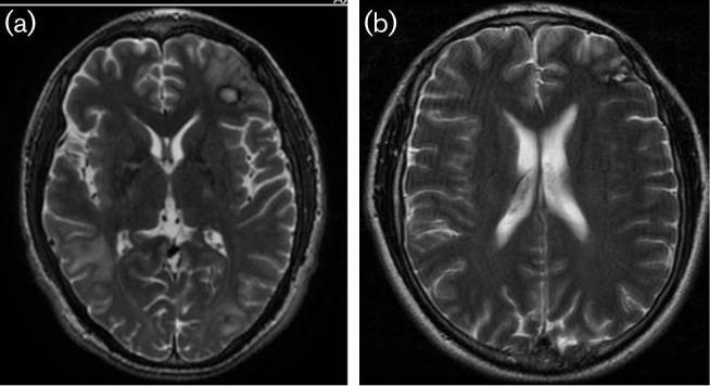
This diagnostic image pair displays axial T2-weighted magnetic resonance imaging (MRI) of the brain, comparing a pathological state with post-treatment resolution. Figure (a) illustrates multiple intracranial lesions consistent with granulomatous amoebic encephalitis, specifically Balamuthia mandrillaris. Key findings include an area of biopsy-related signal change in the left frontal region, extensive hyperintense perilesional oedema associated with a mass in the right temporal lobe, and hyperintense satellite lesions within the left frontal and parietal lobes. These lesions demonstrate characteristic T2-hyperintensity and mass effect. Figure (b) shows a follow-up MRI of the same patient taken two years into treatment, demonstrating significant clinical improvement. There is a complete resolution of the previously observed central nervous system (CNS) lesions, disappearance of the associated vasogenic oedema, and restoration of normal ventricular and sulcal anatomy. This comparison serves as a clinical timeline for the radiological monitoring of rare parasitic encephalitis under intensive therapy.
PMID: 42158383
Granulomatous Amoebic Encephalitis (GAE)
Definition
Causative Organisms
| Organism | Host preference | Notes |
|---|---|---|
| Acanthamoeba spp. | Predominantly immunocompromised | Most common overall (33% of cases) |
| Balamuthia mandrillaris | Both immunocompromised AND immunocompetent | Accounts for ~63% of confirmed GAE cases; higher mortality |
| Sappinia pedata | Any host | Single documented case of GAE |
- Medical Microbiology 9e
Epidemiology
- Rare worldwide; cases reported predominantly from North America and Asia
- Overall mortality: 76.4% (146/191 cases in the most recent 2026 systematic review of 201 confirmed cases)
- Immunosuppression documented in only ~30% of all cases - meaning immunocompetent individuals are not spared, especially with Balamuthia
- Deaths significantly more frequent with Balamuthia infection
Risk Factors & Transmission
- Acanthamoeba: Inhalation of cysts or skin contact (wounds, ulcers) → hematogenous spread to CNS. Found ubiquitously in soil, freshwater, seawater, air, and dust. Risk factors include HIV/AIDS, organ transplantation, malignancy, diabetes, corticosteroid use.
- Balamuthia: Soil or water exposure; exact route unclear but likely inhalation or skin → hematogenous dissemination. Can affect healthy individuals.
- Both organisms do NOT require a host to survive and have NO insect vector.
Pathogenesis
- Amoebae (usually as cysts) enter via respiratory tract or skin breaks
- Hematogenous spread to the CNS
- In the brain: organisms elicit a granulomatous inflammatory response around both trophozoites and cysts
- Progressive focal necrosis, edema, and encephalomalacia
- Both trophozoites and cysts can be found in brain tissue (this is diagnostically key - differentiating GAE from PAM, where only trophozoites are seen)
Clinical Features
| Feature | Description |
|---|---|
| Onset | Subacute to chronic (weeks to months) |
| Fever | Low-grade |
| Headache | Present |
| Focal neurologic deficits | Common |
| Behavioral/personality changes | Present |
| Visual disturbances | Reported |
| Seizures | Occur as disease progresses |
| Elevated ICP | Late finding |
| Coma | Terminal event |
- Bradley and Daroff's Neurology in Clinical Practice; Kaplan & Sadock's Comprehensive Textbook of Psychiatry
Diagnosis
Imaging
- MRI/CT: space-occupying lesions in 78% of cases - can mimic tumor, abscess, or other neurological conditions
- Ring-enhancing lesions with perilesional edema; may be multifocal
- MRI is preferred - shows T2-hyperintense lesions with mass effect
CSF
- Profile consistent with aseptic meningitis (lymphocytic pleocytosis, elevated protein, normal or low glucose)
- Hypoglycorrhachia (low CSF glucose) is a strong correlate of non-survival
- Standard Gram stain and culture negative
Definitive Diagnosis
- Brain biopsy is typically required - will show trophozoites AND cysts within granulomatous inflammation (the cysts are the hallmark distinguishing GAE from PAM)
- PCR (offered by CDC) - can be performed on CSF or tissue
- Culture of biopsy material
- Serology has limited utility
Pathology
Treatment
Acanthamoeba GAE
- Pentamidine
- Azole (fluconazole, itraconazole, or voriconazole)
- Sulfonamide (sulfadiazine or TMP-SMX)
- Flucytosine
- Miltefosine (FDA-approved antileishmanial, now recommended as part of multidrug regimens) - has improved survival
Balamuthia GAE
- Combination of TMP-SMX + azithromycin + flucytosine + fluconazole has been used
- Miltefosine is now recommended as part of multidrug regimens
- Surgical resection of accessible lesions may complement medical therapy
Key Point on Miltefosine
- Goldman-Cecil Medicine; Bradley and Daroff's Neurology
Prognosis
- Overall case fatality rate: ~76% (2026 systematic review, PMID 42158383)
- Balamuthia GAE carries even higher mortality than Acanthamoeba GAE
- Early diagnosis and aggressive multidrug therapy (particularly with miltefosine) are the best determinants of survival
- Hypoglycorrhachia on CSF is a marker of poor prognosis
Comparison: GAE vs PAM
| Feature | GAE | PAM |
|---|---|---|
| Causative organism | Acanthamoeba, Balamuthia, Sappinia | Naegleria fowleri |
| Host | Mainly immunocompromised (but Balamuthia also immunocompetent) | Healthy young individuals |
| Onset | Subacute/chronic (weeks-months) | Acute (days) |
| Exposure | Soil, skin contact, inhalation | Freshwater (swimming/diving) |
| Histology | Trophozoites + cysts (granulomas) | Trophozoites only |
| CSF | Lymphocytic, aseptic pattern | Polymorphonuclear, hemorrhagic |
| Mortality | ~76% | >95% |