
What is heart
The heart is a muscular organ that pumps blood throughout the body via the circulatory system. It sits in the chest (mediastinum), between the lungs, slightly left of center. It has four chambers - two atria (upper) and two ventricles (lower) - and beats roughly 60-100 times per minute at rest.
Searching sources...
Reading File
Finding Sources
Searching the Web
heart anatomy structure function overview
Finding Sources
Finding Sources
Reading File
Finding Sources
Reading File
Reading File
Reading File
Loading Image
Loading Image
Here is a thorough overview of the heart, drawn from authoritative medical textbooks:
The Heart
What Is the Heart?
The heart is a muscular pump that maintains unidirectional flow of blood throughout the body. It lies obliquely, about two-thirds into the left side of the thoracic cavity, in the middle mediastinum - the space enclosed by the sternum, vertebral column, diaphragm, and lungs. It is surrounded by a tough fibrous sac called the pericardium, through which the great vessels enter and leave. - Histology: A Text and Atlas, p. 1050
Four Chambers
The heart has four chambers - the right and left atria (upper) and the right and left ventricles (lower). Valves guard the exits of the chambers, preventing backflow of blood. An interatrial septum and an interventricular septum separate the right and left sides of the heart. - Histology: A Text and Atlas, p. 1051
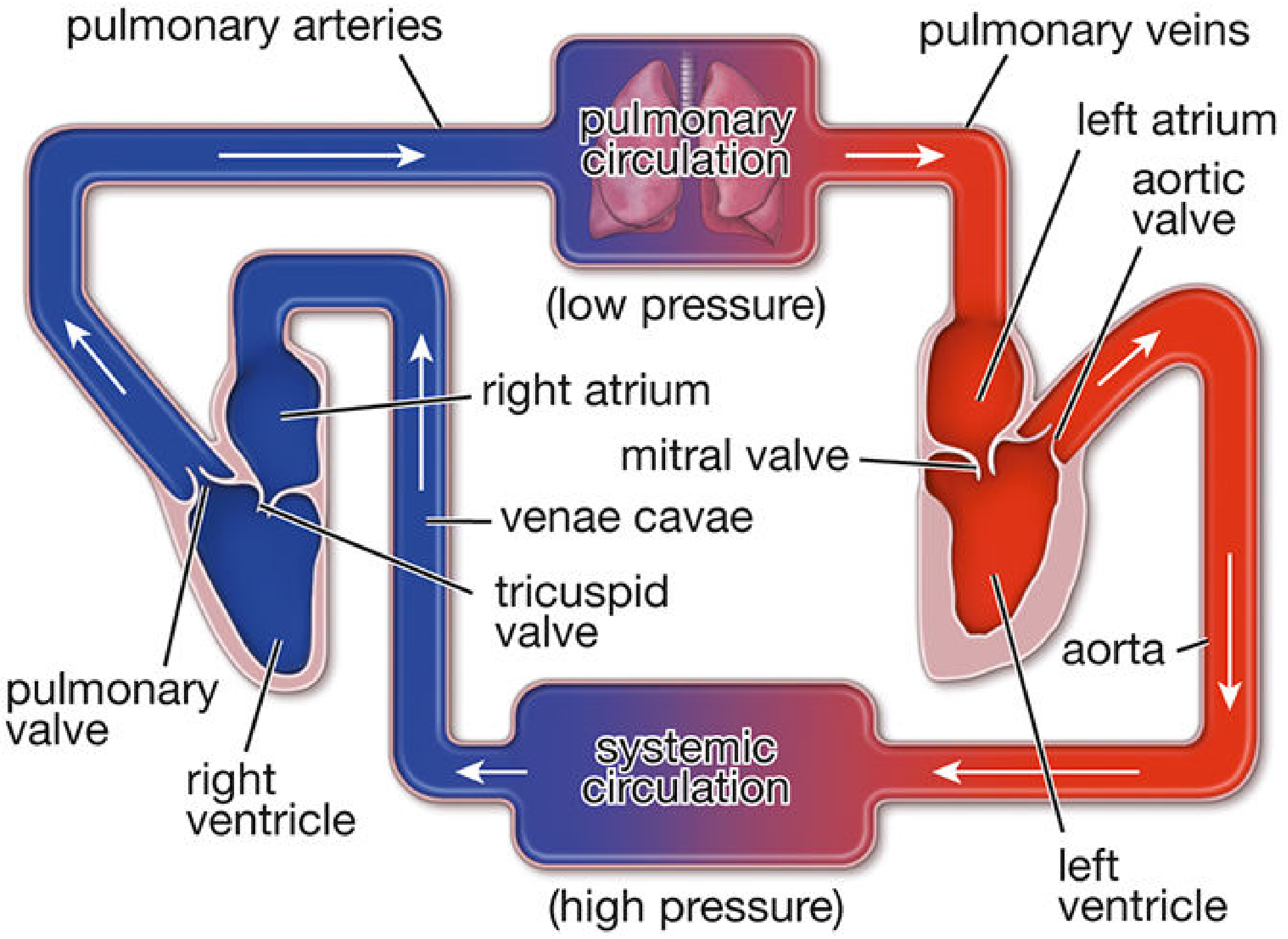
Two Pumps, Two Circuits
The heart functionally acts as two pumps working in parallel:
| Side | Receives | Pumps To |
|---|---|---|
| Right pump (right atrium + right ventricle) | Deoxygenated blood from the body via the superior and inferior venae cavae | Lungs via pulmonary arteries (low pressure) |
| Left pump (left atrium + left ventricle) | Oxygenated blood from the lungs via 4 pulmonary veins | The entire body via the aorta (high pressure) |
Because more force is needed to pump blood through the whole body than through just the lungs, the muscular wall of the left ventricle is thicker than that of the right. - Gray's Anatomy for Students, p. 230
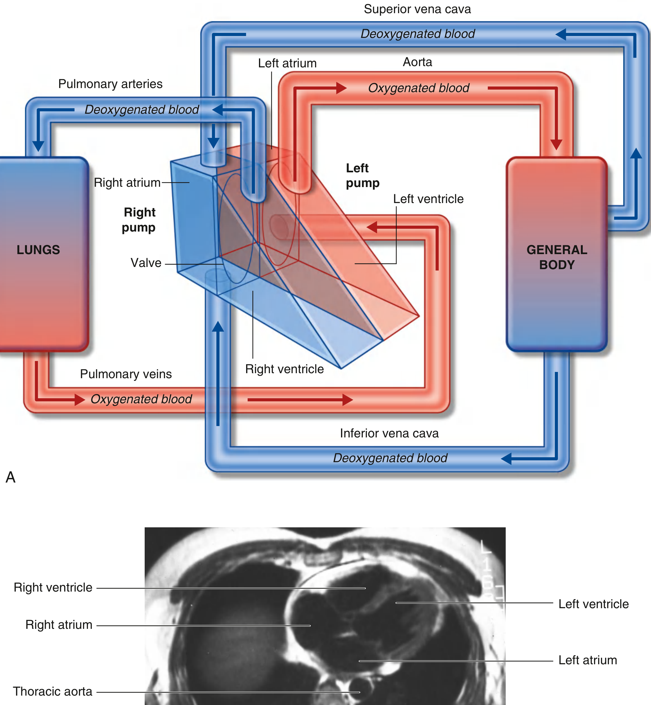
Four Valves
Four valves ensure blood flows in only one direction:
- Tricuspid valve - between right atrium and right ventricle
- Pulmonary valve - at the exit of the right ventricle into the pulmonary artery
- Mitral valve (bicuspid) - between left atrium and left ventricle
- Aortic valve - at the exit of the left ventricle into the aorta
Structural Components
The heart contains: - Histology: A Text and Atlas, p. 1051
- Cardiac muscle (myocardium) - specialized muscle that contracts rhythmically and involuntarily to propel blood
- Fibrous skeleton - four fibrous rings surrounding valve orifices, two fibrous trigones connecting the rings, and the membranous parts of the septa; this provides structural support and electrically insulates the atria from the ventricles
- Conduction system - a network of specialized cells (SA node, AV node, Bundle of His, Purkinje fibers) that generates and coordinates the electrical impulses driving each heartbeat
- Coronary arteries - the blood vessels that supply the heart muscle itself with oxygen and nutrients
Key Facts at a Glance
| Feature | Detail |
|---|---|
| Location | Middle mediastinum, chest |
| Weight | ~250-350 g in adults |
| Resting heart rate | 60-100 beats/minute |
| Output at rest | ~5 liters/minute |
| Outer covering | Pericardium (fibrous sac) |
| Wall layers | Epicardium, myocardium, endocardium |
In short, the heart is the central engine of the circulatory system - a double pump that simultaneously sends deoxygenated blood to the lungs for oxygen pickup and oxygenated blood to every tissue in the body.
This is a shared conversation. Sign in to Orris to start your own chat.