I have comprehensive information from multiple authoritative textbooks. Now I'll write the complete, detailed, simplified answers for all three topics.
ANSWER 1: OMC Variations and Their Role in Chronic Rhinosinusitis (CRS)
What is the Osteomeatal Complex (OMC)?
The OMC is a functional concept, not a single anatomical structure. It is a region in the middle meatus of the lateral nasal wall where several anatomical components converge to form the common drainage pathway for the anterior sinuses (maxillary, frontal, and anterior ethmoid sinuses).
Think of the OMC as a narrow corridor. If anything blocks this corridor - whether from infection, swelling, or an anatomical variation - multiple sinuses get obstructed at once, explaining why sinusitis commonly involves more than one sinus simultaneously.
Components of the OMC
| Structure | Role |
|---|
| Uncinate process | Thin curved bone; forms medial wall of infundibulum |
| Ethmoidal infundibulum | 3D cleft; final common drainage channel |
| Hiatus semilunaris | 2D slit between uncinate process and ethmoid bulla |
| Ethmoid bulla | Largest anterior ethmoid cell |
| Middle turbinate (MT) | Medial boundary of the middle meatus |
| Frontal recess | Drainage channel of frontal sinus |
| Maxillary sinus ostium | Natural opening of maxillary sinus into infundibulum |
The maxillary sinus drains into the ethmoidal infundibulum. The anterior ethmoid cells and frontal sinus drain into the middle meatus via the frontal recess. Any obstruction here = simultaneous blockage of all three drainage pathways.
OMC Anatomical Variations and How They Cause/Worsen CRS
Blockage of the OMC is one of the primary anatomic factors driving CRS. Below are the major variations:
1. Concha Bullosa (Pneumatized Middle Turbinate)
- What it is: Air pneumatizes into the middle turbinate, making it bulky and enlarged
- Incidence: Very common - seen in ~34% of patients
- How it causes CRS: The enlarged turbinate narrows the middle meatus and directly compresses the OMC region, reducing drainage from the maxillary and anterior ethmoid sinuses
- Types: Bulbous (body only), lamellar (vertical lamella only), or extensive (both parts pneumatized)
- A large concha bullosa can push the nasal septum to the opposite side, creating bilateral obstruction
2. Paradoxical Middle Turbinate
- What it is: The middle turbinate curves in the opposite (lateral) direction instead of the normal medial curve
- How it causes CRS: The reversed curvature directly impinges on the uncinate process and infundibulum, narrowing the OMC
3. Septal Deviation
- What it is: The nasal septum curves to one side
- How it causes CRS: A high septal deviation (especially at the level of the middle meatus) directly compresses the OMC structures, reduces airflow, and impairs mucociliary clearance in that region
- Can also narrow the contralateral nasal passage, leading to bilateral effects
4. Uncinate Process Variations
The uncinate process is the "key" structure of the OMC. Its variations strongly impact frontal and maxillary drainage:
| Variation | Effect |
|---|
| Pneumatized uncinate | Bulky uncinate narrows the infundibulum |
| Lateralized uncinate (attaches to skull base/LP) | Closes off the frontal recess; frontal sinus drainage blocked; termed "terminal recess" |
| Medialized uncinate (attaches to MT) | Frontal sinus may drain directly into middle meatus; infundibulum may be blind-ending |
| Deflected/bent uncinate | Direct OMC narrowing |
| Paradoxical uncinate | Reversal of normal curve narrows hiatus semilunaris |
The attachment of the uncinate top determines where the frontal sinus drains. This is surgically critical because it determines the approach during FESS (Functional Endoscopic Sinus Surgery).
5. Agger Nasi Cell
- What it is: The most anterior ethmoid cell, creating a bony mound just anterior to the middle turbinate attachment
- Incidence: Present in ~98.5% of CT scans - most constant ethmoid cell
- How it causes CRS: A large, well-pneumatized agger nasi cell narrows the frontal recess anteriorly, blocking frontal sinus drainage
- A partially resected agger nasi cell can leave the dome in the frontal recess, causing iatrogenic frontal sinus obstruction after surgery
6. Haller Cells (Infraorbital Ethmoid Cells / IOC)
- What it is: Anterior ethmoid cells that pneumatize along the floor of the orbit, above the maxillary sinus ostium
- Incidence: ~10-30% of patients
- How it causes CRS: When large, they encroach directly on the natural ostium of the maxillary sinus, obstructing its drainage
- The lateral wall of the Haller cell may be attached to the infraorbital nerve canal, requiring careful surgical dissection
- Direct link to maxillary sinusitis: Recurrent or refractory maxillary sinusitis with a large Haller cell is a classic FESS indication
7. Large Ethmoid Bulla
- What it is: The ethmoid bulla is the largest and most constant anterior ethmoid cell; occasionally it is over-pneumatized
- How it causes CRS: A very prominent ethmoid bulla can reduce the space in the middle meatus and limit access to the infundibulum, narrowing the OMC
- Its posterior wall forms part of the retrobullar recess; if it contacts the basal lamella, it obliterates this space
8. Accessory Maxillary Ostia and Recirculation Phenomenon
- What it is: An additional ostium in the posterior fontanelle of the medial maxillary wall
- How it causes CRS: Mucus from the maxillary sinus exits through the natural ostium, re-enters through the accessory ostium, then re-exits through the natural ostium again in a continuous loop - this is called mucus recirculation
- This perpetuates chronic inflammation and infection in the maxillary sinus
- Surgery must incorporate the accessory ostium with the natural ostium to break this cycle
9. Frontoethmoidal Cells (Kuhn Type Cells)
- Ethmoid cells that pneumatize into or around the frontal recess
- Type 1: Single cell above agger nasi
- Type 2: Two or more cells above agger nasi
- Type 3: Cell extending >50% into the frontal sinus
- Type 4: Cell extending >50% of frontal sinus height (most likely to cause obstruction)
- Large type 3 or type 4 cells significantly narrow or completely obstruct the frontal sinus drainage pathway, leading to frontal sinusitis
The Cascade Mechanism - How OMC Obstruction Causes CRS
Anatomical variation → OMC narrowing
         ↓
Mucociliary clearance impaired
         ↓
Mucus stasis → bacterial colonization
         ↓
Mucosal edema → further OMC obstruction
         ↓
Sinus hypoxia → altered flora → anaerobic growth
         ↓
Repeated acute episodes → chronic inflammation
         ↓
CRS (neutrophilic or eosinophilic, with or without polyps)
Key fact from KJ Lee's: "Blockage of the osteomeatal complex, sphenoethmoidal recess, frontal recess, or other natural drainage pathways by anatomic abnormalities can cause or prolong chronic sinusitis" - Anatomic variations listed include: septal deviation, turbinate hypertrophy, middle turbinate concha bullosa, paradoxical turbinates, prominent agger nasi cell, Haller cells, prominent ethmoid bulla, pneumatization and inversion of uncinate process, hypoplastic sinuses, and accessory maxillary ostia with recirculation.
Other Contributing Factors to CRS (Beyond Anatomy)
- Mucociliary impairment: Cilia beat the mucus layer toward natural ostia. When impaired (e.g., in primary ciliary dyskinesia, CF, viral infection), mucus stagnates
- Bacterial biofilms: Polymicrobial communities adhere to sinus mucosa, evade antibiotics, and perpetuate chronic low-grade infection
- Allergy and eosinophilic inflammation: TH-2 driven response (CRSwNP) - eosinophil infiltration, elevated IL-5, IL-13, histamine cause persistent mucosal swelling
- Immunodeficiency: Primary (IgA deficiency, common variable immunodeficiency) or acquired states impair local defense
- Fungal elements: Allergic fungal rhinosinusitis (AFS) - 8-12% of CRS
(Source: KJ Lee's Essential Otolaryngology; Cummings Otolaryngology Head and Neck Surgery; Scott-Brown's ORL Vol 1)
ANSWER 2: Onodi Cell (Sphenoethmoidal Cell)
What is an Onodi Cell?
An Onodi cell (also called a sphenoethmoidal cell or SEC) is a posterior ethmoid air cell that has pneumatized posteriorly beyond the sphenoid sinus, extending over its superolateral aspect.
Put simply - it is an ethmoid cell that has "sneaked behind" the sphenoid sinus, sitting on top of and lateral to it. On casual inspection, it looks like it is part of the sphenoid sinus, which is where the danger lies.
Named after Adolf Onodi (Hungarian anatomist, 1857-1920).
Key Anatomical Facts
| Feature | Detail |
|---|
| Location | Superolateral to the sphenoid sinus |
| Relationship | Onodi cell is above and lateral; sphenoid sinus is below and medial |
| Incidence | ~30% of individuals |
| Optic nerve | Projects along the superolateral wall of the Onodi cell (not the sphenoid) when present |
| Internal carotid artery | May also course along the Onodi cell wall |
| CT identification | Coronal CT shows a horizontal septation within or superior to the sphenoid sinus, posterior to the bony choanal arch |
| Drains into | Superior meatus (as a posterior ethmoid cell) |
Why the Onodi Cell is Clinically Critical
1. Optic Nerve at Risk
When an Onodi cell is present, the optic nerve deviates to run along the wall of the Onodi cell rather than the sphenoid sinus. During endoscopic sphenoid surgery or posterior ethmoidectomy, a surgeon who does not recognize the Onodi cell may inadvertently:
- Open the Onodi cell thinking it is the sphenoid sinus
- Injure the optic nerve while trying to reach the sphenoid
- Result: Immediate or delayed blindness/vision loss
2. Internal Carotid Artery (ICA) Involvement
The ICA can also course through or indent the wall of the Onodi cell. Unrecognized entry into this cell carries risk of catastrophic ICA hemorrhage.
3. Mucocele Risk
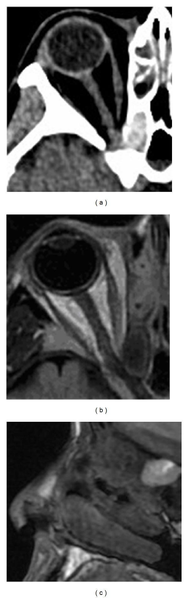
As shown in clinical imaging, when an Onodi cell develops a mucocele (fluid-filled, expanding cyst), it can:
- Compress the adjacent optic nerve
- Lead to compressive optic neuropathy with progressive visual loss
- Compress the orbital apex - causing orbital apex syndrome
How to Identify on CT (Pre-operatively Mandatory)
Coronal CT: Look for a horizontal bony septation that appears to divide the "sphenoid sinus" - the true sphenoid sinus lies inferiorly/medially and the Onodi cell lies superiorly/laterally.
Axial CT: The Onodi cell sits posterior to the ethmoid complex and lateral to the sphenoid sinus; the optic nerve canal may be seen running through its superolateral wall.
Key rule: Always identify Onodi cells pre-operatively before any sphenoid or posterior ethmoid surgery. Failure to recognize this variation is a major cause of optic nerve injury in FESS.
Surgical Significance in FESS
- The sphenoethmoidal recess (where the sphenoid ostium opens) lies medial to the Onodi cell
- The true sphenoid ostium should be found on the superior nasal septum / anterior face of the sphenoid, typically 1-1.5 cm above the posterior choana and 7 cm from the anterior nasal spine
- When an Onodi cell is present, the surgeon must navigate past it (laterally) to find the true medial sphenoid ostium below
- The Onodi cell itself may also be diseased and require drainage (if opacified on CT)
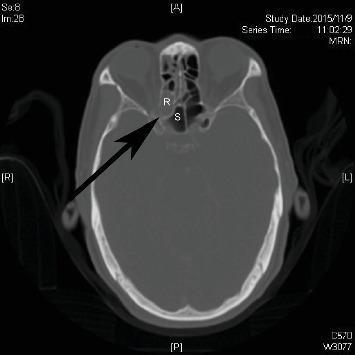
Axial CT: Right sphenoethmoidal (Onodi) cell (R) is opacified; the arrow points to the right optic canal, showing how close the optic nerve is to this cell. (S = sphenoid sinus)
Onodi cell mucocele on CT and MRI: Shows the expanding fluid-filled lesion compressing the optic nerve at the orbital apex - a potentially blinding complication
(Source: KJ Lee's Essential Otolaryngology; Cummings ORL; Scott-Brown's ORL Vol 1)
ANSWER 3: Endoscopic Anatomy of the Nose and Paranasal Sinuses
Overview: Why Endoscopic Anatomy?
Endoscopic sinus surgery (FESS) requires the surgeon to navigate the nose using a 0°, 30°, or 45° Hopkins rod endoscope. The anatomy seen through the endoscope looks very different from textbook diagrams. Understanding it is the foundation of safe and effective surgery. As Messerklinger showed in 1978, the OMC is the key region, and its endoscopic visualization drives modern rhinology.
Part A: The Nasal Cavity
Medial Wall (Nasal Septum)
- Formed by the perpendicular plate of ethmoid (superior, bony) and vomer (inferior/posterior, bony); the anterior portion is the quadrilateral cartilage
- The septum divides the nose into two nasal cavities
- Kiesselbach's plexus (Little's area): on the anterior septum - the most common site of anterior epistaxis (anastomosis of SPA posterior septal branch, AEA, greater palatine artery, and alar branch of superior labial artery)
Lateral Nasal Wall (Endoscopically Critical)
The lateral wall carries the turbinates and meatal spaces that house the sinus drainage pathways.
The 3 Turbinates (from inferior to superior):
| Turbinate | Bone | Key Points |
|---|
| Inferior turbinate | Independent bone | Largest turbinate; regulates nasal airflow and resistance; important for humidification |
| Middle turbinate (MT) | Part of ethmoid | The key surgical landmark; everything important is medial or lateral to it |
| Superior turbinate | Part of ethmoid | Marks the sphenoethmoidal recess (sphenoid sinus drains here) |
| Supreme turbinate | Occasionally present | Small, above superior turbinate |
The 3 Meatal Spaces:
| Meatus | Location | What Drains Here |
|---|
| Inferior meatus | Below inferior turbinate | Nasolacrimal duct (tear drainage) |
| Middle meatus | Below middle turbinate | Frontal sinus, maxillary sinus, anterior ethmoid cells - OMC region |
| Superior meatus | Below superior turbinate | Posterior ethmoid cells, occasionally sphenoid sinus |
| Sphenoethmoidal recess | Above and medial to superior turbinate | Sphenoid sinus ostium |
Part B: The Middle Meatus in Detail (Heart of FESS)
What You See Endoscopically:
Inserting a 0° endoscope, after passing the inferior turbinate, you see the middle turbinate medially. Lateral to it (in the middle meatus) from anterior to posterior, you encounter:
1. Agger Nasi Region
- A bony mound/bulge at the anterior end of the middle turbinate attachment
- Marks the beginning of the frontal recess region
- Agger nasi cell (present in 98.5%) sits here
2. Uncinate Process
- A thin curved bone that hangs from the lateral nasal wall
- Like a "sickle-shaped" thin bony leaf projecting medially
- Creates the hiatus semilunaris (a 2D slit) between itself and the ethmoid bulla
- Superior attachment determines frontal sinus drainage:
- Attaches to skull base/lamina papyracea → frontal sinus drains into the infundibulum (medial to the uncinate)
- Attaches to middle turbinate → frontal sinus drains into middle meatus (lateral to the uncinate)
3. Hiatus Semilunaris
- The two-dimensional (2D) crescentic slit between the free edge of the uncinate process (anteroinferior) and the ethmoid bulla (posterosuperior)
- Acts as the "gateway" into the ethmoidal infundibulum
- Endoscopically seen as a dark crescent-shaped opening in the middle meatus
4. Ethmoidal Infundibulum
- The 3D funnel-shaped space deep to the hiatus semilunaris
- Bounded: anteriorly by uncinate process, posteriorly by ethmoid bulla, laterally by lamina papyracea, medially opens into middle meatus via hiatus semilunaris
- The maxillary sinus natural ostium opens into it posteriorly
5. Ethmoid Bulla
- Largest and most constant anterior ethmoid cell
- Appears as a smooth rounded bulge posterior to the hiatus semilunaris
- Posterior to it lies the retrobullar recess (space between bulla and basal lamella of MT)
- Above it lies the suprabullar recess
- Together these = Sinus lateralis
6. Basal Lamella of Middle Turbinate
- The key dividing structure between anterior and posterior ethmoid
- Runs obliquely from anterosuperior to posteroinferior
- Everything anterior to it = anterior ethmoid (drains to middle meatus)
- Everything posterior to it = posterior ethmoid (drains to superior meatus)
- Surgically: once you perforate the basal lamella, you are in the posterior ethmoid
Part C: Individual Sinus Endoscopic Anatomy
1. Maxillary Sinus
- Natural ostium: Located in the posterosuperior part of the medial wall (superior aspect of fontanelle), opens into the infundibulum
- Endoscopic view: After uncinectomy, the natural ostium is visualized in the anterosuperior part of the medial maxillary wall
- Relations: Roof = orbital floor (infraorbital nerve canal runs here); floor = alveolar process; posterior wall = pterygopalatine fossa
- Fontanelle: The membrane-only area of the medial maxillary wall (no bone, just mucosa) - inferior to the natural ostium; site where accessory ostia appear
2. Anterior Ethmoid
- Multiple small cells (smaller but numerous) anterior to the basal lamella of MT
- Key cells: agger nasi, ethmoid bulla, suprabullar cells, frontoethmoidal cells
- Lamina papyracea: The thin bone forming the lateral wall of ethmoid cells = medial wall of orbit. Do NOT breach this - orbital fat herniation or orbital hematoma results
3. Posterior Ethmoid
- Fewer cells but larger
- Located posterior to basal lamella, drains to superior meatus
- Key landmark: Ground lamella of superior turbinate separates posterior ethmoid from sphenoid
- Onodi cells are here (see Answer 2)
4. Frontal Sinus
- Drains via the frontal recess (not a true ostium but an hourglass-shaped drainage pathway)
- The frontal recess is bounded: anteriorly by agger nasi cell, posteriorly by ethmoid bulla/suprabullar cells, medially by middle turbinate, laterally by lamina papyracea
- Agger nasi cell = most important structure to open for frontal sinus access
- Endoscopically approached with a 70° or 45° angled scope
- The drainage pathway can be further narrowed by frontoethmoidal (Kuhn type) cells
5. Sphenoid Sinus
- Ostium: Located on the anterior face of the sphenoid sinus, in the sphenoethmoidal recess - approximately 1-1.5 cm above the posterior choana, 7 cm from the anterior nasal spine
- Approach: Medial to superior turbinate (trans-sphenoethmoidal recess approach)
- Dangerous neighbors:
- Optic nerve: Runs in the superolateral wall (may bulge into sinus, dehiscent in 6-8%)
- Internal carotid artery (ICA): Runs along posterolateral wall (may bulge into sinus, dehiscent in 4-8%)
- Pituitary gland: Superior wall
- Cavernous sinus: Lateral wall (contains CN III, IV, V1, V2, VI, ICA)
- Optic chiasm: Roof/superior wall
Part D: Key Surgical Landmarks - Safe Zones
| Landmark | Surgical Significance |
|---|
| Fovea ethmoidalis (ethmoid roof) | Slopes ~15° from anterior to posterior and lateral to medial; posteromedial corner is lowest and most vulnerable to iatrogenic CSF leak |
| Lateral lamella of cribriform | Formed by ethmoid bone; thinnest and weakest skull base bone; penetrating it causes CSF leak and anosmia |
| Lamina papyracea | Medial orbital wall; breaching causes orbital complications |
| Anterior ethmoid artery (AEA) | Runs through bulla lamella; commonly in or below the skull base near bulla lamella attachment; severing it causes intraorbital hematoma |
| Keros classification | Depth of olfactory fossa: Type 1 (1-3mm), Type 2 (4-7mm), Type 3 (8-16mm); Type 3 = highest risk of skull base perforation |
Summary Diagram of Endoscopic Navigation
NASAL CAVITY (lateral wall, from anterior to posterior):

Agger nasi region
     ↓
Axilla of MT (MT attachment to lateral wall)
     ↓
Uncinate process (+ hiatus semilunaris opens here)
     ↓
Infundibulum (behind hiatus semilunaris)
     ↓
Ethmoid bulla (first prominent cell you see posterior to hiatus)
     ↓
Retrobullar recess / Sinus lateralis
     ↓
Basal lamella of MT (anterior ethmoid ends here)
     ↓
Posterior ethmoid cells (larger)
     ↓
Sphenoethmoidal recess (sphenoid ostium here, medial to superior turbinate)
     ↓
Sphenoid sinus face
Paranasal Sinus Development (Quick Reference)
| Sinus | Development |
|---|
| Ethmoid | First to develop in utero; most developed at birth |
| Maxillary | Present at birth; adult size by late childhood |
| Sphenoid | Pneumatization begins ~1 year postnatally; adult size by age 12 |
| Frontal | Last to start and finish; continues into early adulthood |
| Size order (largest to smallest) | Maxillary > Frontal > Sphenoid > Ethmoid |
(Sources: Cummings Otolaryngology Head and Neck Surgery, Chapter 44; KJ Lee's Essential Otolaryngology, Chapter 26; Scott-Brown's Otorhinolaryngology Head & Neck Surgery Vol 1)