
Intra fusal muscle fiber and extra fusal muscle fiber
Reading File
Intrafusal fibers are specialized muscle fibers inside the muscle spindle (a sensory organ), while extrafusal fibers are the ordinary skeletal muscle fibers that make up the bulk of a muscle and generate its force. Intrafusal fibers are innervated by gamma motor neurons and detect stretch; extrafusal fibers are innervated by alpha motor neurons and produce contraction.
Searching sources...
Finding Sources
Finding Sources
Reading File
Reading File
Reading File
Loading Image
Loading Image
Here is a complete, textbook-based comparison of intrafusal and extrafusal muscle fibers.
Intrafusal vs. Extrafusal Muscle Fibers
Overview
A skeletal muscle contains two functionally distinct populations of muscle fibers that work together to produce and monitor movement:
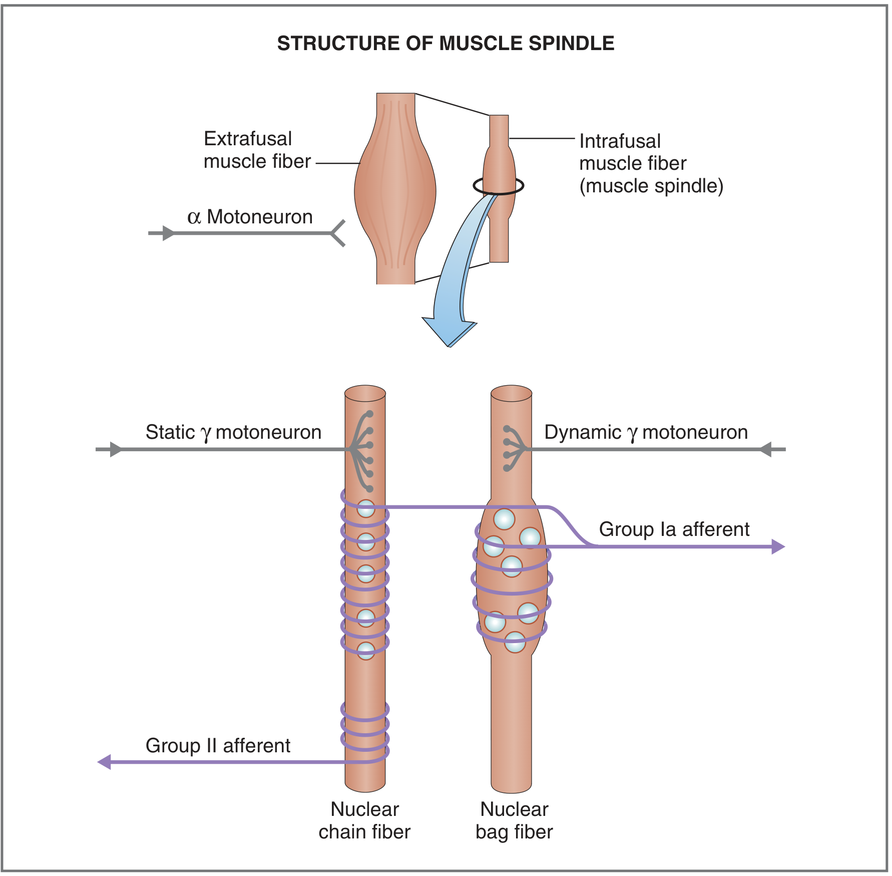
Extrafusal Muscle Fibers
| Feature | Detail |
|---|---|
| Location | Outside the muscle spindle capsule; form the bulk of the muscle |
| Function | Force generation - responsible for all voluntary muscle contraction |
| Size | Relatively large, typically 20-100 µm in diameter |
| Innervation (Motor) | Alpha (α) motor neurons |
| Arrangement | Arranged in parallel with intrafusal fibers |
| Sensory receptors | Golgi tendon organs (GTOs) are placed in series with extrafusal fibers at the muscle-tendon junction |
When an alpha motor neuron fires, the extrafusal fibers contract, shortening the whole muscle. - Costanzo Physiology 7th Ed., p.108
Intrafusal Muscle Fibers
| Feature | Detail |
|---|---|
| Location | Inside the muscle spindle capsule, running parallel to extrafusal fibers |
| Function | Sensory - detect muscle length and rate of stretch (proprioception) |
| Size | Small, only 8-25 µm wide |
| Innervation (Motor) | Gamma (γ) motor neurons |
| Innervation (Sensory) | Group Ia afferents (primary endings) + Group II afferents (secondary endings) |
| Force contribution | Negligible - too small to generate significant contractile force |
Intrafusal fibers are encapsulated in sheaths forming muscle spindles, which run in parallel with the extrafusal fibers. - Costanzo Physiology 7th Ed., p.108
Types of Intrafusal Fibers
There are two subtypes within every muscle spindle:
| Nuclear Bag Fiber | Nuclear Chain Fiber | |
|---|---|---|
| Nuclei arrangement | Clustered in a central "bag" region | Arranged in a row ("chain") |
| Size | Larger | Smaller |
| Number per spindle | ~2 | ~5-6 |
| Sensory ending | Group Ia (primary) - dynamic response | Group II (secondary) - static length |
| Motor innervation | Dynamic γ motoneuron | Static γ motoneuron |
| Detects | Rate of change of muscle length (velocity) | Steady-state muscle length |
The α-γ Coactivation Principle
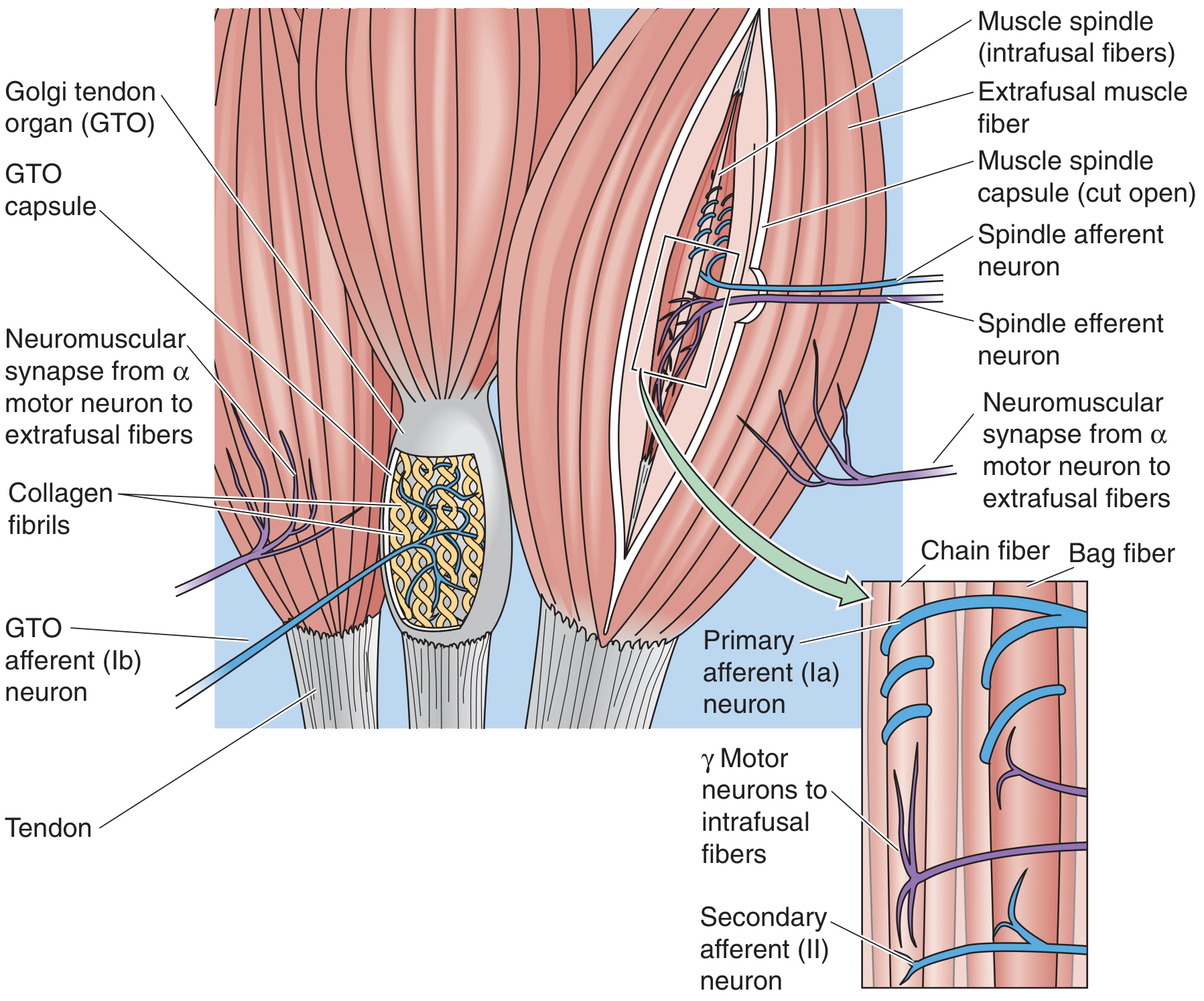
When alpha motor neurons fire to contract the extrafusal fibers, the muscle shortens - this would make the muscle spindle go slack and silence the Ia afferents, losing length information. To prevent this, gamma motor neurons are co-activated simultaneously with alpha motor neurons. Gamma activation causes the intrafusal fiber poles to contract, keeping the central (equatorial) sensory region taut and the Ia axons active throughout the contraction. This mechanism keeps the muscle spindle sensitive across the full range of muscle lengths. - Neuroscience: Exploring the Brain, 5th Ed., p.1288-1290
Side-by-Side Comparison
| Property | Extrafusal Fiber | Intrafusal Fiber |
|---|---|---|
| Location | Outside spindle | Inside spindle capsule |
| Primary role | Contraction / force | Proprioception / length sensing |
| Motor neuron | α (alpha) | γ (gamma) |
| Sensory afferent | None (but GTOs nearby) | Ia + Group II |
| Size | Large (20-100 µm) | Small (8-25 µm) |
| Force production | Yes - nearly all | Negligible |
| Arrangement relative to each other | Parallel to intrafusal | Parallel to extrafusal |
| Arrangement with GTOs | In series | Not involved |
| Abundance | Vast majority of muscle | Few (specialized) |
Quick Memory Aid
- Extrafusal = Executive - does the actual work of contraction, commanded by α neurons
- Intrafusal = Inspector - monitors length and stretch inside the spindle, commanded by γ neurons
Sources: Costanzo Physiology 7th Ed., p.108-109; Neuroscience: Exploring the Brain 5th Ed., p.1288-1290; Medical Physiology (Boron & Boulpaep), p.575-576
This is a shared conversation. Sign in to Orris to start your own chat.