
tell me the best treatment for urticaria and is there any role of antioxidant in this and tell me the stages of urticaria and best possible medicine and the safest option for long term management of this for a patient with history of 6 years
chronic urticaria treatment guidelines antihistamine
antioxidants urticaria oxidative stress treatment
chronic urticaria classification stages treatment guidelines 2024 2025
PMID: 41649409
PMID: 41008654
chronic urticaria treatment algorithm wheal angioedema
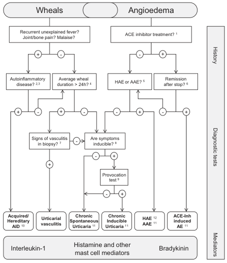
Summary : This flowchart provides a diagnostic algorithm for distinguishing between different causes of wheals and angioedema, based on patient history, diagnostic tests, and mediators involved. flowchart: # Nodes : • Wheals (header, rounded rectangle) • Angioedema (header, rounded rectangle) • Recurrent unexplained fever? Joint/bone pain? Malaise? (rectangle) • ACE inhibitor treatment? (rectangle) • Autoinflammatory disease? (rectangle, with superscript references 2,3) • Average wheal duration > 24h? (rectangle, with superscript reference 4) • HAE or AAE? (rectangle, with superscript reference 5) • Remission after stop? (rectangle, with superscript reference 6) • Signs of vasculitis in biopsy? (rectangle, with superscript reference 7) • Are symptoms inducible? (rectangle, with superscript reference 8) • Provocation test (rectangle, with superscript reference 9) • Acquired/Hereditary AID (rectangle, with superscript reference 10) • Urticarial vasculitis (rectangle) • Chronic Spontaneous Urticaria (rectangle, with superscript reference 11) • Chronic Inducible Urticaria (rectangle, with superscript reference 11) • HAE (rectangle, with superscript reference 12) • AAE (rectangle, with superscript reference 11) • ACE-Inh induced AE (rectangle, with superscript reference 11) • Interleukin-1 (footer, rectangle) • Histamine and other mast cell mediators (footer, rectangle) • Bradykinin (footer, rectangle) • History (vertical label, right margin) • Diagnostic tests (vertical label, right margin) • Mediators (vertical label, right margin) # Connectors : • Arrows connect each decision node to the next step based on a positive (+) answer. • For Wheals: – If recurrent unexplained fever/joint pain/malaise is present, proceed to autoinflammatory disease. – If not, check average wheal duration > 24h. – If duration > 24h, check for signs of vasculitis in biopsy. – If vasculitis present, diagnose urticarial vasculitis. – If not, diagnose acquired/hereditary AID. – If duration ≤ 24h, check if symptoms are inducible. – If inducible, perform provocation test. – If positive, diagnose chronic inducible urticaria. – If not inducible, diagnose chronic spontaneous urticaria. • For Angioedema: – If ACE inhibitor treatment, check for remission after stopping. – If remission, diagnose ACE-Inh induced AE. – If not, consider HAE or AAE. – If no ACE inhibitor, check for HAE or AAE. – If not, check if symptoms are inducible. – If inducible, perform provocation test. – If positive, diagnose chronic inducible urticaria. – If not, diagnose chronic spontaneous urticaria. • Final diagnoses are linked to mediators at the bottom: Interleukin-1, Histamine and other mast cell mediators, Bradykinin. # Layout : • Two main columns: Wheals (left) and Angioedema (right). • Decision nodes flow vertically with branches for positive answers. • Diagnostic categories at the bottom, aligned with their respective mediators. • Vertical sidebars for History, Diagnostic tests, and Mediators. # Analysis : • The flowchart systematically differentiates between causes of wheals and angioedema using clinical history and diagnostic tests. • It highlights the importance of symptom duration, inducibility, biopsy findings, and medication history. • The chart links each diagnosis to its primary mediator, aiding in targeted therapy. • The structure ensures that rare causes (e.g., autoinflammatory diseases, ACE inhibitor-induced angioedema) are considered early in the diagnostic process.
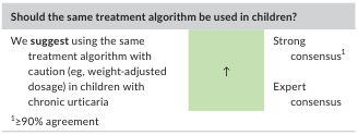
Summary : This figure presents a consensus recommendation regarding whether the same treatment algorithm should be used in children with chronic urticaria, including the level of agreement among experts. consensus recommendation panel: Question & Recommendation : • Question: "Should the same treatment algorithm be used in children?" • Recommendation: "We suggest using the same treatment algorithm with caution (e.g., weight-adjusted dosage) in children with chronic urticaria." Consensus Level : • Strong consensus (≥90% agreement) • Both expert consensus and strong consensus are indicated. Visual Elements : • Green highlighted box with upward arrow, signifying positive or affirmative recommendation. • Footnote: "1 ≥90% agreement" Analysis : • The panel strongly supports using the same treatment algorithm for children as for adults, provided dosage is adjusted for weight and caution is exercised. The recommendation is based on high expert agreement (≥90%).
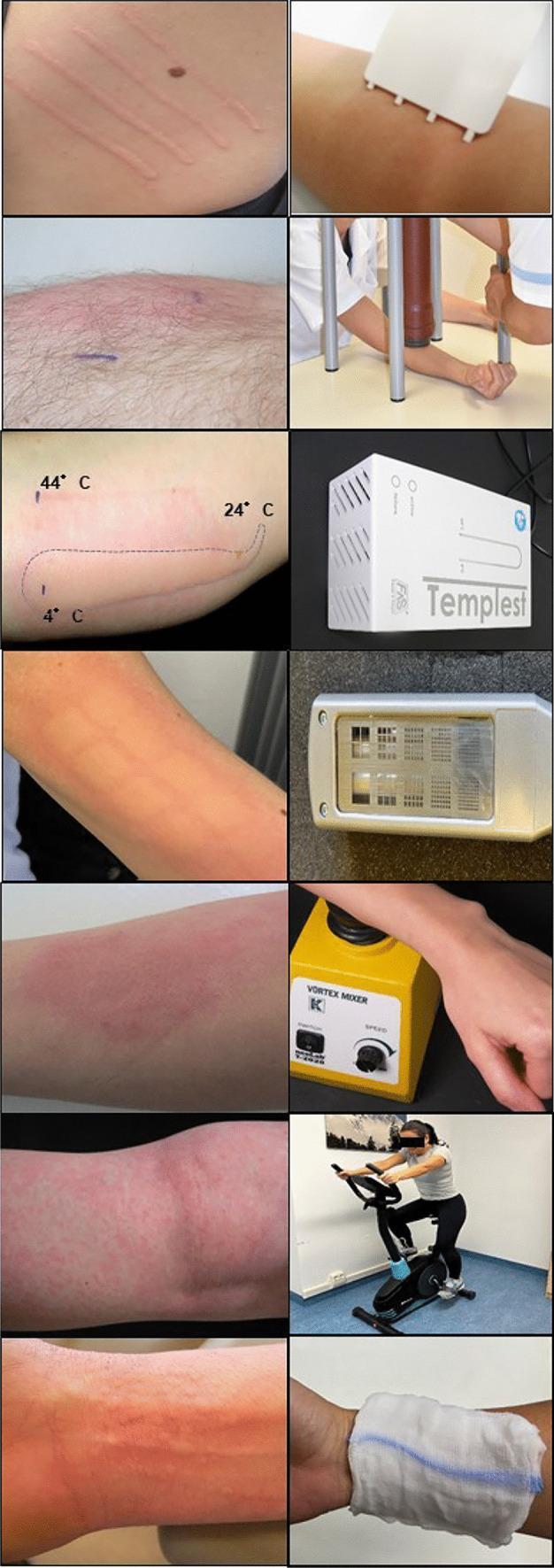
A multi-panel clinical photograph grid demonstrating standardized provocation tests and resulting skin manifestations for Chronic Inducible Urticaria (CIndU). The left column displays various cutaneous reactions on the volar forearm, including: linear wheals and flares characteristic of symptomatic dermographism; a localized erythematous response to temperature testing (marked with 4 C to 44 C gradients); grid-like macular erythema following solar provocation; diffuse edematous plaques from vibratory testing; and punctate, folliculocentric wheals suggestive of cholinergic urticaria. The right column illustrates the corresponding diagnostic modalities and tools, including: a plastic FricTest device for mechanical friction; a weight-rod suspension system for delayed pressure urticaria; the TempTest electronic temperature gradient device; a hand-held UVA/UVB light source for solar urticaria; a laboratory vortex mixer for vibratory angioedema; a static bicycle ergometer for cholinergic urticaria provocation; and a wet towel application for aquagenic urticaria testing. This collection serves as a dermatological guide for identifying subtype-specific wheal morphology and the clinical application of standardized provocation protocols.
Urticaria: Classification, Treatment, Antioxidant Role & Long-Term Management
1. What Is Urticaria? (Quick Pathophysiology)
- Goldman-Cecil Medicine, p. 4595-4603
2. Classification / "Stages" of Urticaria
By Duration
| Type | Definition |
|---|---|
| Acute urticaria | Episodes lasting ≤ 6 weeks |
| Chronic urticaria (CU) | Recurrence over > 6 weeks |
By Etiology (Chronic)
| Subtype | Description |
|---|---|
| Chronic Spontaneous Urticaria (CSU) | No identifiable trigger; accounts for >75% of chronic cases |
| Chronic Inducible Urticaria (CIndU) | Specific reproducible physical/chemical triggers (see below) |
Chronic Inducible Subtypes
| Type | Trigger |
|---|---|
| Dermatographism (symptomatic) | Firm stroking |
| Delayed pressure urticaria | Sustained pressure |
| Cold urticaria | Cold exposure |
| Cholinergic urticaria | Heat, exercise, emotional stress |
| Solar urticaria | Sunlight (visible or UV) |
| Aquagenic urticaria | Water contact |
| Vibratory urticaria | Vibration |
3. Treatment: Stepwise Algorithm
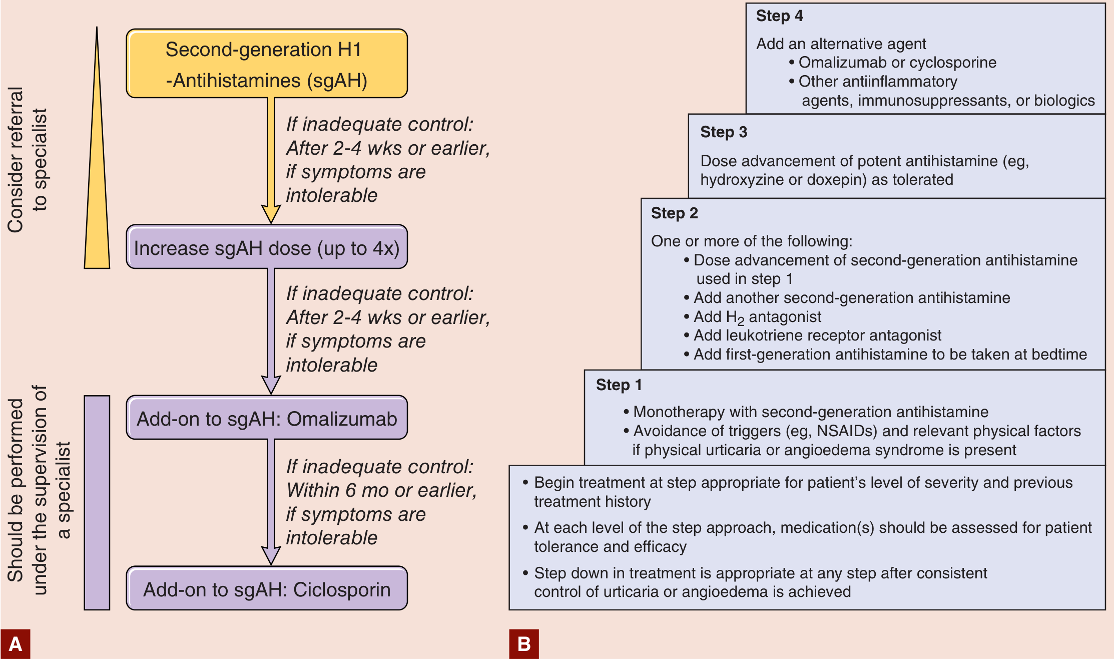
Step 1: Second-Generation H1 Antihistamines (First-Line)
| Drug | Dose | Key Features |
|---|---|---|
| Cetirizine | 10 mg once daily | Low sedation, fast onset |
| Levocetirizine | 5 mg once daily | Most potent 2nd-gen H1 blocker |
| Fexofenadine | 180 mg once daily | Non-sedating, safest for long-term |
| Loratadine | 10 mg once daily | Non-sedating, minimal drug interactions |
| Desloratadine | 5 mg once daily | Active metabolite of loratadine |
- Goldman-Cecil Medicine, p. 4345
Step 2: Add-On Options (if Step 1 fails)
-
H2 antagonists: famotidine or ranitidine (conventional doses) - minor synergistic benefit
-
Leukotriene receptor antagonist: montelukast 10 mg daily - useful in aspirin/NSAID-intolerant patients
-
Add a first-generation antihistamine at bedtime (e.g., hydroxyzine 25-50 mg) for nocturnal itch
-
Harrison's Principles of Internal Medicine 22E, p. 2852
Step 3: Omalizumab (Anti-IgE Biologic) - Gold Standard for Refractory CSU
- Dose: 300 mg subcutaneously every 4 weeks
- Onset: Many patients respond within 1-4 weeks
- Efficacy: 36-40% achieve complete symptom control (UAS7 = 0) in Phase III trials (ASTERIA I and II)
- Safety: Excellent long-term safety profile; safe in pregnancy; low anaphylaxis risk
- FDA approved since 2014 for CSU in patients ≥12 years unresponsive to H1-antihistamines
Step 4: Cyclosporine / Refractory Agents (Specialist Only)
| Agent | Notes |
|---|---|
| Cyclosporine 2.5-5 mg/kg/day | Effective but requires BP/renal monitoring, avoid >1-2 years |
| Hydroxychloroquine | Anti-inflammatory; slower onset |
| Dapsone | Useful if biopsy shows neutrophilic infiltrate |
| Sulfasalazine | Used in delayed pressure urticaria |
| Mycophenolate mofetil | Off-label, moderate evidence |
| Tacrolimus | Used in refractory autoimmune urticaria |
-
Remibrutinib (BTK inhibitor): A 2025 systematic review/meta-analysis (PMID: 41005705) shows significant improvement in UAS7 and quality of life; currently in late-phase trials for CSU
-
Dupilumab (anti-IL-4/IL-13): Phase III LIBERTY-CSU CUPID trials show efficacy in antihistamine-refractory CSU
-
A 2025 clinical practice guideline specifically for H1-antihistamine-resistant CSU (PMID: 41182242) recommends this escalation framework
-
Goldman-Cecil Medicine, p. 4345; Fitzpatrick's Dermatology, p. 732
4. Role of Antioxidants in Urticaria
Oxidative Stress in Urticaria
- Chronic urticaria patients show elevated markers of oxidative stress - increased reactive oxygen species (ROS), lipid peroxidation products, and reduced antioxidant enzyme activity (superoxide dismutase, catalase, glutathione peroxidase)
- Mast cell activation itself generates oxidative stress, creating a feedback loop
Nutraceutical/Antioxidant Evidence
- Promising results seen with supplementation in some conditions, associated with:
- Reduction in oxidative stress markers
- Enhancement of endogenous antioxidant systems
- Anti-inflammatory effects
- However, the review explicitly notes: further large-scale, unbiased studies with broader oxidative stress biomarkers are still needed
Practically Relevant Antioxidants
| Antioxidant | Mechanism | Evidence Level |
|---|---|---|
| Vitamin C | ROS scavenging, antihistamine potentiation | Weak; some small trials |
| Vitamin D | Immune modulation, mast cell regulation | Deficiency correction shown to reduce disease activity |
| Vitamin E | Lipid peroxidation inhibitor | Preliminary data in skin allergies |
| Quercetin | Flavonoid; inhibits mast cell histamine release | Preclinical promising; limited human data |
| Omega-3 fatty acids | Anti-inflammatory, reduce PGE2 | Adjunctive benefit reported |
| Polyphenols (green tea, turmeric) | NF-kB inhibition, mast cell stabilization | Largely preclinical |
5. Best & Safest Options for Long-Term Management (6-Year History)
Immediate Assessment
- Confirm whether CSU or CIndU (or both) - physical provocation testing
- Check: thyroid antibodies (TPO/anti-TG), ANA, CBC, ESR/CRP, Vitamin D level
- Assess disease activity with UAS7 score (Urticaria Activity Score over 7 days) to guide treatment decisions and monitor response
Long-Term Management Hierarchy (Safest Options First)
- Non-sedating 2nd-generation antihistamine (fexofenadine 180 mg or levocetirizine 5 mg daily) - fexofenadine is arguably the safest for indefinite use due to minimal CNS and cardiac effects. These can be continued for years with no organ toxicity.
- Avoidance of triggers: NSAIDs (flare in ~30%), opioids, alcohol, tight clothing
- Omalizumab 300 mg SC every 4 weeks - this is the single best option for long-term management of a 6-year refractory CSU patient. Safety data from real-world registries (5+ years) shows no significant long-term organ toxicity, no immunosuppression, no dependency. It can be continued until complete remission is achieved. After 1 year of symptom control, a trial of dose spacing (every 6-8 weeks) or cessation can be attempted.
- "For recurrent chronic spontaneous urticaria, omalizumab therapy can be very effective" - Goldman-Cecil Medicine, p. 4754
- Cyclosporine - effective but nephrotoxic, hypertensive, and immunosuppressive; limit to the shortest effective course (ideally <12 months at a time)
- Avoid chronic oral corticosteroids - long-term steroid use in urticaria causes significant systemic toxicity (osteoporosis, metabolic syndrome, adrenal suppression)
- Vitamin D supplementation (if deficient, target 40-60 ng/mL)
- Stress reduction (stress is a known CSU flare trigger via CRH-mast cell axis)
- Low pseudoallergen diet trial in CSU (some patients benefit; eliminate for 3 months and reassess)
- Antihistamine at bedtime (hydroxyzine 25 mg) for nocturnal flares - short-term/PRN basis
Summary Table for Your 6-Year Patient
| Priority | Drug/Approach | Safety Profile | Duration |
|---|---|---|---|
| 1st line | Fexofenadine 180 mg daily (up to 4x if needed) | Excellent - indefinite use safe | Ongoing |
| 2nd line | + Montelukast 10 mg daily | Good - long-term safe | Ongoing |
| 3rd line (BEST OVERALL) | Omalizumab 300 mg SC q4 weeks | Excellent - safest biologic long-term | Until remission |
| 4th line | Cyclosporine 2.5-3 mg/kg/day | Moderate - requires monitoring, time-limited | ≤12 months |
| Adjunctive | Vitamin D optimization | Safe | Indefinite |
| Avoid | Chronic oral corticosteroids | High long-term toxicity | Avoid |
| Avoid | NSAIDs, opioids | Flare triggers | Avoid |
Key Takeaways
- Chronic urticaria lasting 6 years is refractory disease - the patient almost certainly requires specialist evaluation and likely omalizumab
- Omalizumab 300 mg SC every 4 weeks is both the most effective and the safest long-term pharmacological option per the 2024 International Guideline and 2025 network meta-analysis
- Second-generation antihistamines remain the daily backbone - fexofenadine or levocetirizine for long-term use
- Antioxidants have adjunctive supportive value (especially Vitamin D) but are not disease-modifying primary therapies; the evidence base is still building
- Monitor disease activity with UAS7 scoring and consider a step-down in therapy after 12 months of complete control
Note: Emerging data (2025) on BTK inhibitors like remibrutinib and dupilumab show promise for antihistamine-refractory patients; discuss these options with a dermatologist or allergist if omalizumab response is suboptimal. A 2025 systematic review (PMID: 41005705) confirms remibrutinib's efficacy and safety in CSU.
what are the test to confirm in urticaria and the role of test
chronic urticaria diagnostic workup laboratory evaluation
urticaria diagnostic tests role confirmation guidelines 2024 2025 EAACI
urticaria diagnostic algorithm provocation test dermographism
A multi-panel clinical photograph grid demonstrating standardized provocation tests and resulting skin manifestations for Chronic Inducible Urticaria (CIndU). The left column displays various cutaneous reactions on the volar forearm, including: linear wheals and flares characteristic of symptomatic dermographism; a localized erythematous response to temperature testing (marked with 4 C to 44 C gradients); grid-like macular erythema following solar provocation; diffuse edematous plaques from vibratory testing; and punctate, folliculocentric wheals suggestive of cholinergic urticaria. The right column illustrates the corresponding diagnostic modalities and tools, including: a plastic FricTest device for mechanical friction; a weight-rod suspension system for delayed pressure urticaria; the TempTest electronic temperature gradient device; a hand-held UVA/UVB light source for solar urticaria; a laboratory vortex mixer for vibratory angioedema; a static bicycle ergometer for cholinergic urticaria provocation; and a wet towel application for aquagenic urticaria testing. This collection serves as a dermatological guide for identifying subtype-specific wheal morphology and the clinical application of standardized provocation protocols.
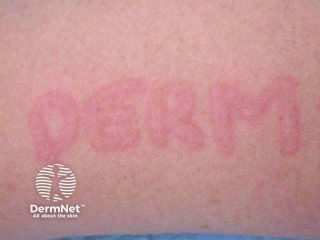
This clinical dermatology photograph depicts a positive dermographism (dermatographic urticaria) response on the skin surface. The imaging modality is clinical photography (Dermatology); technique: close-up, macro shot under consistent lighting to visualize subtle vascular changes. The primary subject is the skin with linearly arranged erythematous wheals produced by light stroking (dermatographism test). Anatomical context: skin, epidermis and superficial dermis. The appearance shows elongated, blanching, reddish plaques along the stroke lines with well-defined margins; the wheals are transient and pruritic, reflecting superficial dermal edema with edema around superficial dermal vessels. Notable features: linear, symmetric or irregular parallel streaks, sharp borders, uniform red-pink color, rapid onset within minutes after mechanical stimulation and resolution thereafter. Diagnostic significance: classic demonstration of physical urticaria due to mast cell degranulation and histamine-mediated increased vascular permeability; supports differential diagnoses including urticaria mechanica, cholinergic urticaria, contact urticaria, and systemic mastocytosis in rare cases. Clinical correlation: correlate with history of itching, trigger exposure to friction, clothing, or pressure; management includes nonsedating antihistamines and trigger avoidance; useful for patient education and medical training. This image is not stained or derived from histology. Annotations underscore intact integument with no pigmentary alteration beyond transient erythema; suitable for clinical teaching, case libraries, and AI training datasets.
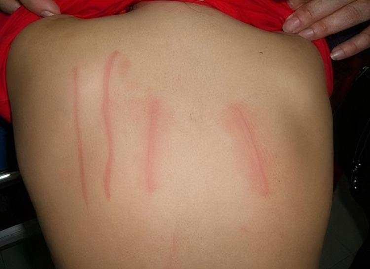
Clinical photograph of a patient's back demonstrating symptomatic dermographism, a form of physical urticaria. The image shows several erythematous, linear wheals (skin writing) arranged vertically across the mid-to-lower back. These lesions are characterized by raised, edematous, pink-to-reddish tracks that follow the path of mechanical friction or pressure applied to the skin. The surrounding skin appears normal, without generalized rash or inflammation, highlighting the localized hypersensitivity reaction. This visual serves as a classic diagnostic example of inducible urticaria, often assessed using a calibrated dermographometer to determine the pressure threshold required to elicit a wheal-and-flare response. The presentation is highly relevant for dermatology and allergy/immunology education, specifically for distinguishing between spontaneous and inducible urticarial conditions.
Diagnostic Tests in Urticaria: What to Order and Why
Key Principle First
"Extensive laboratory investigations add little to making a final diagnosis. Laboratory tests and provocation tests should be performed based on careful history taking and inspection."
- Fitzpatrick's Dermatology, p. 725 (citing EAACI Guidelines)
- Identify or exclude an underlying cause
- Determine the subtype (especially CSU vs. CIndU vs. urticarial vasculitis)
- Assess disease severity and activity
- Rule out dangerous mimics
Tests by Urticaria Type
A. Acute Urticaria
| Situation | Test Indicated |
|---|---|
| History suggests food/drug allergy | Skin prick test + serum specific IgE |
| Anaphylaxis component | Serum tryptase (within 1-4 hours of reaction) |
| Recurring acute episodes | Consider workup for infection (viral panel) |
- Fitzpatrick's Dermatology, p. 726
B. Chronic Spontaneous Urticaria (CSU) - Core Workup
1. Complete Blood Count (CBC) with Differential
- Eosinophilia - suggests parasitic infection or drug reaction as a trigger
- Lymphocytosis - may indicate viral/EBV trigger
- Neutropenia/anemia - may point to autoimmune disease
- Raised basophils - associated with autoimmune CSU
- Thrombocytopenia - signals systemic disease (lupus, lymphoma)
2. Erythrocyte Sedimentation Rate (ESR) and C-Reactive Protein (CRP)
- Elevated in urticarial vasculitis, systemic autoimmune disease, infection
- Both are disease activity biomarkers in CSU - elevated CRP correlates with more severe disease
- Normal ESR/CRP in a whealing patient makes urticarial vasculitis less likely
- CRP and IL-6 are included in the biomarker panel for CSU disease severity
3. Thyroid Function Tests + Anti-Thyroid Antibodies (Anti-TPO, Anti-TG)
-
CSU is strongly associated with autoimmune thyroid disease (Hashimoto's thyroiditis)
-
10-30% of CSU patients have positive thyroid autoantibodies
-
Treating underlying hypothyroidism can reduce urticaria activity in some patients
-
Anti-TPO antibody positivity also predicts a more difficult-to-treat, autoimmune-type CSU
-
Dermatology 2-Volume Set 5e, p. 47
4. Total IgE and Allergen-Specific IgE (RAST/ImmunoCAP)
- Elevated total IgE suggests atopic background or parasitic infection
- Specific IgE identifies food/aeroallergen triggers in IgE-mediated urticaria
- Particularly useful when history clearly suggests a trigger (food, pet, latex, drug)
- Note: Most CSU is NOT IgE-mediated; specific IgE testing has low yield in unselected CSU
5. D-Dimer and Coagulation Markers (F1+2, FDP)
-
D-dimer is elevated in CSU and correlates directly with disease activity and severity (UAS7 score)
-
Prothrombin fragment 1+2 (F1+2) and fibrin degradation products (FDP) reflect the coagulation cascade activation that occurs in CSU via tissue factor on eosinophils
-
Markedly elevated D-dimer predicts poor antihistamine response and may indicate need for omalizumab
-
These are emerging clinical biomarkers, not yet routine everywhere but validated in multiple studies
-
Fitzpatrick's Dermatology, p. 726
6. Autologous Serum Skin Test (ASST)
- Screens for the presence of circulating functional autoantibodies (IgG against FcεRI or IgE) in autoimmune CSU
- Method: 0.05 mL of patient's own serum injected intradermally on the volar forearm; saline is the negative control; histamine is the positive control. A red wheal ≥1.5 mm larger than the saline response at 30 minutes = positive result
- Positive ASST = approximately 40-50% of CSU patients; indicates autoimmune subtype
- Limitations: Not specific for autoantibodies; not predictive of treatment response; safety concerns with using blood products in skin testing have reduced its use in recent years
| ASST Result | Interpretation |
|---|---|
| Positive | Circulating serum factors present; autoimmune type likely |
| Negative | Does not exclude autoimmune urticaria |
- Dermatology 2-Volume Set 5e (Table 18.3)
7. Basophil Activation Test (BAT) / Histamine Release Assay
- Patient's serum is incubated with healthy donor basophils; histamine release or CD63/CD203c expression is measured by flow cytometry
- Detects functional IgG autoantibodies against FcεRI or IgE - more specific than ASST
- A positive BAT with high histamine release may predict a good response to cyclosporine
- Not widely available outside specialist/research centers
8. Antinuclear Antibody (ANA) and Complement Levels (C3, C4, CH50)
- ANA positivity in CSU signals associated autoimmune disease (lupus, Sjögren's)
- Low C4 (chronically depleted) is the hallmark of Hereditary Angioedema (HAE) - essential to measure in isolated angioedema without wheals
- Low C1q + low C4 + low C1 inhibitor = acquired C1-INH deficiency (may signal underlying B-cell lymphoma)
- Normal complement makes HAE very unlikely
9. Skin Biopsy (Punch Biopsy)
- Wheals lasting >24-36 hours (normally urticarial wheals last <24 hours)
- Wheals that leave bruising or discoloration (pigmentation after wheals fade)
- Painful rather than pruritic lesions
- Associated systemic symptoms (fever, arthritis, weight loss)
| Histological Finding | Diagnosis Suggested |
|---|---|
| Perivascular lymphocytic infiltrate ± eosinophils | Typical CSU |
| Dense neutrophilic infiltrate + nuclear debris + fibrinoid necrosis of venules | Urticarial vasculitis |
| Predominantly neutrophilic infiltrate (no vasculitis) | Neutrophilic urticaria (responds to dapsone/colchicine) |
| Dense eosinophilic infiltrate | Eosinophilic cellulitis (Well's syndrome) |
- Fitzpatrick's Dermatology, p. 725
10. Helicobacter pylori Testing (Stool Antigen or Urea Breath Test)
- H. pylori eradication has been shown to improve CSU in some patients
- Recommended in patients with GI symptoms or in geographic areas with high H. pylori prevalence
- Not a routine universal test, but indicated if history suggests
C. Chronic Inducible Urticaria (CIndU) - Provocation Tests
| Urticaria Subtype | Provocation Test | Positive Result |
|---|---|---|
| Symptomatic Dermographism | FricTest device - calibrated friction on volar forearm | Linear wheal along stroke path |
| Cold Urticaria | TempTest - cold stimulator (4°C) on volar forearm for 5 min | Wheal on warming |
| Delayed Pressure Urticaria | Weight-rod device - 7 kg weight for 20 min | Wheal 3-8 hours later |
| Cholinergic Urticaria | Exercise on stationary bicycle; hot bath (42°C for 15 min) | Punctate wheals during/after |
| Solar Urticaria | UV light source - various wavelengths (290-700 nm) | Wheal within 30 min |
| Aquagenic Urticaria | Wet towel at 35°C applied to upper body for 30 min | Perifollicular wheals |
| Vibratory Urticaria | Vortex mixer on forearm for 4 min | Localized edema |
D. Angioedema Without Wheals - Additional Tests
| Test | What It Shows |
|---|---|
| C4 level | Low (chronically) in HAE types 1 & 2; most useful screening test |
| C1 inhibitor antigen | Low in HAE type 1 |
| C1 inhibitor function | Low/absent in HAE type 2 (normal antigen but dysfunctional) |
| C1q | Low in acquired C1-INH deficiency; normal in HAE types 1 & 2 |
| FXII gene mutation | Identifies HAE type 3 (normal complement) |
Disease Activity Scoring (Not a Lab Test, But Essential)
UAS7 (Urticaria Activity Score over 7 days)
- Patient records wheals (0-3) and itch (0-3) daily for 7 days; max score = 42
- Guides when to escalate treatment and monitors response
| UAS7 Score | Disease Activity |
|---|---|
| 0 | Complete control (remission) |
| 1-6 | Well-controlled |
| 7-15 | Mild |
| 16-27 | Moderate |
| 28-42 | Severe |
Summary: Which Tests for Which Patient
| Patient Scenario | Priority Tests |
|---|---|
| Acute urticaria - clear trigger | Specific IgE / skin prick test only |
| Acute urticaria - no trigger | No routine tests needed |
| Chronic urticaria - new onset | CBC, ESR/CRP, TSH + anti-TPO, total IgE |
| Chronic urticaria - refractory (like your 6-year patient) | + ASST or BAT, D-dimer, ANA, complement (C3/C4), H. pylori |
| Wheals lasting >24h, painful | Skin biopsy (to exclude urticarial vasculitis) |
| Angioedema without wheals | C4, C1-INH antigen and function, C1q |
| Suspected physical/inducible | Targeted provocation test for relevant stimulus |
| Systemic symptoms (fever, joint pain) | ANA, ANCA, CBC with differential, ESR, biopsy |
Key Takeaway

tell me the best possible treatment in form of tablets and syrups to cure this elevated level and the reason of evevation with test to perform
elevated AST ALT transaminase causes evaluation management
elevated AST ALT SGOT SGPT causes diagnosis treatment tablets 2024
https://www.ncbi.nlm.nih.gov/books/NBK482489
nonalcoholic fatty liver disease treatment lifestyle pharmacotherapy
Lab Report Analysis: Mr. Amit Bansal, 49 years, Male
Abnormal Values Summary
| Test | Result | Reference Range | Status | Degree |
|---|---|---|---|---|
| AST (SGOT) | 74 U/L | <50 U/L | 🔴 HIGH | 1.5x upper limit |
| ALT (SGPT) | 82 U/L | <50 U/L | 🔴 HIGH | 1.6x upper limit |
| Total Bilirubin | 1.43 mg/dL | 0.30-1.20 mg/dL | 🟡 MILDLY HIGH | Just above normal |
| Direct Bilirubin | 0.28 mg/dL | <0.20 mg/dL | 🟡 MILDLY HIGH | Borderline |
| Globulin | 3.60 g/dL | 2.0-3.5 g/dL | 🟡 MILDLY HIGH | Borderline |
| AST:ALT Ratio | 0.90 | <1 | Normal (important!) | - |
Interpretation of the Pattern
- ALT > AST = hepatocellular (liver cell) pattern of injury
- This pattern strongly suggests MASLD (Metabolic-Associated Steatotic Liver Disease), formerly NAFLD (Non-Alcoholic Fatty Liver Disease) OR early viral/drug-induced hepatitis
- An AST:ALT ratio >2 would suggest alcoholic liver disease - which is NOT the case here
- The elevation is mild (less than 5x the upper limit of normal) - this is the most common, manageable range
- StatPearls/NIH - Liver Function Tests
Causes of This Pattern (Ranked by Likelihood)
Most Likely (Mild elevation, ALT > AST)
| # | Cause | Why Likely |
|---|---|---|
| 1 | MASLD/NAFLD (Fatty Liver Disease) | Most common cause of mild transaminase elevation; 49-year-old male; accounts for ~75% of mild AST/ALT elevation globally |
| 2 | Medications / hepatotoxic drugs | Many common drugs raise liver enzymes - NSAIDs, statins, antifungals, antiepileptics, ayurvedic/herbal supplements |
| 3 | Chronic Viral Hepatitis B or C | Can cause persistent mild elevation; often asymptomatic for years |
| 4 | Alcohol-related liver disease | Even modest, regular alcohol intake; ratio <1 makes this less likely but not excluded |
| 5 | Autoimmune hepatitis | Should be considered in persistent elevation |
Less Likely (but must rule out)
| Cause | Reason to Consider |
|---|---|
| Thyroid disease (hypothyroidism) | Can elevate AST via muscle involvement |
| Celiac disease | Causes transaminase elevation, often missed |
| Wilson's disease | In younger adults with unexplained hepatitis |
| Hemochromatosis | Iron overload; elevated ferritin clue |
Tests to Perform to Find the Root Cause
Tier 1 - Essential (Order Now)
| Test | What It Detects | Why Important |
|---|---|---|
| Hepatitis B Surface Antigen (HBsAg) | Active Hepatitis B infection | Common cause of persistent mild ALT elevation; treatable |
| Anti-HCV (Hepatitis C Antibody) | Hepatitis C infection | Highly treatable now; often silent for years |
| Hepatitis B Core Antibody (Anti-HBc IgM/IgG) | Past or current HBV | Distinguishes old vs. active infection |
| Fasting Blood Sugar (FBS) + HbA1c | Diabetes/prediabetes | Metabolic syndrome drives fatty liver |
| Fasting Lipid Profile (Total cholesterol, TG, LDL, HDL) | Dyslipidemia | High triglycerides = strong fatty liver marker |
| Ultrasound Abdomen | Fatty infiltration, liver size, gallstones | Most important imaging; shows echogenic (bright) liver in fatty liver disease |
| Serum Ferritin + Transferrin Saturation | Hemochromatosis | Iron overload can silently damage the liver |
Tier 2 - If Tier 1 is Negative (Rule Out Less Common Causes)
| Test | What It Detects |
|---|---|
| ANA + Anti-smooth muscle antibody (ASMA) | Autoimmune hepatitis |
| TSH (Thyroid Stimulating Hormone) | Hypothyroidism (can raise AST/CK) |
| Serum Ceruloplasmin | Wilson's disease (copper accumulation) |
| Anti-tissue transglutaminase IgA (Anti-tTG) | Celiac disease |
| Creatine Kinase (CK/CPK) | Muscle disease - if AST elevated but ALT normal |
Tier 3 - If Chronic Hepatitis Confirmed
| Test | Purpose |
|---|---|
| HBV DNA (Viral Load) | Quantify HBV if HBsAg positive |
| HCV RNA (Viral Load) | Confirm and quantify HCV if anti-HCV positive |
| Fibroscan or FIB-4 Score | Non-invasive liver stiffness assessment (fibrosis staging) |
| Liver biopsy | Gold standard for staging if non-invasive tests inconclusive |
⚠️ Important Note About Bilirubin
- Gilbert's Syndrome - benign, hereditary, benign; bilirubin rises with fasting, stress, illness; no treatment needed; requires only confirmation
- Mild hemolysis - add Peripheral Blood Smear + Reticulocyte Count
Treatment
IMPORTANT DISCLAIMER
Step 1 - Lifestyle (The Most Powerful Treatment for MASLD)
| Intervention | Target | Evidence |
|---|---|---|
| Weight loss 7-10% | If overweight | Reduces liver fat and normalizes enzymes in most patients |
| Mediterranean diet | Daily | Rich in olive oil, vegetables, fish, legumes; reduces liver inflammation |
| Avoid alcohol completely | Total | Even moderate alcohol worsens liver enzyme elevation |
| Physical activity | 150-200 min/week moderate aerobic exercise | Independently reduces liver enzymes even without weight loss |
| Stop hepatotoxic drugs/supplements | Immediately if identified | Ayurvedic herbs, NSAIDs, high-dose paracetamol especially risky |
| Reduce refined sugars and fructose | High-fructose corn syrup, soft drinks | Direct driver of hepatic fat accumulation |
Step 2 - Tablets (Hepatoprotective Agents)
A. Silymarin (Milk Thistle / Silybum marianum)
| Brand Name (India) | Dose | Frequency |
|---|---|---|
| Livolin Forte, Silybon 70/140, Legalon | 140 mg | 3 times daily with meals |
B. Ursodeoxycholic Acid (UDCA)
| Brand Name (India) | Dose | Frequency |
|---|---|---|
| Udiliv 300, Ursocol 300, Udca 300 | 8-10 mg/kg/day (approx 300-600 mg for 70 kg) | 2-3 times daily |
C. Essential Phospholipids (Essentiale / EPCLC)
| Brand Name (India) | Dose | Frequency |
|---|---|---|
| Essentiale Forte, Livogen, Lepofos | 2 capsules (600 mg EPL) | 3 times daily with meals |
D. N-Acetylcysteine (NAC)
| Brand Name (India) | Dose | Frequency |
|---|---|---|
| Mucomix, Fluimucil 600 mg tablets | 600 mg | Once or twice daily |
E. Vitamin E (Alpha-Tocopherol) - If NASH Confirmed
| Dose | Evidence |
|---|---|
| 800 IU/day | Recommended in EASL 2024 guidelines for biopsy-proven NASH in non-diabetics |
Step 3 - Syrups / Liquid Formulations
| Syrup / Liquid | Key Ingredients | Dose | Best For |
|---|---|---|---|
| Liv 52 syrup (Himalaya) | Caper, Chicory, Black nightshade, Tamarisk | 2 tsp twice daily | Fatty liver, mild hepatitis, general hepatoprotection |
| Livomap syrup (Charak) | Bhumyamalaki, Kalmegh, Punarnava | 2 tsp twice daily | Liver enzyme elevation, hepatitis |
| Syrlife/Livomyn syrup | Silymarin + B vitamins + antioxidants | 10-15 mL twice daily | Mild transaminase elevation |
| Cytozen syrup | Silymarin + Vitamin E + Zinc | 10 mL twice daily | NAFLD, oxidative liver injury |
Step 4 - Manage Underlying Cause (Once Confirmed)
| Confirmed Cause | Specific Treatment |
|---|---|
| Hepatitis C | Sofosbuvir + Velpatasvir (Velpanat, Sofocure) - 12 weeks; >95% cure rate |
| Hepatitis B (active) | Tenofovir (Tenvir 300 mg once daily) or Entecavir 0.5 mg once daily - long-term |
| Diabetes + Fatty Liver | Pioglitazone 30 mg or GLP-1 agonists (semaglutide) shown to reduce liver fat |
| Dyslipidemia | Statins are actually SAFE in mild transaminase elevation; pravastatin or rosuvastatin preferred |
| Drug-induced | Stop offending drug; liver enzymes normalize in 4-12 weeks |
| Gilbert's Syndrome | No treatment needed; benign condition |
Monitoring Plan
| Time | Action |
|---|---|
| After 4 weeks | Repeat AST, ALT, bilirubin |
| After completing investigations | Treat identified cause specifically |
| If no improvement in 3 months | Refer to gastroenterologist/hepatologist |
| If enzymes rise >5x normal at any point | Urgent hepatology referral |
Summary for Mr. Amit Bansal
- Get Hepatitis B (HBsAg), Hepatitis C (Anti-HCV), fasting lipids, blood sugar, and Ultrasound Abdomen done immediately
- Start lifestyle modifications - this is more powerful than any tablet
- Start Silymarin 140 mg three times daily (Silybon/Livolin Forte) as a safe, evidence-based hepatoprotectant
- Add Liv 52 syrup or Essentiale Forte as adjunctive support
- Stop all alcohol, NSAIDs, and unnecessary supplements completely
- Repeat LFT in 4-6 weeks to track response
⚠️ This analysis is for educational purposes. Please consult a physician or gastroenterologist before starting any medication. Specific treatment requires identifying the underlying cause first.