
Explain to me in detail from Braunwald and latest guidelines considering I a DrNB Cardiology Resident how to calculate and tell the origin of a VPC and the axis in a Ventricular Tachycardia from ECG only
Reading File
Now let me run parallel searches across Braunwald and PubMed simultaneously.
Finding Sources
Finding Sources
Searching PubMed
"ventricular tachycardia" AND "ECG localization" AND "site of origin"
Searching the Web
VPC VT ECG localization algorithm site of origin Braunwald 2024 guidelines
Finding Sources
Finding Sources
Searching PubMed
"ventricular tachycardia" AND "12-lead ECG" AND "localization"
Reading Web Page
https://thoracickey.com/how-to-localize-ventricular-tachycar…
Now let me read the actual Braunwald sections and Harrison's sections on VT localization:
Reading File
Reading File
Reading File
Reading File
Finding Sources
Finding Sources
Reading File
Finding Sources
Reading File
Finding Sources
Reading File
Reading File
Reading File
Reading File
Excellent - found the key Fuster/Hurst diagram description. Let me get that important image and read more of this section:
Loading Image
Reading File
Loading Image
Now let me get the Harrison's Figure 259-4 which is the key VT origin diagram, and the RVOT detailed ECG criteria from Braunwald block 7:
Loading Image
Reading File
I now have comprehensive data from Braunwald, Fuster/Hurst's The Heart, and Harrison's. Let me compile the full, expert-level answer.
ECG Localization of VPC/VT Origin: A DrNB Cardiology Reference
Sources: Braunwald's Heart Disease (10th ed.), Fuster & Hurst's The Heart (15th ed.), Harrison's Principles of Internal Medicine (22nd ed.), 2022 ESC Guidelines for Ventricular Arrhythmias
The Core Principle: 3 Spatial Planes on the 12-Lead ECG
The 12-lead ECG defines the site of ventricular origin in three orthogonal planes. This is the framework from which all localization flows (Fuster & Hurst, Fig. 37-8):
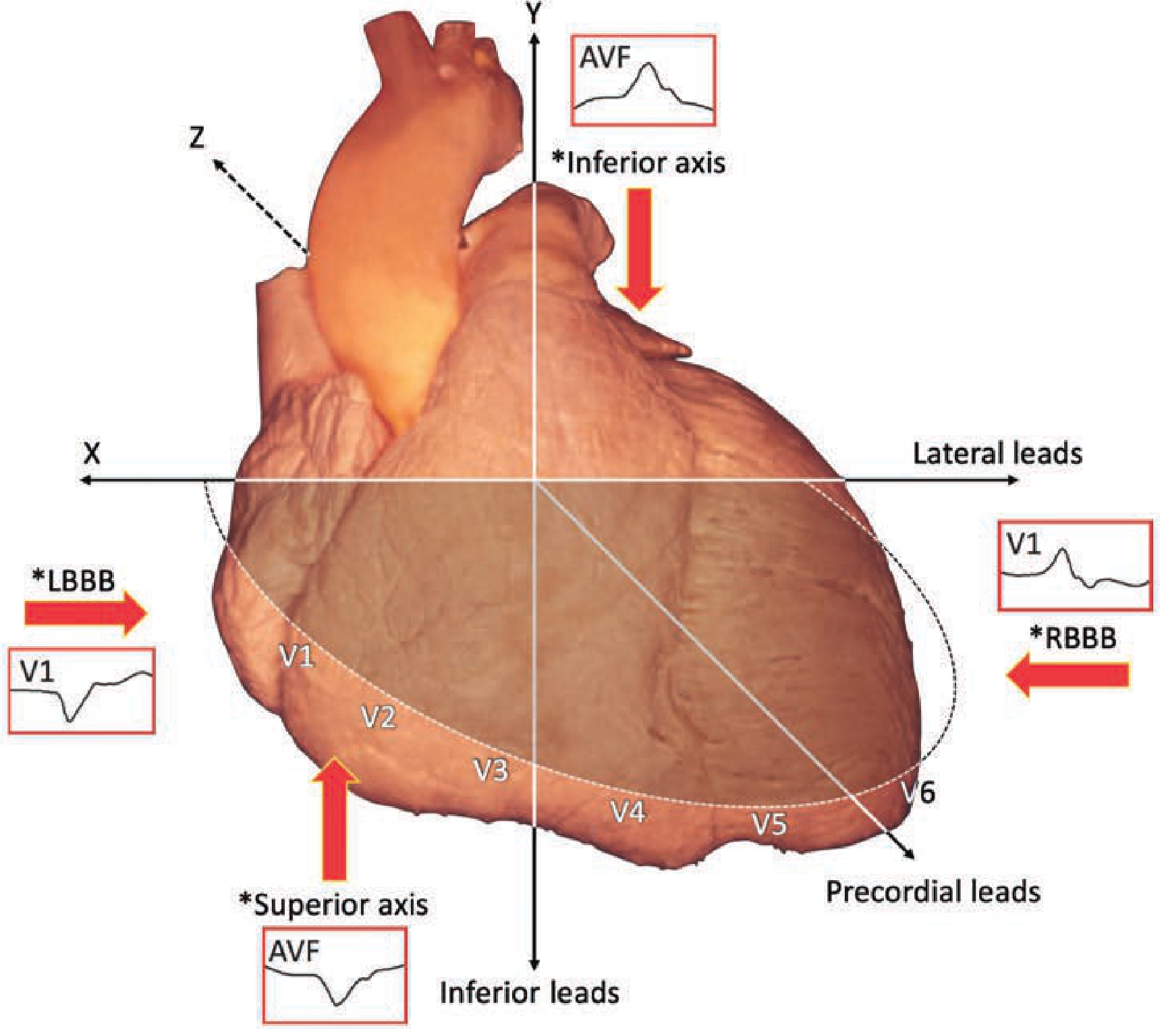
| Plane | Determined by | Interpretation |
|---|---|---|
| X-Y (Coronal) | V1 morphology (LBBB vs RBBB) | Left vs Right ventricular or septal origin |
| Y-axis (Frontal) | Limb lead axis (inferior vs superior) | Superior vs inferior wall origin |
| Z-axis (Axial) | Precordial transition | Basal vs apical origin |
Step 1 - Determine the Bundle Branch Block Pattern (V1)
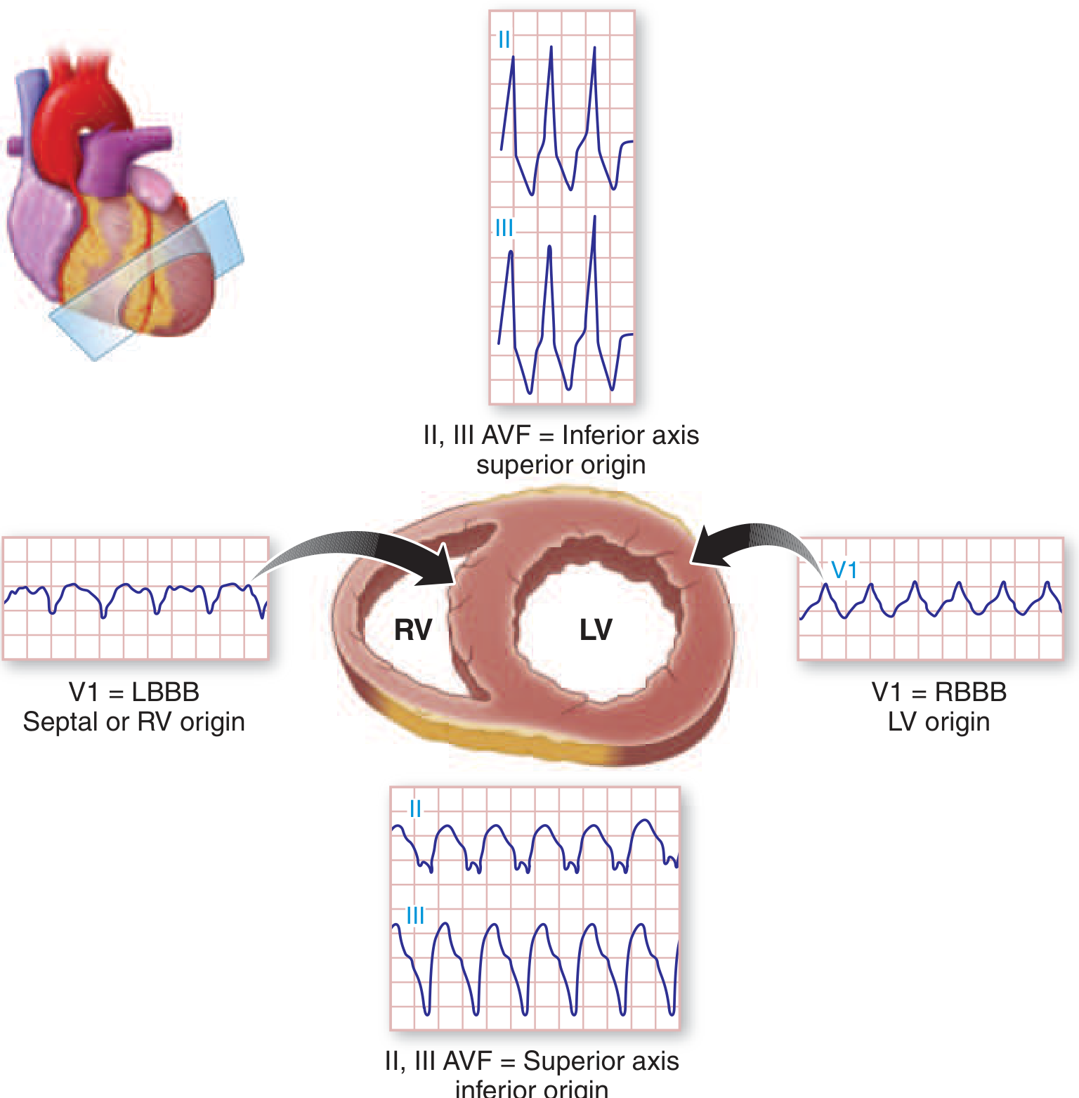
This is the single most important lead. It tells you which ventricle activated last - and the ectopic beat originates in the contralateral ventricle or septum.
LBBB Pattern in VPC/VT (rS or QS in V1)
- The left ventricle activates late → depolarization travelling from right to left
- Origin is in the RV, septum (right side), or RVOT
- The classic example: RVOT VT - LBBB + inferior axis
RBBB Pattern in VPC/VT (rsR', qR, or dominant R in V1)
- The right ventricle activates late → origin in the LV free wall or LV outflow tract
- Classic: Idiopathic LV fascicular VT (Belhassen/verapamil-sensitive) - RBBB + left superior axis
As Harrison's (22nd ed.) states explicitly:
"Arrhythmias that originate from the right ventricle or septum result in late activation of much of the left ventricle, thereby producing a prominent S wave in V1 referred to as a left bundle branch block-like configuration. Arrhythmias that originate from the free wall of the left ventricle have a prominent positive deflection in V1, thereby producing a right bundle branch block-like morphology in V1."
Step 2 - Determine the Frontal Axis (Limb Leads)
Inferior Axis (positive R in II, III, aVF)
- Initial activation of the cranial/superior portion of the ventricle
- Exit site is basal or outflow tract (apical activation happens last → inferior forces)
- LBBB + inferior axis = RVOT origin (classic)
Superior Axis (negative in II, III, aVF; S or QS in aVF)
- Initial activation of the inferior wall
- Exit site is the inferior, apical, or diaphragmatic surface
- RBBB + superior axis = LV posterior fascicle / posteroinferior wall (Belhassen VT)
Right Superior Axis (negative I, II, III, aVF)
- Exit from RV anterior free wall or apical septum
- Seen in ischemic VT from anterior MI (RBBB + right superior axis)
Step 3 - Determine the Precordial Transition (Z-plane / Basal-to-Apical)
The precordial transition tells you the anterior-posterior depth of the origin.
| Transition | Meaning |
|---|---|
| Early (V1-V2) | Posteriorly located origin (posterior LV free wall, posterior LVOT, aortic cusps) |
| Mid (V3-V4) | Mid-ventricular origin |
| Late (V5-V6) or no transition | Anterior origin (anterior RV, anterior RVOT) |
For LBBB morphology VTs specifically:
- Transition V3 or earlier → raises strong suspicion for LVOT, aortic sinus cusp, or left-sided origin (despite LBBB appearance)
- Transition V4-V6 → RVOT (right-sided)
- The V2 transition ratio (Betensky criterion): If V2-transition ratio ≥0.6 → LVOT origin (86% specificity)
The 3-Dimensional Framework Applied to Specific Origins
A. RVOT (Right Ventricular Outflow Tract) VT/PVC
Classic ECG pattern:
- LBBB morphology in V1
- Inferior axis (tall R in II, III, aVF)
- Late precordial transition (V3-V5)
- Tall, monophasic R waves in inferior leads
- Predominantly negative or isoelectric in aVL
- Lead I: positive (free wall) or negative/isoelectric (septal origin)
"Outflow origin PVCs and VT have a characteristic pattern on the 12-lead ECG, with an LBBB morphology and inferior axis." - Fuster & Hurst (15th ed.)
RVOT Septum vs Free Wall:
- Septal RVOT: isoelectric or negative lead I, narrower QRS, earlier transition
- Free wall RVOT: positive lead I, broader QRS
Distinguishing RVOT from LVOT (when both give LBBB + inferior axis):
| Feature | RVOT | LVOT / Aortic Cusp |
|---|---|---|
| V1 | rS or QS (pure LBBB) | Small but definite r wave (S:r ratio <1.5) |
| R/S in V1 | R/S <30% | R/S ≥30% |
| R-wave duration index V1 or V2 | <50% | ≥50% |
| Precordial transition | V3-V5 | V1-V3 (early) |
| aVL | Negative (deep S or QS) | Positive or isoelectric |
| Lead I | Variable | Often positive R |
| V2 transition ratio (Betensky) | <0.6 | ≥0.6 |
B. LVOT / Aortic Sinus Cusp VTs
These originate from structures in close anatomical proximity: the aortomitral continuity (AMC), anterior mitral annulus, aortic sinus cusps (LCC, RCC, NCC), and the LV summit.
Left Coronary Cusp (LCC):
- RBBB-like or transitional in V1 (dominant R)
- Inferior axis
- Negative aVL (R-wave in aVL rules out LCC, RCC, or junction origin)
RCC vs LCC distinction:
- R-wave amplitude ratio III/II > 0.9 → LCC
- R-wave amplitude ratio III/II ≤ 0.9 → RCC
Non-Coronary Cusp (NCC):
- Deeper S in aVL, later transition than LCC/RCC
C. LV Fascicular (Verapamil-Sensitive / Belhassen) VT
This is idiopathic reentrant VT involving the LV posterior fascicle (most common form).
ECG signature:
- RBBB pattern in V1 (dominant R or rsR')
- Left superior axis (negative in II, III, aVF; left axis deviation)
- Relatively narrow QRS (<140 ms) - reflects near-normal conduction via Purkinje system
- QRS often <120 ms in the posterior fascicular form
- Sensitive to IV verapamil (terminates the tachycardia)
- Inducible by atrial pacing
- Occurs in young adults with structurally normal hearts
Anterior fascicular VT (less common): RBBB + right inferior axis
Upper septal VT (uncommon): narrow QRS, RBBB, relatively normal axis
"Left fascicular reentrant tachycardia...the most common form involves the LV posterior fascicle giving rise to an RBBB-like tachycardia with a superior left axis." - Braunwald (block7, line 3028)
D. Bundle Branch Reentry VT (BBR-VT)
ECG signature:
- LBBB morphology (most common): circuit goes anterograde down RBB, retrograde up LBB
- RBBB morphology (rare reverse circuit)
- Often rapid (>200 bpm)
- Often occurs in patients with pre-existing LBBB or interventricular conduction delay during sinus rhythm
- Associated with severe LV dysfunction (DCM, ischemic CMP)
- Ablation of the RBB is curative
E. Ischemic/Scar-Related VT (CAD)
Braunwald states explicitly:
"In almost all patients who have VT associated with ischemic heart disease, the arrhythmia, regardless of its configuration on the surface ECG, arises in the left ventricle or on the left ventricular side of the interventricular septum."
ECG framework for scar VT (Segal/Miller algorithm):
LBBB pattern VTs from ischemic substrate:
- Exit from the septum (LV side)
- Superior axis → basal or mid-septal location
- Inferior axis → mid-septal location
- Anterior MI: LBBB VTs from apical septum; inferior axis → anterior wall, superior axis → inferior wall
RBBB pattern VTs from ischemic substrate:
- Exit from the lateral or free wall (away from septum)
- Positive lead I + superior axis → mid-posterior location
- Negative lead I + superior axis → apical-posterior location
- Inferior axis + RBBB → anterior wall; Lead I positive → basal; Lead I negative → mid-anterior
F. Inferior Wall / Diaphragmatic VT (RBBB + Superior Axis)
- RBBB + extreme left axis (negative II, III, aVF) → inferior LV
- Inferior MI-related VTs often RBBB with right superior axis (antero-apical septum)
G. Papillary Muscle VTs
- Posteromedial papillary muscle: RBBB + superior axis + S waves V4-V6
- Anterolateral papillary muscle: RBBB + inferior rightward axis
- Often associated with mitral valve prolapse; may trigger VF
Summary Algorithm: Step-by-Step Approach
Step 1: Is V1 → LBBB or RBBB?
  ├── LBBB → RV, septum, or RVOT
  │     Step 2: Axis in inferior leads (II, III, aVF)?
  │       ├── INFERIOR axis → RVOT / basal outflow tract origin
  │       │     Step 3: Lead I & transition?
  │       │       ├── Transition V4+ , Lead I –  → Septal RVOT
  │       │       ├── Transition V4+ , Lead I +  → Free wall RVOT
  │       │       └── Early transition V1-V3, R in V1 → LVOT / Aortic cusp
  │       └── SUPERIOR axis → Inferior LV, inferior septum, apical
  │
  └── RBBB → LV free wall or LVOT
        Step 2: Axis?
          ├── LEFT SUPERIOR axis, narrow QRS → Posterior fascicle (Belhassen VT)
          ├── LEFT SUPERIOR axis, broad QRS → Posteroinferior LV free wall
          ├── INFERIOR axis → Anterior LV or anterolateral wall
          └── RIGHT SUPERIOR axis → Antero-apical septum (ischemic)

Step 3: Precordial transition zone
  ├── Early (V1-V2) → Posterior/basal LV, aortic cusp
  ├── V3-V4 → Midventricular
  └── Late/absent → Anterior/apical
Special ECG Clues at a Glance
| ECG Finding | Implication |
|---|---|
| Lead aVR - monophasic R | Strong VT indicator (Vereckei criterion) |
| Lead I positive + LBBB + inferior axis | Free wall RVOT |
| Lead I negative + LBBB + inferior axis | Septal RVOT or LVOT |
| Negative aVL + LBBB + inferior axis | RVOT (aVL should be negative in RVOT) |
| Positive aVL + LBBB + inferior axis | LVOT / LCC origin |
| III/II R-wave ratio >0.9 | LCC > RCC |
| RBBB + left superior axis, QRS <140 ms | Posterior fascicular VT (verapamil!) |
| RBBB + left superior axis, broad QRS | Posterior LV free wall |
| LBBB + rapid rate + dilated CMP | Bundle branch reentry VT (ablate RBB) |
| RBBB + right superior axis + DCM/ischemic | Antero-apical scar VT |
| Precordial concordance (all positive) | LV posterior free wall (RBBB-like) |
| Precordial concordance (all negative) | Apex/anterior LV or RVOT far free wall |
| Precordial transition at V3 in LBBB VT | Likely LVOT not RVOT → use V2 transition ratio |
Notes on Axis Calculation
The frontal axis is calculated in the standard way from the limb leads. For VT:
- Inferior axis (0° to +90°): dominant R in II, III, aVF → origin is superior (outflow tracts, base)
- Superior/left axis (-30° to -90°): S wave dominant in II, III, aVF → origin is inferior wall, apex, or fascicles
- Extreme axis ("northwest axis," +180° to -90°): negative in all inferior leads AND negative lead I → apex, anterior septum, or ischemic scar
- Right axis (>+90°): left lateral LV origin or anterolateral scar
One mnemonic: "Inferior axis = superior exit, superior axis = inferior exit" - because the activation front moves away from the origin (Fuster & Hurst, Fig. 37-8 caption).
2022 ESC Guideline Context
The 2022 ESC Guidelines for Ventricular Arrhythmias categorize idiopathic VT origins with specific ECG characteristics:
- RVOT VT: LBBB + inferior axis → catheter ablation Class I recommendation as first-line for symptomatic patients
- Left fascicular VT: RBBB + superior axis → catheter ablation Class I recommendation
- Non-RVOT, non-fascicular idiopathic VTs: Consider CMR even with normal echo given the importance of identifying substrate
Sources
- Braunwald's Heart Disease (Zipes et al.), Ch. 64 & 67 - Ventricular Tachycardia, Location and Ablation
- Fuster & Hurst's The Heart, 15th ed., Ch. 37 - Ventricular Tachycardia in Structural Heart Disease; Outflow Tract VT; Fascicular VT; Fig. 37-8
- Harrison's Principles of Internal Medicine, 22nd ed., Ch. 259 - Fig. 259-4, site of VT origin based on QRS morphology
- 2022 ESC Guidelines for Management of Ventricular Arrhythmias (Zeppenfeld et al.)
- Josephson ME & Callans DJ: "Using the 12-lead ECG to localize the site of origin of ventricular tachycardia" (referenced in Hurst's)
- Betensky et al. JACC 2011;57:2255 - V2 transition ratio for RVOT vs LVOT discriminationThis is a comprehensive synthesis directly from Braunwald, Fuster/Hurst's The Heart (15th ed.), and Harrison's (22nd ed.) with the key diagrams fetched. Here's a quick orientation of what was covered:
What Was Covered
The 3-Plane Framework
Every VPC/VT is localized by three simultaneous ECG assessments:
- V1 (coronal plane) - LBBB = RV/septal origin; RBBB = LV origin
- Frontal axis (limb leads) - Inferior axis = superior exit (outflow/base); Superior axis = inferior wall exit
- Precordial transition (Z-plane) - Early (V1-V2) = posterior/basal; Late (V5-V6) = anterior/apical
Key Entities with ECG Signatures
| VT Type | V1 | Axis | Transition | Clue |
|---|---|---|---|---|
| RVOT | LBBB | Inferior | V3-V5 | Lead I for septal vs free wall |
| LVOT / Aortic cusp | LBBB but with r in V1, or RBBB | Inferior | V1-V3 (early) | V2 transition ratio ≥0.6; positive aVL |
| LCC | RBBB or transitional | Inferior | Early | III/II ratio >0.9 |
| Posterior fascicular (Belhassen) | RBBB | Left superior | Mid | QRS <140 ms; verapamil-sensitive |
| Bundle branch reentry | LBBB | Variable | Variable | Rapid, pre-existing LBBB, DCM |
| Ischemic scar VT | Variable | Depends on exit | Variable | Multiple morphologies, broad QRS |
| Papillary muscle | RBBB | Superior (posteromedial) or inferior right (anterolateral) | Mid | S waves V4-V6 for posteromedial |
The mnemonic that ties it all together: "Inferior axis = superior exit, Superior axis = inferior exit" - activation moves away from origin, so what you see is the opposite of where it starts.
This is a shared conversation. Sign in to Orris to start your own chat.