
Atypical or virl pneumonia Medical and Chest PT assessment
"atypical pneumonia" AND "management"
atypical pneumonia chest X-ray viral mycoplasma
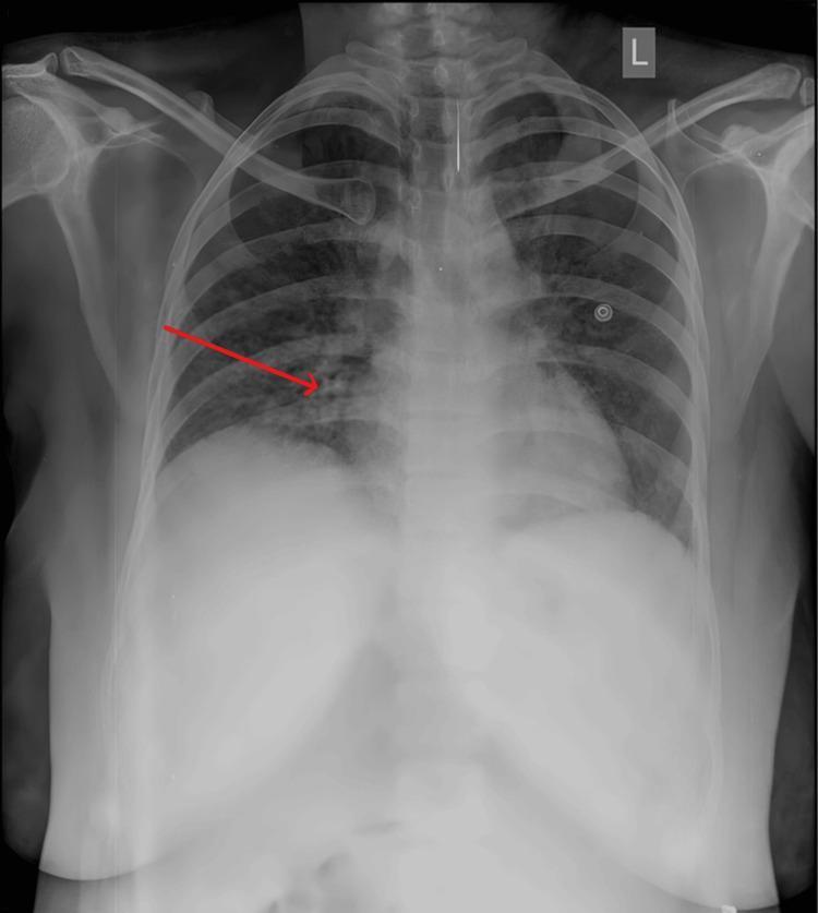
A posterior-anterior (PA) view chest X-ray of an adult patient, presenting key features of atypical viral pneumonia, such as that caused by COVID-19. The image shows bilateral, multifocal ground-glass opacities and patchy consolidations. A red arrow specifically highlights a region of increased density and reticular interstitial markings in the right mid-to-lower lung field. The opacities are peripheral and basal-predominant, which is characteristic of viral inflammatory processes. The cardiac silhouette appears within normal size limits, and the costophrenic angles remain relatively clear, suggesting a lack of significant pleural effusion. Visible medical apparatus includes external monitoring electrodes on the chest wall. The diaphragm is slightly elevated, likely due to the patient's physiological state or positioning. This radiological study is used in medical education to illustrate the diagnostic presentation of respiratory infections and the importance of identifying focal opacities in acute clinical settings.
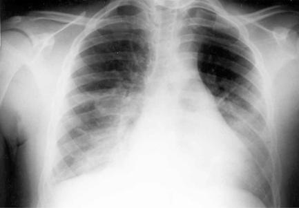
A posterior-anterior (PA) view diagnostic chest X-ray illustrating pulmonary manifestations in a patient with status asthmaticus and concurrent Mycoplasma pneumoniae infection. The radiographic findings are characterized by bilateral, diffuse, and patchy interstitial infiltrates, most prominent in the basilar regions of both lung fields. There is a general decrease in pulmonary transparency at the bases, indicating areas of consolidation or fluid accumulation consistent with atypical pneumonia. The cardiac silhouette appears within normal limits for size and morphology, and the visible mediastinal structures are unremarkable. The skeletal framework, including the ribs and clavicles, shows no acute abnormalities. This image serves as a clinical example of secondary pulmonary infection complicating an acute exacerbation of reactive airway disease, highlighting the characteristic 'walking pneumonia' presentation on plain film radiography.
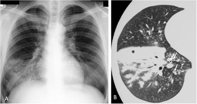
A multi-modal diagnostic comparison illustrating Mycoplasma pneumoniae pneumonia. Image A is a posterior-anterior chest x-ray showing increased density and reticulonodular infiltrates primarily localized in the right lower lobe, signifying an active infectious process. Image B is a high-resolution computed tomography (HRCT) scan axial slice of the lung parenchyma. The HRCT demonstrates a dense region of lobar consolidation, marked with an asterisk (*), indicating alveolar filling. Additionally, arrows (↑) highlight significant bronchovascular bundle thickening, characterized by increased interstitial markings and peribronchial cuffing. The combination of these radiological findings reflects the classic presentation of atypical pneumonia, emphasizing the correlation between plain film radiography and the superior anatomical detail provided by cross-sectional CT imaging for evaluating pulmonary inflammation and consolidation.
atypical viral pneumonia chest physiotherapy assessment clinical findings 2025
Atypical (Viral) Pneumonia - Medical & Chest PT Assessment
1. What is Atypical Pneumonia?
- The organism does not stain on routine Gram or Ziehl-Neelsen stains
- It is not cultured on standard media
- It is resistant to beta-lactam antibiotics (because these organisms lack a cell wall)
2. Causative Organisms
| Category | Organisms |
|---|---|
| Mycoplasma | Mycoplasma pneumoniae (most common - up to 15% of outpatient CAP) |
| Chlamydophila | C. pneumoniae, C. psittaci (psittacosis) |
| Bacteria (special) | Legionella spp., F. tularensis, C. burnetii (Q fever), B. anthracis |
| Viruses | Influenza A/B, RSV, parainfluenza, metapneumovirus, adenovirus, coronavirus (SARS-CoV-2, MERS), rhinovirus, VZV, CMV, EBV, Hantavirus, measles |
| Fungi (atypical presentation) | Histoplasma, Coccidioides, Pneumocystis jirovecii |
| Rickettsia | Coxiella burnetii (Q fever) |
3. Pathogen-Specific Clinical Features (Clues to Diagnosis)
Mycoplasma pneumoniae ("Walking Pneumonia")
- Insidious onset over days: malaise, fever, prominent headache
- Mild sore throat, nonproductive dry cough (hallmark)
- No GI symptoms (unlike Legionella)
- Epidemics every 4-8 years; seen in schools, households, military camps
- Extrapulmonary clues: bullous myringitis, hemolytic anemia, rash (erythema multiforme), arthralgia, cervical lymphadenopathy, myocarditis, diarrhea, neurologic symptoms
- CXR: patchy infiltrates, hilar adenopathy, occasional pleural effusion
Legionella ("Legionnaires' Disease")
- 1-10% of CAP; can be severe, frequently requiring ICU
- Classical: fever + GI symptoms (diarrhea, vomiting, abdominal pain) + pneumonia
- Relative bradycardia (pulse does not rise proportionately with fever - a key clue)
- Hyponatremia, confusion/altered mental status
- No seasonality (prominent in summer when others decline)
- Risk: smokers, COPD, transplant, immunosuppressed
- Diagnose with urinary Legionella antigen
- CXR: patchy infiltrate, hilar adenopathy, pleural effusions
Chlamydia pneumoniae
- Sub-acute illness: sore throat, mild fever, nonproductive cough
- Rales or rhonchi on exam
- CXR: patchy subsegmental infiltrate; may be linked to adult-onset asthma
Q Fever (Coxiella burnetii)
- Transmitted via infected animals (dried urine, unpasteurized milk)
- Acute: lobar infiltrates + pleural effusion, or hepatitis
- Chronic form: endocarditis (nearly 100% mortality if untreated)
- Treatment: doxycycline or respiratory quinolone
Viral Pneumonia (Influenza, COVID-19, etc.)
- Abrupt onset: fever, myalgia, headache, followed by respiratory symptoms
- Dry cough, progressive dyspnea
- Can range from mild to acute respiratory failure
- Secondary bacterial pneumonia complicates ~20% of viral CAP
- Neuraminidase inhibitors (oseltamivir) are first-line for influenza (most effective early)
- CXR/CT: bilateral ground-glass opacities, peripheral/basal-predominant infiltrates (viral pattern)
4. Typical vs. Atypical Pneumonia - Differentiating Features
| Feature | Typical Pneumonia | Atypical Pneumonia |
|---|---|---|
| Onset | Acute, sudden | Gradual, insidious |
| Cough | Productive, purulent/bloody | Dry, nonproductive |
| Fever | High grade, with rigors/chills | Moderate, without rigors |
| Chest pain | Pleuritic (common) | Retrosternal (Mycoplasma) or absent |
| WBC | Neutrophilia | Normal or mildly elevated |
| Procalcitonin | >0.25 µg/L | ≤0.1 µg/L |
| Extrapulmonary features | Uncommon | Frequent (headache, myalgia, GI, rash, etc.) |
| CXR | Lobar consolidation with air bronchograms | Patchy/interstitial infiltrates, bilateral |
| Response to beta-lactams | Yes | No |
| Pathogen ID | Gram stain + culture | PCR, serology, urinary antigen |
Important caveat: Neither clinical symptoms alone nor CXR appearance are sensitive or specific enough to definitively classify etiology or guide therapy. Treat empirically based on syndrome. (Murray & Nadel, p. 985)
5. Investigations
| Test | Purpose |
|---|---|
| CXR | Initial imaging - patchy/interstitial infiltrates in atypical |
| HRCT chest | Better characterization if CXR inconclusive; shows GGO, bronchovascular thickening |
| PCR (NPS/BAL) | Gold standard for viral identification |
| Urinary Legionella antigen | Rapid, sensitive for Legionella spp. |
| Cold agglutinins | Positive in ~50% of Mycoplasma (hemolytic anemia clue) |
| Serology (IgM/IgG) | Mycoplasma, Chlamydia, Q fever |
| FBC | Lymphocytosis (viral), normal or mild leukocytosis (atypical), leukopenia (severe viral) |
| CRP / Procalcitonin | Procalcitonin ≤0.1 µg/L favors atypical/viral |
| ABG / SpO2 | Assess respiratory failure |
| LFTs, U&E | Hepatitis (Q fever, Legionella), hyponatremia (Legionella) |
6. Medical Treatment
- Macrolide: azithromycin, clarithromycin (first-line outpatient)
- Doxycycline: second-line or first-line if macrolide resistance suspected
- Respiratory fluoroquinolone: levofloxacin, moxifloxacin (for severe disease or inpatient)
- Oseltamivir: influenza pneumonia (start within 48 hours ideally)
- Supportive: O2 supplementation, IV fluids, antipyretics
- No beta-lactams - atypical organisms lack a cell wall
7. Chest Physiotherapy (PT) Assessment
A. Subjective Assessment (History)
- Onset and duration of cough, dyspnea, sputum production
- Character of cough: dry/nonproductive (atypical) vs. productive
- Fever history, fatigue, myalgia, GI symptoms
- Breathlessness: MRC dyspnea scale, onset at rest vs. exertion
- Exercise tolerance - functional limitation (6MWT equivalent)
- Past medical history: COPD, asthma, immunosuppression, smoking
- Vaccination status: influenza, pneumococcal
- Medications: especially inhalers, antivirals, antibiotics already started
B. Objective Assessment
Vital Signs
| Parameter | Normal | Concern |
|---|---|---|
| RR | 12-20 /min | >20 (tachypnea) = pneumonia severity marker |
| SpO2 | ≥95% | <94% requires O2 therapy |
| HR | 60-100 | Tachycardia (typical) / relative bradycardia (Legionella, Mycoplasma) |
| Temp | 36.5-37.5°C | Fever present, usually moderate in atypical |
| BP | — | Hypotension = severe/septic |
Inspection
- Respiratory rate, rhythm, depth
- Use of accessory muscles (scalene, sternomastoid, intercostals)
- Pursed-lip breathing
- Nasal flaring, cyanosis (central/peripheral)
- Chest symmetry - lag/splinting on affected side (with consolidation)
- Barrel chest (underlying COPD)
Palpation
- Chest expansion: reduced on affected side
- Tactile fremitus: increased over consolidation (pneumonia), decreased over effusion/collapse
- Tracheal deviation (if tension or large effusion)
- Tenderness (pleuritic component)
Percussion
- Dull or flat: consolidation (pneumonia), pleural effusion
- Hyperresonant: pneumothorax or emphysema
Auscultation (key for atypical pneumonia)
| Finding | Significance |
|---|---|
| Fine crackles (late inspiratory) | Alveolar consolidation / fluid filling |
| Coarse crackles | Secretions in larger airways |
| Bronchial breath sounds | Over area of consolidation |
| Reduced breath sounds | Consolidation, effusion, or collapse |
| Wheezes | Bronchospasm (may occur in viral - RSV, influenza) |
| Bronchophony / egophony | Consolidation confirmed (e-to-a change = egophony) |
| Pleural rub | Pleurisy (uncommon in atypical but possible) |
In atypical pneumonia, auscultation findings may be minimal or absent despite significant radiographic change - this is the classic discordance. The patient may look less ill than expected from the CXR ("walking pneumonia").
Cough Assessment
- Effectiveness: ability to generate and clear sputum
- Cough strength (peak cough flow)
- In atypical pneumonia: usually dry, ineffective, non-productive
Sputum Assessment
- Quantity, color, consistency
- Atypical: scant or absent sputum (dry cough)
- Viral: clear/white mucoid if any
C. Functional Assessment
- Exertional desaturation (6-minute walk test or equivalent)
- ADL performance - how far can the patient walk/manage self-care?
D. Severity Scoring (for PT decision-making)
- CURB-65: Confusion, Urea >7 mmol/L, RR ≥30, BP <90/60, Age ≥65
- Score 0-1: outpatient; Score 2: hospital; Score 3+: ICU consideration
8. Chest PT Goals and Interventions
| Goal | Intervention |
|---|---|
| Improve ventilation | Positioning (high Fowler's, lateral decubitus affected side up) |
| Airway clearance | Active Cycle of Breathing Technique (ACBT), huff coughing |
| Reduce work of breathing | Breathing control, paced breathing, pursed-lip breathing |
| Prevent atelectasis | Incentive spirometry, deep breathing exercises |
| Improve oxygenation | O2 therapy + monitored exercise |
| Mobilize secretions | Percussion/vibrations (if secretions present - less relevant in dry atypical) |
| Early mobilization | Supported sitting, standing, ambulation as tolerated |
Note: In pure dry-cough atypical/viral pneumonia, secretion clearance techniques are less relevant than ventilation exercises and positioning. Chest percussion is most useful when sputum retention is confirmed.
Summary
- Murray & Nadel's Textbook of Respiratory Medicine - pp. 985, 401
- Fishman's Pulmonary Diseases and Disorders - p. 2151
- Tintinalli's Emergency Medicine - p. 483
- Textbook of Family Medicine 9e - p. 305
- Frameworks for Internal Medicine - p. 418