
Middle ear space
Reading File
The middle ear space (tympanic cavity) is an air-filled cavity in the temporal bone containing the ossicular chain, lined by mucous membrane, connected to the nasopharynx via the Eustachian tube.
Searching sources...
Finding Sources
Finding Sources
Reading File
Reading File
Reading File
Reading File
Reading File
Reading File
Reading File
Loading Image
Reading File
I now have comprehensive content from multiple authoritative sources. Here is a detailed overview of the middle ear space.
Middle Ear Space (Tympanic Cavity)
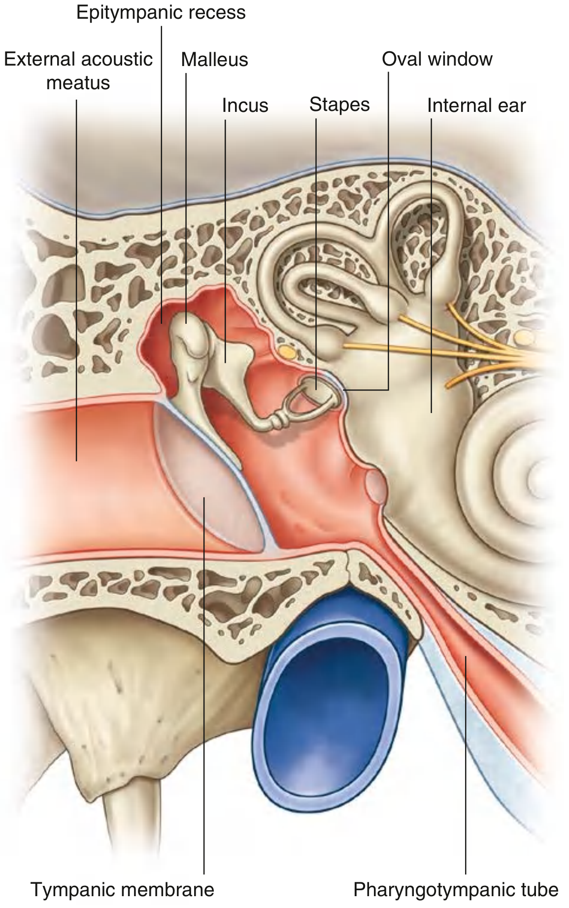
Definition and Overview
The middle ear cleft consists of three communicating parts:
- The tympanic cavity (the middle ear space proper)
- The Eustachian (pharyngotympanic) tube
- The mastoid air cell system
The tympanic cavity is an irregular, air-filled space within the temporal bone, bounded by the tympanic membrane laterally and the osseous labyrinth (inner ear) medially. It is lined throughout by mucous membrane continuous with that of the mastoid cells and Eustachian tube.
(Scott-Brown's Otorhinolaryngology, Vol. 2, p. 574)
Subdivisions
The middle ear is described as two parts:
- Tympanic cavity proper - immediately adjacent to the tympanic membrane (mesotympanum)
- Epitympanic recess (attic) - the space superior to the level of the tympanic membrane, housing the heads of malleus and incus
(Gray's Anatomy for Students, p. 1098)
Additional surgical subdivisions include:
- Protympanum - anterior to the tympanic membrane, near the Eustachian tube orifice
- Hypotympanum - below the level of the tympanic annulus
- Posterior epitympanum and anterior epitympanic space, separated by the cog (a bony crest projecting from the tegmen tympani anteriorly to the malleus head) - surgically important as a site for residual cholesteatoma
Walls of the Tympanic Cavity
Roof - Tegmental Wall
A thin layer of bone (the tegmen tympani) separates the middle ear from the middle cranial fossa. The petrosquamous suture in children can provide a route for spread of infection into the extradural space. Veins draining to the superior petrosal sinus pass through this suture.
Floor - Jugular Wall
A thin bone separates the hypotympanum from the jugular bulb. This bone can be deficient (in ~2% of cases), leaving the jugular bulb covered only by mucosa - a hazard when raising inferior tympanomeatal flaps. The inferior tympanic canaliculus (Jacobson's canal) perforates the floor, carrying the tympanic branch of CN IX (Jacobson's nerve) and preganglionic parasympathetic fibres toward the otic ganglion.
Lateral Wall - Membranous Wall
Formed almost entirely by the tympanic membrane. The upper part (epitympanic recess level) is formed by the bony squamous temporal bone. The tympanic membrane is slightly oval (9-10 mm longest diameter), angled ~55° to the floor of the meatus. It has:
- Pars tensa - the larger inferior portion with a tympanic annulus, three layers (epidermis, lamina propria with radial and circular fibres, inner mucosa)
- Pars flaccida - superior triangular portion above the anterior and posterior malleolar folds, lacking a firm annulus, within the notch of Rivinus
Medial Wall - Labyrinthine Wall
Separates the tympanic cavity from the inner ear. Key structures:
- Promontory - a rounded elevation over the basal turn of the cochlea, carrying the tympanic plexus (from CN IX tympanic branch + caroticotympanic sympathetic fibres)
- Oval window (fenestra vestibuli) - posterosuperior to the promontory, kidney-shaped, closed by the stapes footplate and annular ligament; connects to the vestibule
- Round window (fenestra cochleae) - posteroinferior to the promontory, closed by the secondary tympanic membrane; allows pressure release of cochlear fluid
- Prominence of the facial canal - ridge produced by CN VII passing through the temporal bone, posterosuperior to the oval window
- Prominence of the lateral semicircular canal - broader ridge above and posterior to the facial canal
- Processus cochleariformis - a bony shelf at which the tensor tympani tendon turns laterally to attach to the malleus
Anterior Wall
Narrow (medial and lateral walls converge anteriorly). Contains:
- Lower third: thin bone covering the internal carotid artery (can be dehiscent in ~2%), perforated by caroticotympanic nerves (sympathetic)
- Middle third: tympanic orifice of the Eustachian tube (~5 × 4 mm)
- Just above: canal for the tensor tympani muscle
- Upper third: may contain the supratubal recess, separated from the anterior epitympanic space by the tensor fold
- Exit foramen for the chorda tympani nerve
Posterior Wall - Mastoid Wall
Wider superiorly than inferiorly. Contains:
- Aditus ad antrum - large irregular opening from the posterior epitympanum leading to the mastoid antrum
- Fossa incudis - houses the short process of the incus and its suspensory ligament
- Pyramid (pyramidal eminence) - hollow conical projection housing the stapedius muscle; its tendon inserts into the head of the stapes
- Facial recess - groove between the pyramid/facial nerve and the tympanic annulus; important surgical corridor
- Sinus tympani - medial to the facial nerve, between the ponticulus and subiculum; a common site for residual disease
Contents of the Middle Ear
Ossicular Chain
Three articulated bones transmit vibrations from the tympanic membrane to the oval window:
| Bone | Attachment | Joint |
|---|---|---|
| Malleus | Handle to tympanic membrane (umbo); head in epitympanum | - |
| Incus | Connects malleus and stapes | Synovial joint with malleus |
| Stapes | Footplate in oval window via annular ligament | Synovial joint with incus |
The ossicular chain provides a pressure gain of ~22× due to the area difference between the tympanic membrane and oval window, plus lever action.
Muscles
- Tensor tympani - in a bony canal along the medial wall; tendon hooks around the processus cochleariformis and attaches to the malleus handle. Supplied by CN V3 (medial pterygoid nerve). Stiffens the ossicular chain.
- Stapedius - smallest skeletal muscle in the body; housed in the pyramid, inserts into the stapes head. Supplied by CN VII. The stapedial (acoustic) reflex protects against loud sounds.
Nerves
- Chorda tympani (branch of CN VII) - enters through the posterior wall, runs between the handle of the malleus and the long process of the incus across the medial surface of the tympanic membrane, and exits via the anterior wall into the infratemporal fossa. Carries taste from anterior 2/3 of tongue and preganglionic parasympathetics to submandibular ganglion.
- Tympanic plexus - on the promontory; formed mainly by the tympanic branch of CN IX (Jacobson's nerve). Gives the lesser petrosal nerve (preganglionic parasympathetics to parotid gland via otic ganglion).
Communications
| Direction | Structure | Connects To |
|---|---|---|
| Anterior | Pharyngotympanic (Eustachian) tube | Nasopharynx |
| Posterior/superior | Aditus ad antrum | Mastoid antrum → mastoid cells |
The pharyngotympanic tube is 1/3 bony (near middle ear) and 2/3 cartilaginous (near nasopharynx). It equalises pressure across the tympanic membrane and is normally closed, opening during swallowing/yawning. It is shorter and more horizontal in children, explaining their higher susceptibility to otitis media.
Lining
The tympanic cavity, Eustachian tube, and mastoid cells are lined by a continuous mucous membrane. This continuity explains how upper respiratory tract infections can ascend via the Eustachian tube to cause otitis media, and why middle ear infection can spread to cause mastoiditis.
Clinical Relevance
- Otitis media - inflammation commonly spreading from the nasopharynx via the Eustachian tube; most frequent in young children
- Cholesteatoma - keratinizing squamous epithelium in the middle ear; commonly forms in the pars flaccida region or the posterior epitympanic recess; can erode ossicles, tegmen, and facial canal
- Mastoiditis - infection spreading from the middle ear through the aditus; can progress to intracranial complications given the thin tegmen tympani
- Jugular bulb anomaly - high-riding or dehiscent jugular bulb risks injury during myringotomy or tympanomeatal flap elevation
- Facial nerve dehiscence - the facial canal may be dehiscent over the oval window niche in a significant minority, risking injury during middle ear surgery
- Carotid artery dehiscence - ~2% of cases; the internal carotid is exposed in the protympanum, presenting as a pulsatile mass
(Sources: Gray's Anatomy for Students, pp. 1098-1100; Scott-Brown's Otorhinolaryngology, Vol. 2, pp. 574-576; Junqueira's Basic Histology, 17e)
This is a shared conversation. Sign in to Orris to start your own chat.