
Submental sinus tract
submental sinus tract odontogenic cutaneous fistula chin
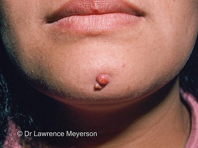
Clinical photography of a single, small nodular lesion in the submental/chin region, illustrating the cutaneous terminus of an odontogenic sinus tract. Modality: Clinical photography; technique: high-resolution frontal close-up, color-calibrated lighting, macro field to document surface contour, color, and surrounding skin. Not applicable staining or contrast. Magnification: close-up macro. Acquisition parameters: standard clinical imaging, consistent white balance. Anatomical location: on the chin, within the submental region, along the lower facial dermis; skin and subcutaneous tissue are involved; midline/central projection. Visual features: a solitary, dome-shaped, erythematous-to-flesh-colored papule approximately a few millimeters in diameter; smooth surface with possible faint crust; no obvious surrounding edema visible in this image; the lesion represents the cutaneous manifestation of a concealed dental infection, at the end of a sinus tract beneath the skin, with potential drainage. Clinical significance: indicates chronic odontogenic infection with cutaneous fistulization; supports diagnosis of odontogenic sinus tract/periapical granuloma. Differential diagnoses include sebaceous cyst, pyogenic granuloma, foreign-body granuloma, basal cell carcinoma, and other skin nodules. Correlate with dental history and radiographs (periapical/CBCT) to locate the tooth and plan root canal therapy or extraction; treat source to resolve tract. Educational use: dermatology and dentistry case illustrating cross-disciplinary diagnostics and fistula tracking.
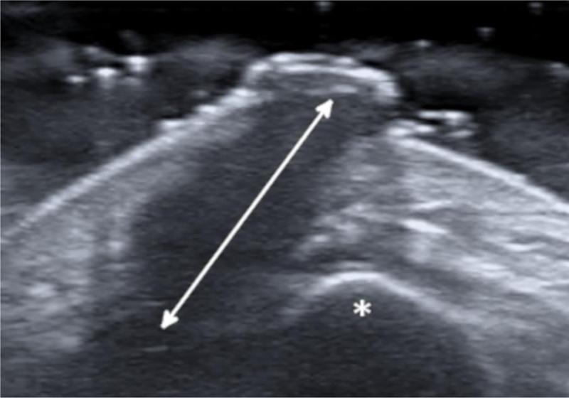
This diagnostic image is a high-frequency (18 MHz) grayscale ultrasonography of the chin and submental region. The scan demonstrates a distinct, wide hypoechoic band (indicated by a double-headed arrow) that represents an inflamed, tortuous sinus tract. This tract originates from the deeper structures and traverses vertically through the subcutaneous tissue layers toward the skin surface. The tract terminates near the mandibular bone, marked with an asterisk, showing a direct anatomical connection between the deep osseous tissue and the superficial soft tissue. The surrounding soft tissues show heterogenous echogenicity, consistent with inflammatory changes. This visual evidence is a key diagnostic feature for an odontogenic cutaneous fistula (OCF), illustrating the pathway of chronic infection from a dental origin through the mandible to a cutaneous exit point. The image is used in medical education to teach the sonographic appearance of fistulous tracts and the importance of identifying deep dental sources in persistent cutaneous nodules of the head and neck.
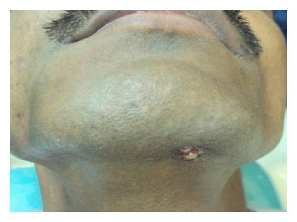
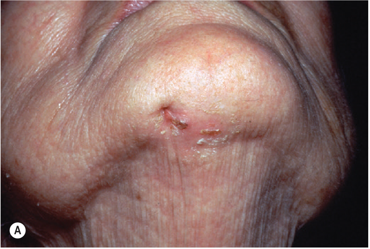
A clinical photograph capturing the inferior view of a patient's submental and chin region. The image demonstrates a non-healing, extraoral draining sinus tract located centrally in the submental area. The lesion is characterized by a small, irregular cutaneous opening with evidence of active seropurulent or yellowish discharge. The immediately surrounding skin exhibits localized erythema, swelling, and hyperpigmentation, consistent with chronic inflammation. This clinical presentation is typical of an odontogenic cutaneous sinus tract, which often results from chronic periapical infections such as dental cysts or abscesses that have tracked through the mandibular bone to the skin surface. The image is a significant diagnostic example in both oral surgery and dermatology for identifying extraoral manifestations of dental pathology, such as odontogenic keratocysts or infected periapical cysts.
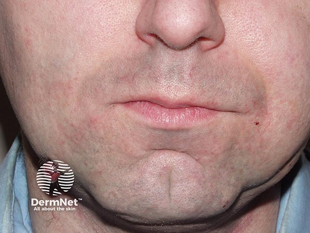
Imaging modality: Clinical photograph. This frontal view of the lower face documents a cutaneous lesion at the end of a dental sinus tract, i.e., an odontogenic cutaneous fistula terminating on the chin/perioral skin. The lesion is a small erythematous papule/nodule with a raised, smooth surface and subtle surrounding erythema. Minimal crust; no evident purulent drainage. Anatomically, it overlies the chin/lower face near the mandible with midline alignment; no atrophy or ulceration is evident. The photograph captures surface morphology without radiographic or histologic data, but the suggested diagnosis is a granulomatous response to a chronic dental infection, where a sinus tract drains via the skin. Clinically this finding indicates an odontogenic source rather than a primary dermatologic process. Pathologically, cutaneous granulomas associated with dental infection reflect granulomatous inflammation along the fistulous tract and may be accompanied by periapical inflammation of pulpal origin. Diagnostic significance lies in directing dental evaluation, radiographic imaging (panoramic/periapical), and source control through root canal therapy or extraction. Differential considerations include odontogenic cutaneous fistula, foreign body granuloma, and chronic dermatitis; clinical correlation with dental status is essential. Management typically requires multidisciplinary care integrating dentistry and dermatology to eradicate infection and close the sinus.
"submental sinus" OR "cutaneous sinus" AND odontogenic
Submental Sinus Tract (Odontogenic / Cutaneous Dental Sinus)
Pathophysiology
- When it drains intraorally through the alveolar mucosa, it is called a parulis (intraoral dental sinus)
- When it penetrates the cortical bone and tunnels through the overlying soft tissue to reach the skin surface, it becomes a cutaneous dental sinus tract
Clinical Presentation
- A soft, erythematous papule or nodule - often mistaken for a cyst, pyogenic granuloma, or furuncle
- A fibrosed depressed scar or an eroded/crusted papule
- An intermittently discharging sinus with seropurulent material
- A practically asymptomatic, palpable cord-like tract leading toward the jaw
Causative Teeth
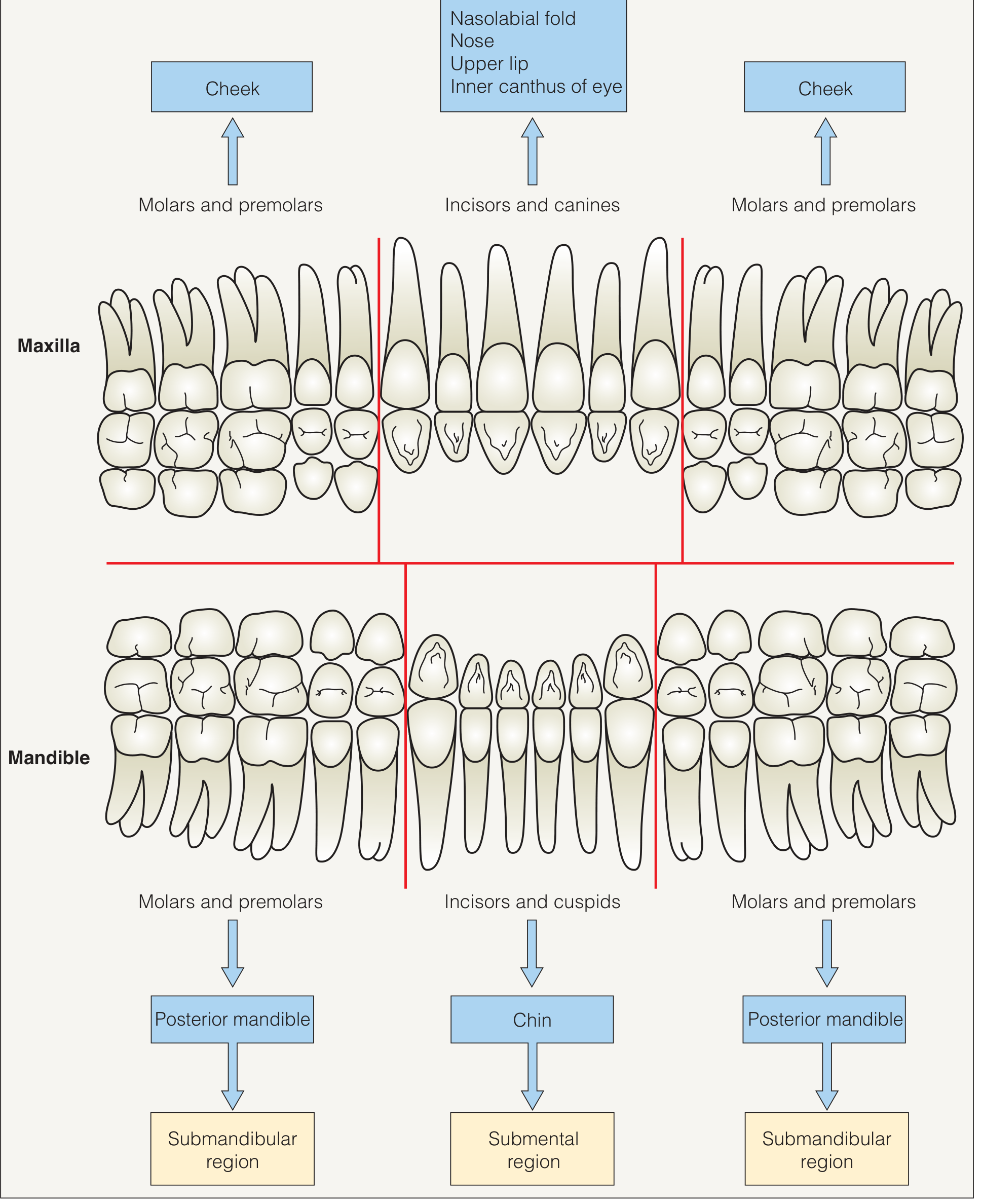
- Most common: mandibular teeth (especially incisors, canines, premolars)
- Less common: maxillary teeth (drain to cheek, nasal fold, upper lip, or inner canthus)
- Bilateral involvement is rare but reported
- The offending tooth often appears clinically innocuous with no obvious pain
Diagnosis
| Step | Finding |
|---|---|
| Dental X-ray (periapical) | Periapical radiolucency (gold standard); often definitively diagnostic |
| Panoramic radiograph | Good overview of all roots |
| CBCT | Best for complex or occult cases |
| Gutta-percha tracing | Insert into sinus tract, take X-ray to trace to offending tooth |
| High-frequency ultrasound | Shows hypoechoic tract connecting mandible to skin surface (see 2024 multicentric ultrasound review) |
| Vitality testing | Offending tooth is non-vital |
Differential Diagnosis
- Pyogenic granuloma - vascular, bleeds easily, not tract-related
- Actinomycosis - sulfur granules on discharge; culture positive
- Infected epidermal/dermoid cyst - no bony connection
- Basal cell carcinoma / SCC - biopsy distinguishes
- Osteomyelitis of the mandible - bone changes on X-ray
- Bisphosphonate-related osteonecrosis of the jaw (BRONJ) - medication history
- Congenital fistulas (thyroglossal duct, branchial cleft) - younger patients, embryological location
- Deep fungal infection (sporotrichosis, actinomycetoma) - culture, histopathology
- Foreign body granuloma
Treatment
- Root canal therapy (endodontic treatment) - preferred if the tooth is restorable; the sinus tract usually closes within 1-4 weeks
- Extraction of the offending non-vital tooth - if non-restorable
- Curettage of periapical tissue / apicectomy - for persistent periapical pathology
- Antibiotics alone are NOT curative - they may temporarily suppress discharge but the sinus returns
Key Teaching Points
- A chronic, non-healing draining papule/nodule on the chin, jaw, or submental region should always prompt dental evaluation
- The causative tooth is often asymptomatic and may look clinically normal
- Dental radiography is diagnostic - look for periapical radiolucency
- Misdiagnosis leads to repeated antibiotic courses and unnecessary dermatological procedures
- Treatment is dental, not dermatological - cure the tooth, cure the sinus
Chronic suppurative otitis media
chronic suppurative otitis media tympanic membrane perforation otoscopy
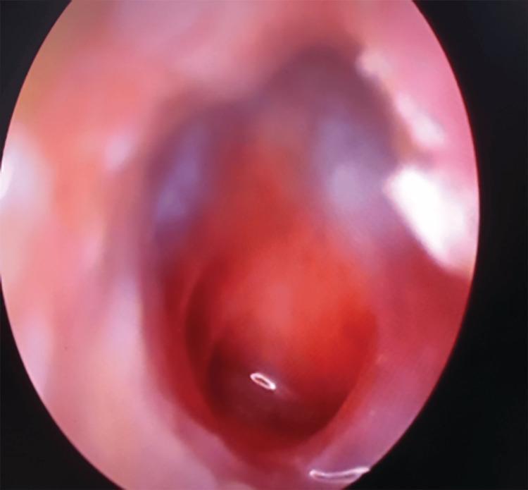
This clinical photograph, obtained via otoscopy/endoscopy, depicts a human tympanic membrane with a large central perforation. The perforation occupies a significant portion of the pars tensa, appearing as a dark, well-demarcated void that reveals the underlying middle ear space. The surrounding tympanic membrane remnant is erythematous and thickened, indicating active or chronic inflammation. The middle ear mucosa visible through the defect appears congested and reddish. Key anatomical landmarks, such as the handle of the malleus and the light reflex, are obscured or distorted due to the extensive tissue loss and inflammatory changes. This visual is characteristic of chronic suppurative otitis media (CSOM) or significant acoustic/mechanical trauma. The image serves as an educational example of middle ear pathology and the visual assessment of tympanic membrane integrity in an otolaryngology context.
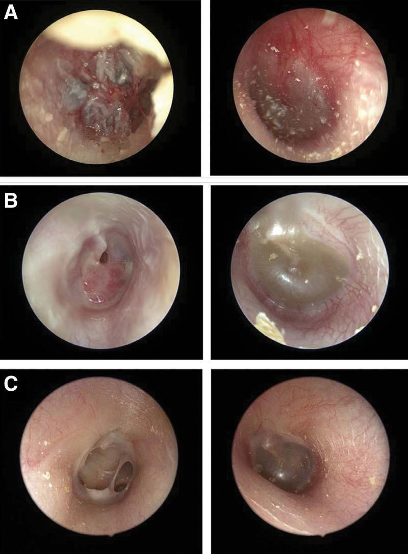
This set of endoscopic otoscopy images documents the bilateral progression and resolution of otitis media. Panel A illustrates acute hemorrhagic otitis media in the right ear with white epithelial thickening and a small perforation, while the left ear shows diffuse hyperemia of the tympanic membrane (TM). Panel B demonstrates the suppurative stage in the right ear with a central TM perforation visible after debridement of purulent material; the left ear shows otitis media with effusion (OME), characterized by a retracted TM and amber-colored middle ear fluid. Panel C shows the chronic phase, with a persistent dry perforation in the right TM lacking acute inflammatory signs, and a resolving or stable state in the left ear. This clinical sequence provides a visual comparison between different stages of inflammatory middle ear disease, including acute infection, suppuration, and persistent perforation, essential for otolaryngology and pediatric diagnostic training.
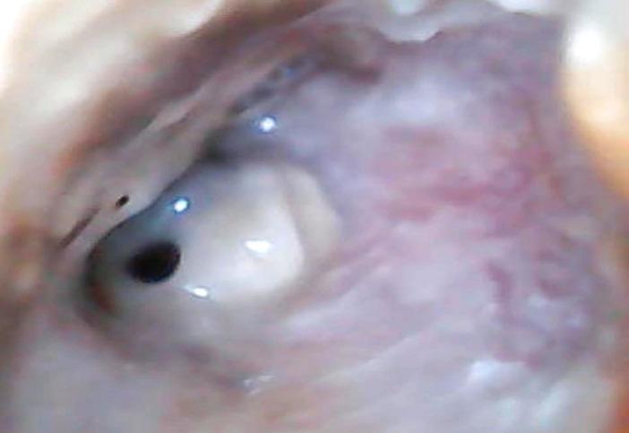
This clinical photograph displays an otoscopic view of the right ear, highlighting pathological findings of the external auditory canal (EAC) and tympanic membrane. The EAC shows significant inflammatory changes, characterized by diffuse erythema and edema of the canal walls. Visible purulent secretions are present within the lumen, suggesting an active infectious process such as chronic suppurative otitis media or acute exacerbation. The tympanic membrane is clearly visualized and exhibits a single, well-defined perforation located in the anterior-inferior quadrant. The perforation appears as a dark, circular opening amidst the remaining translucent, light-reflective portions of the membrane. Normal landmarks like the handle of the malleus are obscured by the surrounding inflammatory tissue and exudate. This image serves as a clinical example of suppurative middle ear disease with associated tympanic membrane rupture and secondary external canal inflammation, relevant for otolaryngology training in diagnostic otoscopy.
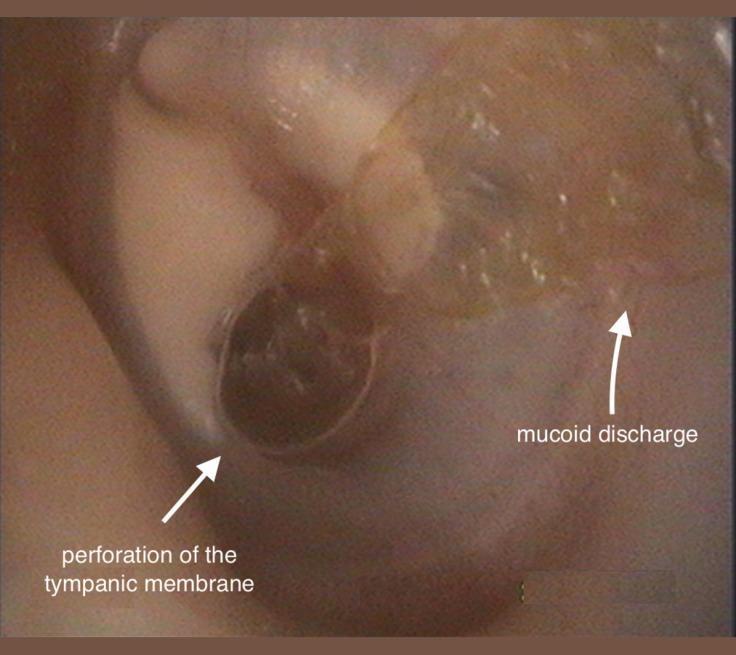
This clinical otoscopic photograph of a left ear demonstrates signs of chronic otitis media (COM). The image clearly shows a large, central perforation of the tympanic membrane, appearing as a dark, circular void where the membrane should be intact. Adjacent to this perforation, there is visible mucoid discharge, which presents as a translucent, glistening, and somewhat viscous fluid resting on the membrane surface. Additionally, white, opaque areas consistent with tympanosclerotic plaques are visible on the remaining portions of the tympanic membrane, indicating previous inflammation or scarring. The external auditory canal appears mildly erythematous. This visual is characteristic of chronic suppurative otitis media, an important educational example for distinguishing types of membrane perforations and associated middle ear pathology. The presence of discharge and structural changes in the membrane are key diagnostic markers used by otorhinolaryngologists to assess disease chronicity and planning for potential tympanoplasty.
"chronic suppurative otitis media" AND management
Chronic Suppurative Otitis Media (CSOM)
Definition
Classification of Chronic Otitis Media
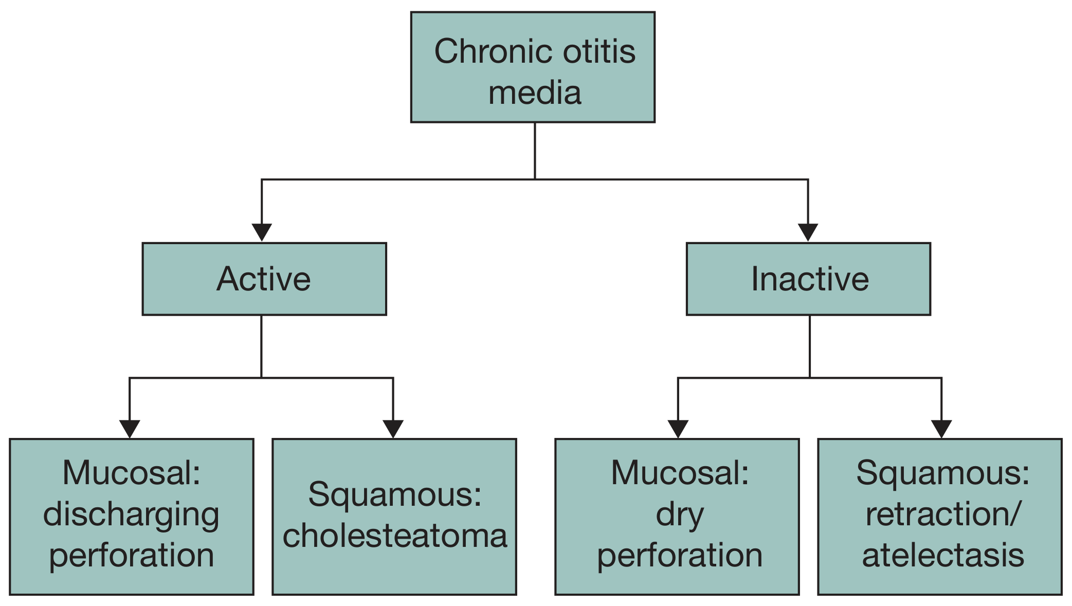
| Type | Subtype | Features |
|---|---|---|
| Active | Mucosal (CSOM) | Discharging perforation; inflamed middle ear mucosa ± granulation tissue |
| Active | Squamous (Cholesteatoma) | Keratinising squamous epithelium in middle ear; bone-eroding |
| Inactive | Mucosal | Dry perforation; no active inflammation |
| Inactive | Squamous | Retraction pocket / atelectasis |
British Classification (Tubotympanic vs Atticoantral)
| Feature | Tubotympanic ("Safe/Benign") | Atticoantral ("Unsafe/Dangerous") |
|---|---|---|
| Perforation site | Pars tensa (central) | Pars flaccida / posterosuperior marginal |
| Cholesteatoma | Absent | Often present |
| Prognosis | Lower complication risk | Higher complication risk |
| Discharge | Mucoid, non-foul | Foul-smelling, scanty |
Pathophysiology
- Persistent middle ear effusion → mucosal oedema → granulation tissue
- Bacterial infection → purulent effusion → chemical mediators → chronic mucosal changes
- Submucosal gland metaplasia → secretory mucosa → perpetuating effusion
- Enzymatic degradation of TM → weakening of collagen skeleton → retraction pockets → perforation
- Retraction pockets → accumulate squamous debris → cholesteatoma formation
- Stable healed scar (tympanosclerosis/myringosclerosis)
- Permanent pars tensa perforation
- Atelectasis (TM draped over medial wall)
- Retraction pocket → cholesteatoma
Microbiology
- Most common organisms: Pseudomonas aeruginosa (~31%), Staphylococcus aureus (~19%), Proteus mirabilis, anaerobes (Peptococcus, Bacteroides, Peptostreptococcus)
- Polymicrobial with both aerobes and anaerobes
- Fungal overgrowth (especially Candida) is surprisingly common - seen in 35% of ears treated with topical ciprofloxacin for 3 weeks
- Bacterial biofilms are a major reason for treatment resistance
Clinical Features
Symptoms
- Intermittent or continuous purulent otorrhoea - sometimes foul-smelling
- Conductive hearing loss (CHL) - variable; >30 dB CHL suggests ossicular erosion
- Mild discomfort; ear may be generally non-painful
- Warning symptoms (suggesting complications):
- Otalgia / headache → suspect intracranial involvement or malignancy
- Vertigo → suspect labyrinthitis or labyrinthine fistula
- Facial weakness → facial nerve involvement
Otoscopic Findings
- TM perforation (central in tubotympanic; posterosuperior/marginal in atticoantral)
- Discharge in the external auditory canal (EAC)
- Congested, thickened middle ear mucosa visible through perforation
- Possible EAC polyps or granulation tissue
- Scutal or ossicular erosion in advanced disease
- Tympanosclerotic plaques (white calcified areas) in healed/inactive disease
Investigations
| Investigation | Purpose |
|---|---|
| Otomicroscopy/Otoendoscopy | Best visualization of TM and middle ear |
| Pure tone audiometry (PTA) | Quantify CHL/SNHL; CHL >30 dB suggests ossicular erosion |
| Tympanometry | Middle ear pressure and compliance |
| CT temporal bone (HRCT) | Extent of disease, ossicular erosion, cholesteatoma, mastoid involvement |
| MRI (DWI) | Non-echo-planar DWI detects cholesteatoma; evaluates intracranial complications |
| Ear swab & culture | Guide antibiotic choice; identify resistant organisms |
Medical Management
Topical Antibiotics (First-line)
- Fluoroquinolone drops (ciprofloxacin, ofloxacin) - most effective and ototopically safe
- Avoid aminoglycoside drops (gentamicin, neomycin) in the presence of perforation - risk of ototoxicity
- Acetic acid drops (1-2%) - antiseptic, useful adjunct
Systemic Antibiotics
- Used for acute exacerbations or when topical therapy fails
- A 2025 Cochrane systematic review on systemic antibiotics for CSOM found limited evidence for oral vs. topical routes
- A concurrent Cochrane review on topical antibiotics found topical fluoroquinolones superior to non-quinolone topical agents and to systemic antibiotics for resolution of discharge
- A Cochrane review on antiseptics vs antibiotics (2025) found antiseptics may be a reasonable alternative where antibiotic resistance is a concern
Water precautions
- Patients should keep ears dry; use cotton wool/vaseline while bathing
Surgical Management
- Cholesteatoma (nearly absolute indication - risk of life-threatening complications)
- Failure of repeated medical treatment
- Symptoms suspicious of complications (vertigo, facial weakness, headache)
- Desire for hearing restoration
- Recurrent otorrhoea with significant impact on quality of life
Surgical Priorities (Shambaugh's three):
- Eradication of disease
- Prevention of recurrence
- Preservation or restoration of hearing
Types of Surgery
| Procedure | Indication | Notes |
|---|---|---|
| Myringoplasty | Isolated TM perforation, inactive disease | Repair of TM only; temporalis fascia or cartilage graft |
| Tympanoplasty | TM perforation ± ossicular involvement | Repair TM + ossiculoplasty if needed |
| Canal Wall Up (CWU) mastoidectomy | Cholesteatoma with intact posterior EAC wall | Lower morbidity, second-look needed; higher recurrence |
| Canal Wall Down (CWD) mastoidectomy | Extensive cholesteatoma, recurrent disease | Creates open cavity; better disease control; cavity maintenance required |
| Mastoid obliteration | Open cavity problems | Fills mastoid with bone pâté or fat |
Complications
Extracranial Complications
- Conductive hearing loss (erosion of ossicles - incus most common)
- Sensorineural hearing loss (suppurative labyrinthitis, toxic damage)
- Labyrinthine fistula - most commonly horizontal semicircular canal; presents with vertigo and positive fistula test
- Facial nerve palsy - acute (infective) or chronic (cholesteatoma erosion of fallopian canal)
- Mastoiditis / subperiosteal abscess
- Petrous apicitis (Gradenigo's syndrome: retrobulbar pain, lateral rectus palsy, otorrhoea)
Intracranial Complications (rare but life-threatening)
- Meningitis (most common intracranial complication)
- Extradural abscess
- Subdural empyema
- Brain abscess (temporal lobe or cerebellum) - lifetime risk in cholesteatoma ~1/200 in a 30-year-old
- Lateral sinus (sigmoid sinus) thrombophlebitis
- Otitic hydrocephalus
- CSF leak / brain hernia (tegmen erosion)
CSOM and Cochlear Implantation
- Staged technique: first eradicate disease (mastoidectomy ± ET obliteration), then implant 2-6 months later when the ear is dry
- Single-stage may be possible with a stable, dry cavity
- Biofilms on CI hardware are a risk; fibrous sealing of the cochleostomy is important intraoperatively
- Fungal colonization (particularly Candida) must be considered post-implantation
Risk Factors for CSOM in Children
Key Teaching Points
- CSOM = perforation + discharge > 2-6 weeks; always consider cholesteatoma ("unsafe ear")
- Foul-smelling discharge from a posterosuperior/marginal perforation = suspect cholesteatoma until proven otherwise
- Otalgia, vertigo, or facial weakness in CSOM = red flags for complications - urgent CT and ENT referral
- Topical fluoroquinolones + aural toilet is the first-line medical treatment; avoid aminoglycosides with perforation
- Surgery targets the disease, not just the perforation - a dry safe ear is the goal