
Pathogenesis of hashimoto thyroiditis
Reading File
Now let me search the medical library for Hashimoto thyroiditis pathogenesis content in parallel.
Finding Sources
Finding Sources
Searching PubMed
"Hashimoto thyroiditis" AND pathogenesis
Excellent sources found. Let me read the most relevant pathology textbooks in parallel.
Reading File
Reading File
Reading File
Excellent content. Let me fetch the pathogenesis diagrams and also check Schwartz's Surgery for the apoptosis mechanism.
Loading Image
Loading Image
Reading File
I now have comprehensive, well-sourced content. Let me compile the full answer.
Pathogenesis of Hashimoto Thyroiditis
Hashimoto thyroiditis (chronic autoimmune/lymphocytic thyroiditis) is the most common cause of hypothyroidism in iodine-sufficient regions. It was first described in 1912 by Hakaru Hashimoto as "struma lymphomatosa." It predominantly affects women (female:male ratio 10:1 to 20:1), peaking between 45-65 years.
Core Mechanism: Breakdown of Self-Tolerance
The central event is a breakdown in immune self-tolerance to thyroid autoantigens. The inciting events are not fully elucidated but likely involve:
- Abnormalities in regulatory T cells (Tregs) that normally suppress autoreactive clones
- Exposure of normally sequestered thyroid antigens to the immune system
This triggers progressive autoimmune destruction of thyrocytes. - Robbins, Cotran & Kumar Pathologic Basis of Disease
Pathogenesis Diagram
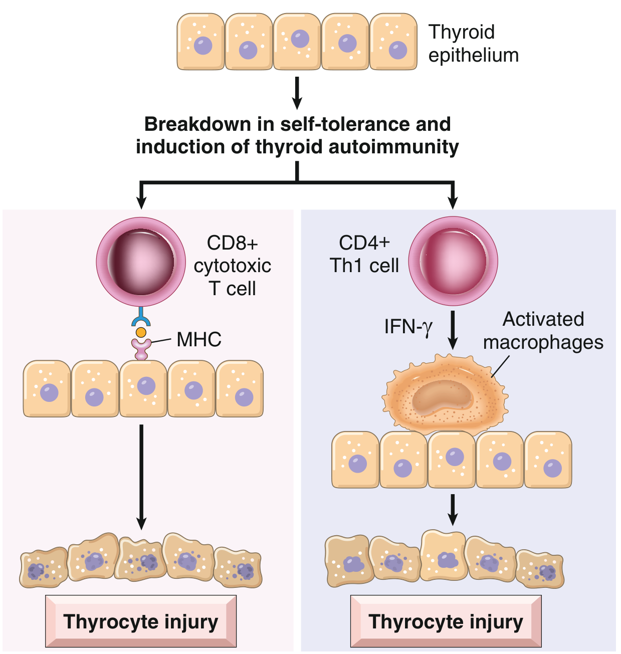
Fig. 18.4 - Pathogenesis of Hashimoto thyroiditis. Breakdown of immune tolerance to thyroid autoantigens results in progressive autoimmune destruction of thyrocytes by infiltrating cytotoxic T cells, locally released cytokines, and other mechanisms. (Robbins & Kumar Basic Pathology)
Four Major Immunologic Mechanisms of Thyrocyte Destruction
1. CD8+ Cytotoxic T Cell-Mediated Killing
- Activated CD4+ T (helper) cells recognize thyroid antigens and recruit CD8+ cytotoxic T cells to the gland
- These cytotoxic T cells directly kill thyroid follicular epithelial cells via MHC-I-restricted antigen recognition
- This is considered the primary effector mechanism
2. Cytokine-Mediated Cell Death (Th1 Pathway)
- Activated CD4+ Th1 cells secrete IFN-γ in the thyroid microenvironment
- IFN-γ recruits and activates macrophages
- Activated macrophages cause bystander damage to thyroid follicles
- This inflammatory milieu sustains progressive follicular destruction
3. Antibody-Mediated Mechanisms
Three main antibody targets:
- Anti-thyroid peroxidase (anti-TPO) antibodies - present in ~95% of patients
- Anti-thyroglobulin (anti-Tg) antibodies - present in ~60% of patients
- Anti-TSH receptor (anti-TSH-R) antibodies - present in ~60% (blocking type, contributing to hypothyroidism)
- Less commonly: anti-sodium/iodine symporter antibodies (~25%)
These antibodies may damage follicular cells via:
- Antibody-dependent cell-mediated cytotoxicity (ADCC) - NK cells kill antibody-coated thyrocytes
- Complement-dependent cytotoxicity - complement fixation leads to membrane attack
- TSH receptor blockade - blocking antibodies impair TSH signaling, reducing thyroid function
Note: It remains debated whether these antibodies are the primary cause or merely a consequence of thyroid injury. - Robbins & Kumar Basic Pathology, Schwartz's Principles of Surgery
4. Apoptosis (Programmed Cell Death)
- Fas-FasL interactions have been implicated as an additional mechanism of thyrocyte destruction
- Apoptosis of thyroid epithelial cells contributes to the progressive loss of functional parenchyma - Schwartz's Principles of Surgery, 11th Edition
Genetic Susceptibility
Hashimoto thyroiditis has a strong genetic component:
- ~40% concordance in monozygotic twins
- ~50% of asymptomatic siblings of affected patients have circulating antithyroid antibodies
Key susceptibility gene polymorphisms include:
| Gene | Function |
|---|---|
| CTLA4 (cytotoxic T lymphocyte-associated antigen-4) | Encodes a T-cell response inhibitor; loss-of-function favors autoimmunity |
| PTPN22 (protein tyrosine phosphatase non-receptor type 22) | Regulates T-cell activation thresholds |
| IL2RA (interleukin-2 receptor α chain, CD25) | Regulates Treg function and T-cell proliferation |
All three are regulators of T-cell responses, confirming that dysregulated T-cell immunity is the central defect. - Robbins, Cotran & Kumar Pathologic Basis of Disease
Environmental Triggers and Associations
- Excess iodine intake - associated with increased incidence of chronic thyroiditis
- Medications: interferon-α, lithium, amiodarone can precipitate or worsen thyroiditis
- Postpartum state - postpartum thyroiditis shares mechanistic parallels (TPOAb positive, preceded by thyrotoxic phase)
- Familial clustering and co-occurrence with Graves' disease and other autoimmune diseases (type 1 diabetes, pernicious anemia, Addison's disease)
Morphological Consequences of the Pathogenesis
The immune-mediated destruction produces characteristic histological changes:
- Diffuse lymphocytic infiltration - lymphocytes, plasma cells, macrophages with germinal center formation
- Hurthle (Oxyphil/Oncocytic) cell metaplasia - follicular epithelial cells develop abundant eosinophilic cytoplasm packed with mitochondria; a metaplastic response to chronic injury
- Follicular atrophy - destruction of normal follicular architecture
- Interstitial fibrosis - connective tissue replacement of destroyed parenchyma (especially in the fibrosing variant)
Unlike Riedel thyroiditis, the fibrosis in Hashimoto thyroiditis does not extend beyond the thyroid capsule. - Robbins, Cotran & Kumar Pathologic Basis of Disease
Summary Flow
Genetic predisposition (CTLA4/PTPN22/IL2RA polymorphisms)
+
Environmental trigger (iodine, drugs, viral?)
↓
Breakdown of Treg-mediated self-tolerance
↓
Autoreactive CD4+ T helper cell activation against thyroid antigens
↙ ↘
CD8+ CTL recruitment Th1 cytokines (IFN-γ)
(direct killing) → macrophage activation
+
Autoantibodies (anti-TPO, anti-Tg, anti-TSHR)
→ ADCC, complement, TSH-R blockade
+
Apoptosis (Fas-FasL)
↓
Progressive thyrocyte destruction → Hurthle cell metaplasia + fibrosis
↓
Hypothyroidism (in ~20-85% depending on population)
Sources:
- Robbins & Kumar Basic Pathology (p. 731) - Primary reference
- Robbins, Cotran & Kumar Pathologic Basis of Disease - Detailed molecular mechanisms
- Schwartz's Principles of Surgery, 11th Edition - Apoptosis and antibody targets
- Current Surgical Therapy, 14th Edition - Clinical correlations
Various types of thyroiditis
Finding Sources
Finding Sources
Good. Now let me read the most comprehensive sources in parallel - Robbins (Basic + Full), Harrison's, and the comprehensive Robbins block covering all thyroiditis types.
Reading File
Reading File
Reading File
Excellent content. Let me now get the subacute, Riedel's, painless/postpartum, and drug-induced thyroiditis sections.
Reading File
Reading File
Excellent. Let me get the Riedel's thyroiditis and drug-induced sections, plus fetch the clinical course image.
Reading File
Reading File
Loading Image
I now have comprehensive content from all major sources. Let me compile the full, well-organized answer.
Types of Thyroiditis
Thyroiditis refers to a diverse group of disorders characterized by some form of thyroid inflammation. They are best classified by onset/duration and etiology:
Classification Overview
| Type | Duration | Etiology | Thyroid Pain | Functional Outcome |
|---|---|---|---|---|
| Acute (suppurative) | Days-weeks | Bacterial/fungal infection | Severe | Usually euthyroid |
| Subacute granulomatous (de Quervain) | Weeks-months | Viral | Yes, severe | Transient hyper → hypo → recovery |
| Subacute lymphocytic (silent/painless) | Weeks-months | Autoimmune | No | Transient hyper → hypo → recovery |
| Postpartum thyroiditis | Weeks-months | Autoimmune | No | Transient hyper → hypo → often recovery |
| Hashimoto (chronic autoimmune) | Chronic, permanent | Autoimmune | No | Progressive hypothyroidism |
| Riedel's thyroiditis | Chronic | IgG4-related disease | No | Variable; may cause hypothyroidism |
| Drug-induced | Variable | Drugs (amiodarone, interferon, ICIs) | Sometimes | Variable |
| Radiation thyroiditis | Days-weeks after RAI | Radiation injury | Yes | Transient, then usually hypothyroid |
- Harrison's Principles of Internal Medicine, 22nd Edition; Current Surgical Therapy, 14th Edition
1. Acute (Suppurative) Thyroiditis
Etiology: Bacterial infection - most commonly Staphylococcus, Streptococcus, Enterobacter. Fungal (Aspergillus, Candida, Histoplasma, Pneumocystis) and mycobacterial infections can occur, especially in immunocompromised patients.
Predisposing factors:
- Children/young adults: piriform sinus fistula (remnant of 4th branchial pouch, predominantly left-sided)
- Elderly: pre-existing goiter, degenerating thyroid malignancy
Clinical features:
- Acute onset with fever, severe thyroid pain referred to the throat or ears
- Tender, asymmetric goiter; erythema over the gland
- Dysphagia, lymphadenopathy, systemic illness
- ESR and WBC elevated; thyroid function usually normal
Investigations: FNA showing polymorphonuclear leukocytes; Gram stain and culture guide therapy
Complications: Abscess formation, tracheal obstruction, septicemia, retropharyngeal abscess, mediastinitis, jugular venous thrombosis
Treatment: Antibiotics; surgical drainage of abscess if required
2. Subacute Granulomatous Thyroiditis (de Quervain / Viral / Giant Cell Thyroiditis)
Etiology: Triggered by viral infection. Viruses implicated: mumps, coxsackievirus, influenza, adenoviruses, echoviruses, SARS-CoV-2. Also reported after COVID-19 vaccination.
Peak incidence: 30-50 years; women affected 3-4x more than men
Pathogenesis: Virus-induced host tissue damage stimulates a cytotoxic T-lymphocyte response against thyroid antigens. Unlike autoimmune thyroiditis, the immune response is virus-initiated and not self-perpetuating, hence self-limited. Strong association with HLA-B35 haplotype.
Histology:
- Early: disrupted follicles, neutrophilic microabscesses
- Later: lymphocytes, activated histiocytes/macrophages, multinucleated giant cells engulfing colloid (characteristic)
- Resolution: fibrosis, then restoration of normal architecture
Clinical Course - Three Phases:
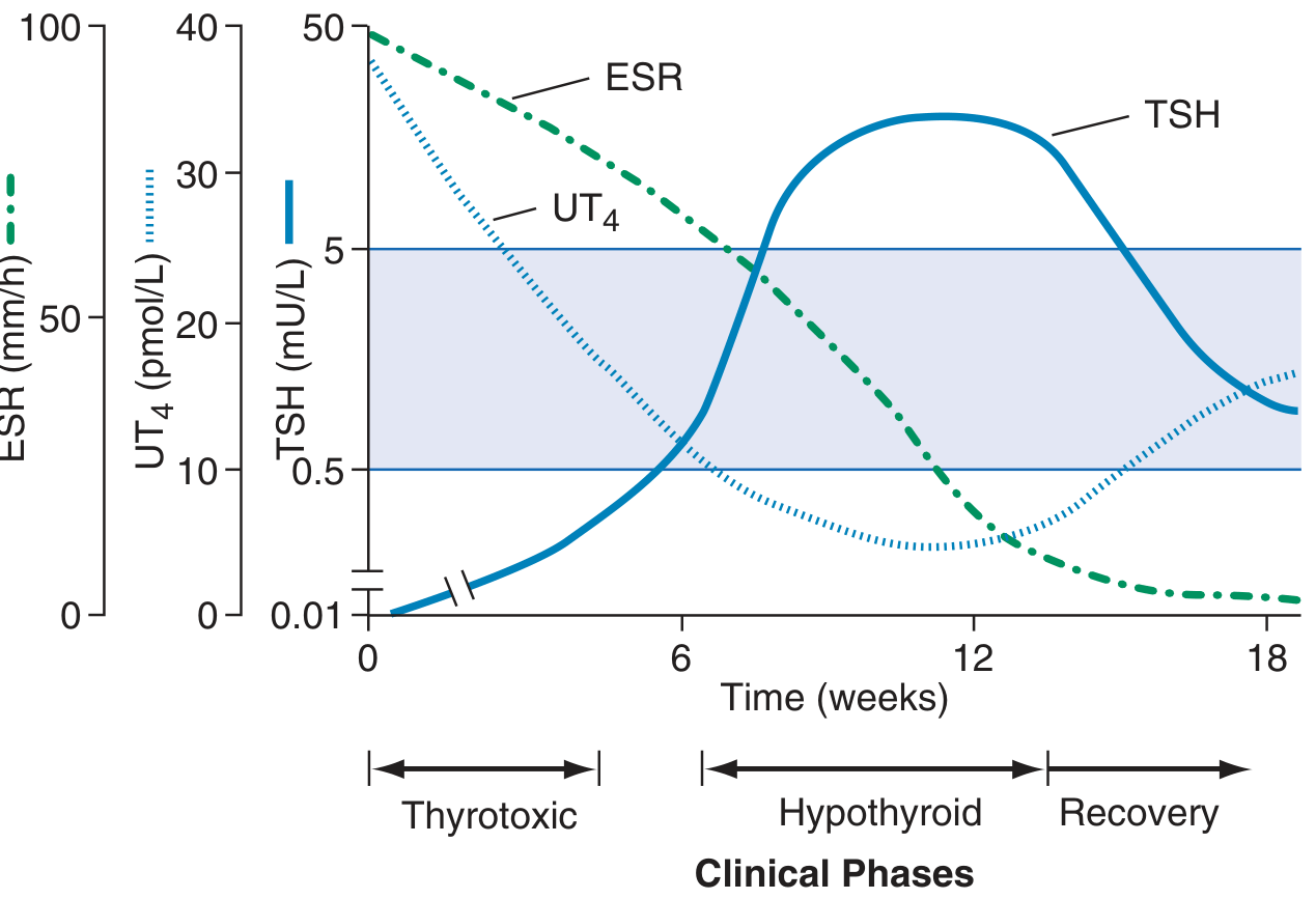
FIGURE 396-3 - Clinical course of subacute thyroiditis. ESR is initially very high; UT4 elevated then falls; TSH suppressed then rises. (Harrison's Principles of Internal Medicine, 22nd Ed.)
| Phase | Duration | T4/T3 | TSH | RAIU | ESR |
|---|---|---|---|---|---|
| Thyrotoxic | ~0-6 wks | ↑ | ↓ | Very low (<5%) | Very high |
| Hypothyroid | ~6-12 wks | ↓ | ↑ | Returns toward normal | Falling |
| Recovery | ~12-18+ wks | Normal | Normal | Normal | Normal |
Key features:
- Exquisitely tender goiter; pain referred to jaw or ear
- Malaise, fever, sore throat; preceded by URTI
- T4:T3 ratio lower than in Graves' disease
- Antithyroid antibodies usually negative
- ESR typically >50 mm/h (often >100)
Treatment:
- NSAIDs/aspirin for pain relief
- Corticosteroids for severe cases
- Beta-blockers for symptomatic thyrotoxicosis
- Thyroid hormone replacement if symptomatic hypothyroidism
- Self-limited in >90%; ~15% develop permanent hypothyroidism
3. Hashimoto Thyroiditis (Chronic Autoimmune / Chronic Lymphocytic Thyroiditis)
Etiology: Autoimmune - breakdown of self-tolerance to thyroid antigens (thyroglobulin, thyroid peroxidase, TSH-R)
Epidemiology: Most common cause of hypothyroidism in iodine-sufficient regions. Prevalence peaks 45-65 years; F:M = 10-20:1. Most common inflammatory thyroid disorder worldwide.
Pathogenesis (summary):
- Genetic predisposition: CTLA4, PTPN22, IL2RA polymorphisms
- Treg dysfunction → autoreactive CD4+ and CD8+ T-cell activation
- CD8+ cytotoxic T cells directly kill thyrocytes
- CD4+ Th1 cells → IFN-γ → macrophage activation and follicular damage
- Autoantibodies (anti-TPO ~95%, anti-Tg ~60%) → ADCC and complement-dependent killing
- Apoptosis (Fas-FasL pathway) contributes additionally
Histology:
- Diffuse lymphoplasmacytic infiltration with germinal center formation
- Hürthle (oxyphil/oncocytic) cell metaplasia of follicular epithelium
- Follicular atrophy
- Variable interstitial fibrosis (does not extend beyond the capsule)
Clinical features:
- Painless diffuse goiter (firm, rubbery)
- Hypothyroidism (slow onset, in ~20-85%)
- "Hashitoxicosis" - transient thyrotoxic phase in some patients
- Goiter may cause compressive symptoms
- Associated with other autoimmune conditions (T1DM, pernicious anemia, Addison's)
- Elevated anti-TPO antibodies (hallmark laboratory finding)
Variants:
- Goitrogenic form (classic - enlarged gland)
- Atrophic form (no goiter, directly hypothyroid)
- Fibrosing variant (extensive fibrosis; can mimic Riedel's thyroiditis)
Complications:
- Permanent hypothyroidism
- Increased risk of thyroid lymphoma (extranodal marginal zone B-cell lymphoma)
- Possible association with papillary thyroid carcinoma (controversial)
4. Silent (Painless) Thyroiditis / Chronic Lymphocytic Thyroiditis
Etiology: Presumed autoimmune - most patients have anti-TPO antibodies or family history of autoimmune disease
Special form - Postpartum thyroiditis: Occurs in ~5% of postpartum females; peaks at ~6 weeks after delivery coinciding with immune rebound after pregnancy-induced immune tolerance
Histology:
- Lymphocytic infiltration with germinal centers
- Patchy follicular disruption and collapse
- No fibrosis, no Hürthle cell metaplasia (distinguishes it from Hashimoto thyroiditis)
- Thyroid grossly normal or mildly enlarged
Clinical features:
- Painless goiter (key distinguishing feature)
- Mild transient hyperthyroidism → hypothyroidism → recovery (similar pattern to de Quervain but without pain)
- RAIU low during thyrotoxic phase (low uptake differentiates from Graves')
- ~30% may evolve into permanent hypothyroidism
5. Riedel's Thyroiditis (Invasive Fibrous Thyroiditis)
Etiology: Now recognized as a manifestation of IgG4-related disease - associated with fibrosis and tissue infiltration by IgG4-positive plasma cells
Characteristics:
- Rare, chronic condition
- Extensive fibrosis involving the thyroid AND contiguous neck structures (muscles, nerves, vessels, trachea, esophagus)
- Fibrosis extends beyond the thyroid capsule (key distinction from Hashimoto's fibrosing variant)
- Hard, fixed thyroid mass clinically simulates thyroid carcinoma
- May be associated with fibrosis at other sites: retroperitoneum (retroperitoneal fibrosis), mediastinum, orbit, bile ducts
Clinical features:
- Progressive compressive symptoms: dyspnea, dysphagia, hoarseness (recurrent laryngeal nerve involvement)
- Hypothyroidism if sufficient thyroid parenchyma is destroyed
- Hypoparathyroidism if parathyroids are encased
Treatment: Corticosteroids (first line for IgG4-RD); tamoxifen; surgical decompression of trachea if needed
6. Drug-Induced Thyroiditis
Several drugs can cause thyroiditis through different mechanisms:
| Drug | Mechanism | Type of Thyroiditis |
|---|---|---|
| Amiodarone | Excess iodine + direct cytotoxic effect | Type 1 (iodine-induced) or Type 2 (destructive/thyroiditis) |
| Interferon-α | Immune activation | Autoimmune (can trigger Hashimoto or Graves') |
| Lithium | Inhibits thyroid hormone release | Hypothyroidism/thyroiditis |
| Tyrosine kinase inhibitors (sunitinib, sorafenib) | Multiple mechanisms | Hypothyroidism |
| Immune checkpoint inhibitors (anti-PD1, anti-CTLA4) | Immune dysregulation | Autoimmune thyroiditis; hyper then hypo pattern |
7. Radiation Thyroiditis
- Occurs days to weeks after radioactive iodine (¹³¹I) treatment or external beam radiation to the neck
- Characterized by acute inflammation and tenderness of the thyroid
- Usually transient; thyroid function eventually declines toward hypothyroidism
- FNA not typically needed (diagnosis is clinical in context of recent radiation)
Comparative Summary: Key Distinguishing Features
| Feature | Acute (Suppurative) | Subacute (de Quervain) | Hashimoto | Silent/Postpartum | Riedel |
|---|---|---|---|---|---|
| Pain | Severe | Severe, referred | None | None | None |
| Fever | Yes | Yes (low-grade) | No | No | No |
| Etiology | Infection | Viral | Autoimmune | Autoimmune | IgG4-RD |
| Anti-TPO | Negative | Negative | Positive (95%) | Often positive | Variable |
| RAIU | Normal | Very low | Low/normal | Low | Variable |
| ESR | High | Very high | Normal/mildly ↑ | Normal | Variable |
| WBC | High | Normal or mild ↑ | Normal | Normal | Normal |
| Histology | PMN infiltrate | Granulomas, giant cells | Lymphocytes, germinal centers, Hürthle cells | Lymphocytes, no Hürthle cells | Dense fibrosis |
| Outcome | Resolution | Self-limited; 15% permanent hypo | Permanent hypothyroidism | 30% permanent hypo | Progressive; compressive |
Sources:
- Harrison's Principles of Internal Medicine, 22nd Edition - Primary reference for clinical classification
- Robbins, Cotran & Kumar Pathologic Basis of Disease - Histopathology and pathogenesis
- Robbins & Kumar Basic Pathology - Key concepts
- Current Surgical Therapy, 14th Edition - Clinical variants and surgical management
Grave disease
Finding Sources
Finding Sources
Reading File
Reading File
Finding Sources
Reading File
Reading File
Finding Sources
Reading File
Reading File
Loading Image
Let me get the Harrison's treatment section and the ophthalmopathy image.
Reading File
Loading Image
I now have comprehensive content from all major sources. Let me compile the full answer.
Graves' Disease
Graves' disease is the most common cause of endogenous hyperthyroidism, first described by Robert Graves in 1835 as "violent and long continued palpitations in females" associated with thyroid enlargement. It affects 1.5-2% of women in the United States and has a peak incidence between 20 and 50 years of age. Women are affected 5-10 times more frequently than men.
The Classic Triad
- Thyrotoxicosis - with diffuse goiter
- Infiltrative ophthalmopathy - exophthalmos/proptosis
- Infiltrative dermopathy - pretibial myxedema (minority of patients)
Pathogenesis
Core Mechanism: TSH Receptor Autoantibodies
Graves' disease is an autoimmune disorder driven by thyroid-stimulating immunoglobulins (TSI), IgG autoantibodies against the TSH receptor. These are produced by lymphocytes in the thyroid gland, bone marrow, and lymph nodes.
- TSI (~90% of patients): Binds to the TSH-R and mimics TSH action - stimulates adenylyl cyclase → ↑cAMP → continuous thyroid hormone synthesis and release, independent of TSH
- TSH-R blocking antibodies: Also present in some patients; cause hypothyroidism. Coexistence of stimulating and blocking antibodies in the same patient explains occasional episodes of hypothyroidism
- TPO and Tg antibodies: Present in up to 80% of patients (coexisting lymphocytic thyroiditis); no direct correlation between TSI level and thyroid hormone levels
Because the TSH-R is continuously stimulated by autoantibodies (rather than by the physiologic TSH pulse), the feedback mechanism is bypassed - TSH falls to undetectable levels while thyroid hormone remains persistently elevated.
Genetic Susceptibility
| Factor | Detail |
|---|---|
| Monozygotic twin concordance | 30-40% |
| Dizygotic twin concordance | <5% |
| Susceptibility genes | CTLA4, PTPN22, IL2RA, TSHR gene variants (identified by GWAS) |
These overlap with Hashimoto thyroiditis risk genes, placing both on a continuum of autoimmune thyroid disease.
Pathogenesis of Ophthalmopathy
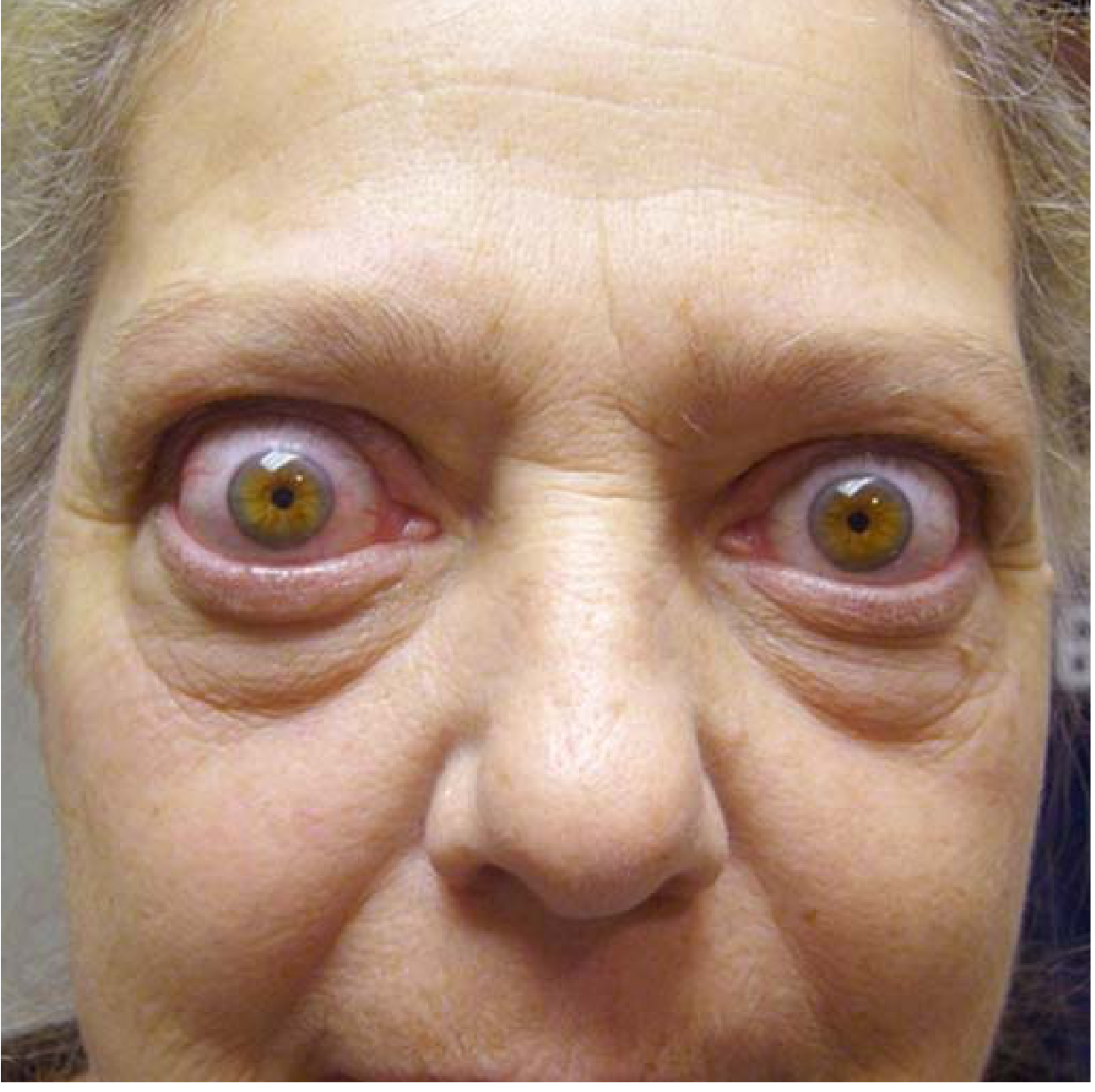
Graves' ophthalmopathy: bilateral exophthalmos with wide staring gaze, periorbital edema, and conjunctival injection. (Robbins & Kumar Basic Pathology)
The TSH receptor is expressed not only in the thyroid but also on orbital fibroblasts and fat cells. The mechanism of retro-orbital expansion:
- Activated CD4+ T cells infiltrate the retro-orbital space and secrete cytokines (IFN-γ, TNF, IL-1)
- Cytokines stimulate orbital fibroblasts → proliferation and synthesis of hydrophilic glycosaminoglycans (hyaluronic acid, chondroitin sulfate) that trap water
- Progressive edema and swelling of extraocular muscles
- Fatty infiltration (increased adipocytes) expands retrobulbar volume
- Mononuclear cell (predominantly T-cell) infiltration of connective tissue
- Late: irreversible fibrosis of extraocular muscles
Additional mechanism: aberrant IGF-1 receptor signaling on orbital fibroblasts amplifies fibroblast activation - the basis for teprotumumab (anti-IGF-1R monoclonal antibody), a targeted treatment for Graves' ophthalmopathy.
Result: Increased intraorbital pressure → proptosis, diplopia, optic neuropathy, corneal injury. Ophthalmopathy may progress independently of thyroid status - can occur even in hypothyroid patients and may worsen despite successful treatment of thyrotoxicosis.
Pathogenesis of Dermopathy
Same mechanism as ophthalmopathy - glycosaminoglycan deposition and lymphocytic infiltration, but affecting the dermis, most commonly over the shins (pretibial myxedema).
Morphology
Thyroid Gland
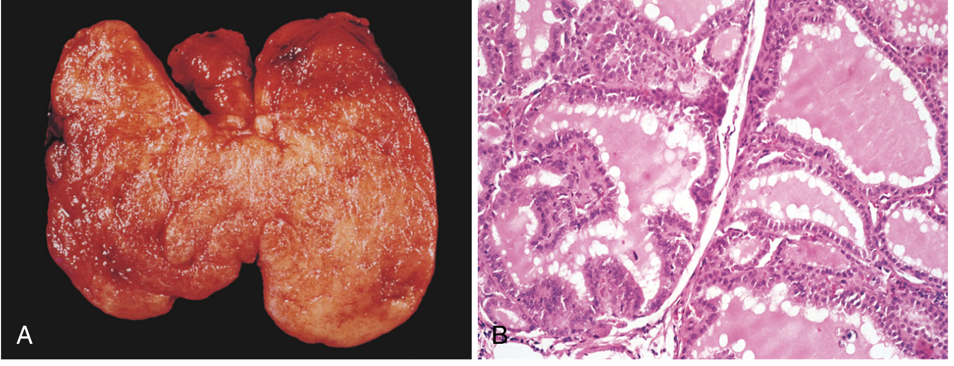
Fig. 24.13 - Graves' disease. (A) Diffuse symmetric enlargement with beefy deep-red parenchyma. (B) Follicles lined by tall columnar epithelium projecting as papillae into the lumen; pale scalloped colloid. (Robbins, Cotran & Kumar Pathologic Basis of Disease)
Gross:
- Symmetrically enlarged (can exceed 80 g; normal ~25 g)
- Soft, beefy deep-red parenchyma; intact capsule
Histology (untreated):
- Follicular epithelial cells are tall and crowded → form small papillae projecting into lumen (note: lack fibrovascular cores, distinguishing from papillary carcinoma)
- Colloid is pale with scalloped margins (being actively resorbed)
- Lymphoid infiltrates (predominantly T cells) with scattered germinal centers throughout the interstitium
Effect of treatment on morphology:
- Iodine pre-treatment: causes involution of epithelium and colloid accumulation (blocks thyroglobulin secretion)
- Propylthiouracil: exaggerates epithelial hypertrophy by stimulating TSH secretion
Extrathyroidal changes:
- Generalized lymphoid hyperplasia; thymic enlargement (especially in young patients)
- Cardiac hypertrophy; ischemic changes if pre-existing coronary artery disease
- Orbital: edema, glycosaminoglycan deposition, T-cell infiltration, muscle fibrosis
- Skin: dermal thickening from glycosaminoglycan deposition and lymphocytic infiltration
Clinical Features
Symptoms and Signs of Thyrotoxicosis
| Symptoms | Signs |
|---|---|
| Hyperactivity, irritability, dysphoria | Tachycardia; atrial fibrillation (elderly) |
| Heat intolerance and sweating | Tremor (fine, postural) |
| Palpitations | Goiter (diffuse, firm, with bruit/thrill) |
| Fatigue and weakness | Warm, moist skin; palmar erythema |
| Weight loss with increased appetite | Proximal myopathy |
| Diarrhea, increased stool frequency | Lid retraction or lag |
| Polyuria | Onycholysis |
| Oligomenorrhea, loss of libido | Gynecomastia (in men) |
Note: In elderly patients, thyrotoxicosis may present with only fatigue and weight loss ("apathetic thyrotoxicosis"), and can be mistaken for depression.
Graves-Specific Features
1. Diffuse goiter - firm, smooth; bruit/thrill audible at inferolateral margins due to increased vascularity
2. Graves' ophthalmopathy (thyroid eye disease, TED):
- Occurs in ~50% of patients; in 10%, may occur without clinical hyperthyroidism
- Lid retraction → staring appearance (also present in other thyrotoxicoses via sympathetic overactivity)
- Proptosis/exophthalmos (forward displacement of eyeball)
- Periorbital and conjunctival edema, chemosis
- Diplopia (restrictive extraocular myopathy)
- Optic neuropathy (severe cases) → vision loss
- Corneal exposure and injury
- Onset is within the year before or after diagnosis of thyrotoxicosis in 75% of patients
3. Pretibial myxedema (thyroid dermopathy):
- Scaly, thickened, indurated skin over the shins
- May appear as pigmented papules or nodules with an "orange peel" texture
- Present in a minority of patients
4. Thyroid acropachy (~0.1-1%): Digital clubbing + soft tissue swelling of digits + periosteal new bone formation
Cardiovascular Complications
- Sinus tachycardia (most common), supraventricular tachycardia, atrial fibrillation
- Bounding pulse, widened pulse pressure
- High-output cardiac failure in elderly or those with pre-existing disease
- Atrial fibrillation converts to sinus rhythm in ~75% after achieving euthyroidism
Other Systemic Effects
- Bone: Increased bone resorption → osteopenia; mild hypercalcemia (up to 20%); increased fracture risk
- Reproductive: Oligomenorrhea/amenorrhea; impaired sexual function; gynecomastia
- Hair/skin: Fine hair; diffuse alopecia (up to 40%, may persist months after treatment)
- Neuromuscular: Hyperreflexia, proximal myopathy; hypokalemic periodic paralysis (especially in Asian males)
Laboratory Findings
| Test | Result | Explanation |
|---|---|---|
| Free T4, free T3 | ↑↑ | Continuous stimulation by TSI |
| TSH | ↓ (often undetectable) | Negative feedback from ↑ thyroid hormones |
| TSI / TRAb | ↑ (positive) | Pathognomonic for Graves' |
| Anti-TPO, Anti-Tg | Often positive (~80%) | Coexisting autoimmunity |
| Radioactive iodine uptake (RAIU) | Diffusely increased | Ongoing TSH-R stimulation |
RAIU is the key scan finding - diffuse uniform uptake distinguishes Graves' from destructive thyroiditis (low uptake) and toxic nodular goiter (patchy/focal uptake).
Treatment
Three modalities reduce thyroid hormone production or thyroid mass:
1. Antithyroid Drugs (Thionamides)
- Methimazole (preferred) or propylthiouracil (PTU) (preferred in first trimester pregnancy, thyroid storm)
- Mechanism: Block thyroid peroxidase → inhibit thyroid hormone synthesis. PTU also blocks peripheral T4→T3 conversion
- PTU has additional advantage of blocking T4→T3 conversion (useful in thyroid storm)
- Remission (~50%) may be achieved after 12-18 months of therapy
- Beta-blockers (propranolol, atenolol) control adrenergic symptoms (tachycardia, tremor, anxiety)
2. Radioiodine Ablation (¹³¹I)
- Destroys thyroid tissue; most common definitive therapy in the US
- Eventually leads to hypothyroidism requiring lifelong thyroid hormone replacement
- Contraindicated in pregnancy and breastfeeding; may worsen pre-existing severe ophthalmopathy
3. Thyroidectomy
- Indicated for: large compressive goiters, patients who cannot receive radioiodine, those failing antithyroid drugs, suspected concurrent malignancy, young patients or those planning pregnancy
- Pre-operative iodine (Lugol's solution) is given to reduce gland vascularity
Treatment of Ophthalmopathy
- Smoking cessation (smoking is a major risk factor for worsening TED)
- Corticosteroids for active/moderate-severe disease
- Orbital radiation
- Teprotumumab (anti-IGF-1R monoclonal antibody): targeted therapy that reduces TSH-R/IGF-1R complex signaling in orbital fibroblasts; approved for active Graves' ophthalmopathy
- Surgical orbital decompression (severe cases)
Relationship to Hashimoto Thyroiditis
Graves' disease and Hashimoto thyroiditis represent opposite ends of an autoimmune thyroid continuum:
- Both involve autoimmune reactions to thyroid antigens with lymphocytic infiltrates and germinal centers
- Both share genetic susceptibility genes (CTLA4, PTPN22, IL2RA)
- Hyperthyroidism can supervene on Hashimoto thyroiditis ("Hashitoxicosis"), and Graves' patients can spontaneously develop hypothyroidism
- Both diseases co-occur in families; both associated with systemic autoimmune diseases (SLE, pernicious anemia, type 1 diabetes, Addison's disease)
Sources:
- Robbins, Cotran & Kumar Pathologic Basis of Disease - Pathogenesis and morphology (primary reference)
- Robbins & Kumar Basic Pathology - Key concepts and histology
- Harrison's Principles of Internal Medicine, 22nd Edition - Clinical features, ophthalmopathy mechanism, treatment
- Cummings Otolaryngology - Ophthalmopathy and surgical management
Classification and explanation of thyroid neoplasms take reference from Robbins
Finding Sources
Finding Sources
Finding Sources
Reading File
Good - found the exact section. Now let me read all the thyroid neoplasm content from both Robbins texts in parallel.
Reading File
Reading File
Excellent - the Robbins Basic Pathology block has all the neoplasm content. The read from offset 2089 got cut off - let me continue reading from where it stopped, and also pull the Robbins Cotran full neoplasm section in parallel.
Reading File
Reading File
The Robbins Cotran (9780443264528) block has the neoplasm content starting around line 1282+. Let me read it plus continue with the Robbins Basic Pathology neoplasm content from where it was truncated.
Reading File
Reading File
Excellent - rich content. Let me continue reading both sources for the full carcinoma descriptions (papillary, follicular, anaplastic, medullary) and fetch key images.
Reading File
Reading File
Let me now fetch the key histology images and read the remaining medullary carcinoma and Robbins Cotran content.
Loading Image
Reading File
Reading File
I have comprehensive content from both Robbins texts. Let me fetch the papillary carcinoma histology image.
Loading Image
I now have all the content needed for a comprehensive answer from Robbins. Let me compile the full response.
Classification and Explanation of Thyroid Neoplasms
(Based on Robbins & Kumar Basic Pathology and Robbins, Cotran & Kumar Pathologic Basis of Disease)
Overview and Classification
Thyroid neoplasms are broadly divided into benign (adenomas) and malignant (carcinomas). Most thyroid cancers (except medullary carcinoma) arise from the follicular epithelium. Benign neoplasms outnumber thyroid carcinomas by approximately 10:1.
THYROID NEOPLASMS
│
├── BENIGN
│ └── Follicular Adenoma
│ ├── Nonfunctional (cold nodule)
│ └── Toxic adenoma (functional/warm nodule)
│
└── MALIGNANT (Carcinomas)
│
├── Derived from FOLLICULAR EPITHELIUM (~95%)
│ ├── 1. Papillary Thyroid Carcinoma (PTC) - >85%
│ ├── 2. Follicular Thyroid Carcinoma (FTC) - 5-15%
│ ├── 3. Noninvasive Follicular Thyroid Neoplasm with
│ │ Papillary-like Nuclear Features (NIFTP)
│ ├── 4. Poorly Differentiated Carcinoma
│ └── 5. Anaplastic (Undifferentiated) Carcinoma - <5%
│
└── Derived from PARAFOLLICULAR C CELLS (~5%)
└── 6. Medullary Thyroid Carcinoma (MTC)
Molecular Pathogenesis Overview
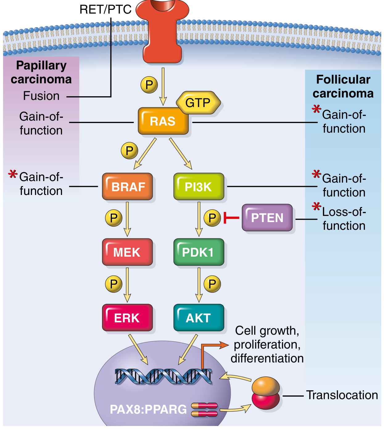
FIG. 18.11 - Genetic alterations in follicular cell-derived thyroid malignancies. Red asterisks mark the most common mutations. Papillary carcinoma activates predominantly the MAPK pathway (BRAF, RAS, RET/PTC fusions); follicular carcinoma activates predominantly the PI3K/AKT pathway (RAS, PIK3CA gain-of-function, PTEN loss-of-function) and PAX8:PPARG translocation. (Robbins & Kumar Basic Pathology)
Part I: Benign Neoplasm - Follicular Adenoma
Definition and General Features
Adenomas of the thyroid are typically discrete, solitary neoplasms derived from follicular epithelium. Most are nonfunctional; a small minority produce thyroid hormones (toxic adenomas), causing clinically apparent thyrotoxicosis.
Follicular adenomas are generally not forerunners of carcinoma, though shared genetic alterations raise the possibility that a subset of follicular carcinomas may arise from preexisting adenomas.
Pathogenesis
- Toxic adenomas: Somatic gain-of-function mutations causing constitutive activation of the TSH receptor signaling pathway:
- TSHR mutations (most common) - stimulate thyrocytes to secrete hormone independent of TSH
- GNAS mutations (Gα-s subunit, less common)
- Present in >50% of toxic adenomas
- Nonfunctional adenomas: Mutations in RAS (<20%) and PTEN - same mutations also seen in follicular carcinoma
Morphology
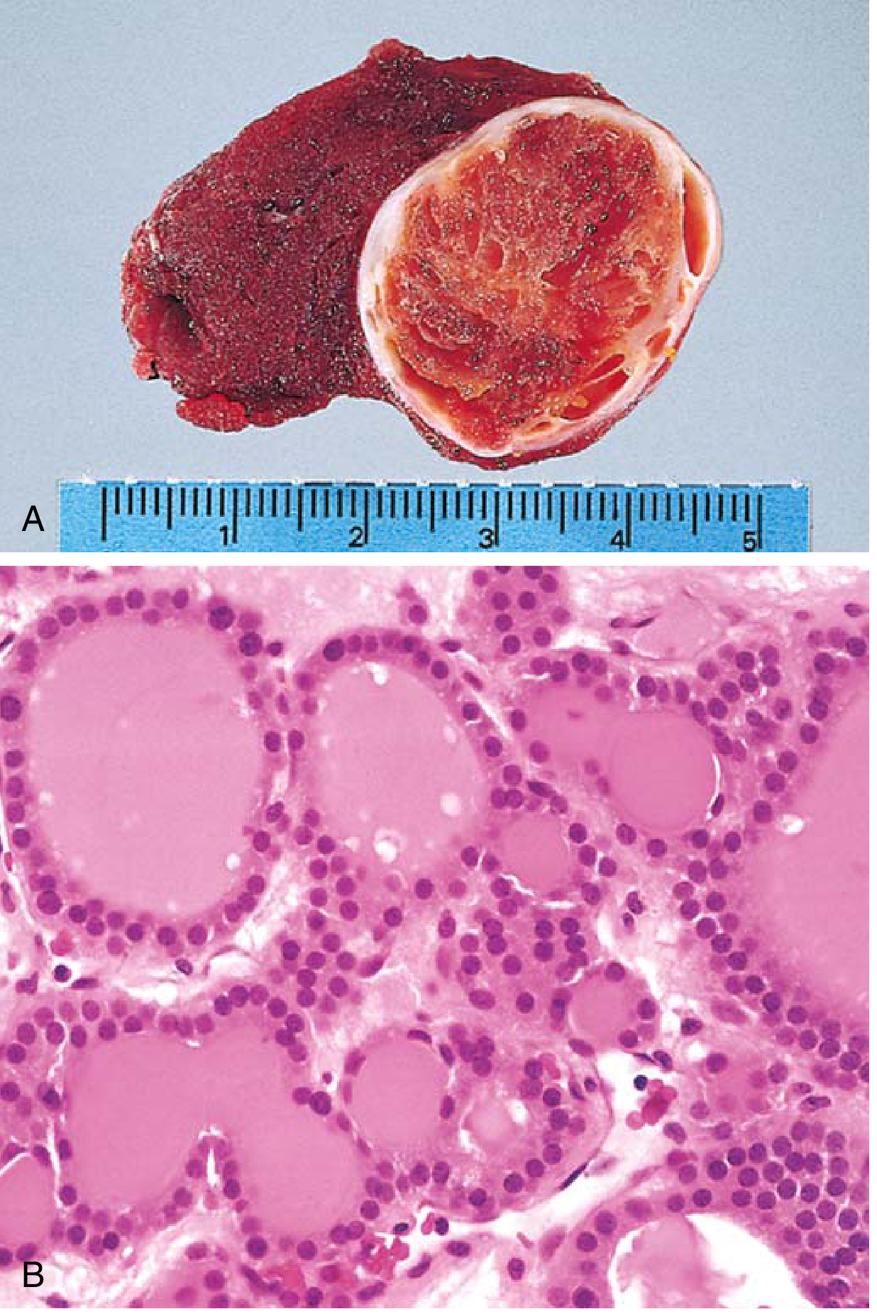
- Gross: Solitary, spherical, well-circumscribed, compresses adjacent thyroid tissue; intact well-defined fibrous capsule
- Microscopy:
- Uniform follicles containing colloid; little variation in cell size, shape, or nuclear morphology
- Mitotic figures rare
- Hürthle (oxyphil) cell change may occur (brightly eosinophilic granular cytoplasm)
- Hallmark: complete intact encapsulation - NO capsular or vascular invasion (this is the critical distinction from follicular carcinoma)
Clinical Features
- Painless solitary nodule discovered on routine examination
- Nonfunctional: cold nodule on radionuclide scan (takes up less iodine than normal parenchyma)
- Toxic adenoma: warm or hot nodule on scan; associated thyrotoxicosis
- Treatment: surgical excision
Part II: Malignant Neoplasms - Thyroid Carcinomas
Frequency
| Type | Frequency |
|---|---|
| Papillary thyroid carcinoma | >85% |
| Follicular thyroid carcinoma | 5-15% |
| Anaplastic (undifferentiated) carcinoma | <5% |
| Medullary thyroid carcinoma | ~5% |
Risk factors for malignancy in a thyroid nodule:
- Solitary nodule (vs. multinodular)
- Patient age <30 years or male sex
- History of ionizing radiation exposure
- Cold (non-functioning) nodule on radionuclide scan
1. Papillary Thyroid Carcinoma (PTC)
Frequency: >85% of all thyroid carcinomas - the most common thyroid malignancy
Pathogenesis
Activation of the MAP kinase (MAPK) pathway is the central molecular event. Three mutually exclusive mechanisms:
| Mechanism | Frequency | Notes |
|---|---|---|
| BRAF point mutation (V600E) | 40-65% | Most common; activates MAPK via B-Raf kinase |
| RAS oncogenic mutations | 10-30% | Also seen in follicular carcinoma |
| RET/PTC gene fusions (chromosomal translocations) | 10-20% | RET gene fused to PTC1/PTC3; dramatically increased post-Chernobyl |
| NTRK1 gene fusions | Smaller subset | Similar to RET/PTC rearrangements |
Because these mutations all activate MAPK signaling, they are mutually exclusive - rarely occur in the same tumor.
Key risk factor: Exposure to ionizing radiation, particularly in the first two decades of life. Marked increase in papillary carcinomas was documented in children exposed after the Chernobyl nuclear disaster (1986).
Morphology
Gross:
- Solitary or multifocal lesions
- May be well-circumscribed and encapsulated OR infiltrate adjacent parenchyma with ill-defined margins
- Papillary foci visible on cut surface
Microscopy - Three Hallmarks:
-
Branching papillae - fibrovascular stalks covered by single-to-multiple layers of cuboidal epithelial cells (crowded, sometimes pleomorphic)
-
Ground-glass (Orphan Annie eye) nuclei - finely dispersed chromatin imparts an optically clear/empty appearance. In addition:
- Intranuclear pseudoinclusions - invaginations of cytoplasm into nucleus
- Nuclear grooves - linear longitudinal grooves in the nuclear membrane
- These nuclear features are diagnostic of PTC even in the absence of papillary architecture
-
Psammoma bodies - concentrically calcified laminated structures, usually within cores of papillae. Almost never found in follicular or medullary carcinomas.
Lymphoid infiltrates may be present; may also contain follicular areas.
Clinical Features
- Most common incidentally detected thyroid mass (greatly increased detection with widespread use of ultrasound)
- Often presents as a thyroid nodule or neck lymphadenopathy
- Lymphatic spread is characteristic - metastases to regional cervical lymph nodes are common (but do not worsen prognosis significantly)
- Hematogenous spread less common
- Cold nodule on scintigraphy
- Elevated thyroglobulin (used for postoperative monitoring)
Prognosis: Excellent - >90% 20-year survival rate. The overall favorable outcome underscores the low mortality despite increasing detection rates.
Treatment: Total thyroidectomy + radioactive iodine (¹³¹I) ablation for residual/metastatic disease + TSH suppression therapy (levothyroxine)
2. Follicular Thyroid Carcinoma (FTC)
Frequency: 5-15% of thyroid carcinomas
Pathogenesis
Frequent mutations in the PI3K/AKT signaling pathway and RAS:
| Alteration | Detail |
|---|---|
| RAS mutations | Gain-of-function; most common |
| PIK3CA mutations | Gain-of-function → activated PI3K |
| PTEN mutations | Loss-of-function → loss of PI3K inhibition |
| PAX8/PPARG fusion | Translocation fusing PAX8 (thyroid transcription factor) to PPARG (nuclear hormone receptor for terminal differentiation) |
Iodine deficiency is linked with a higher frequency of follicular carcinomas (mechanism unknown).
Morphology - Key Distinction from Adenoma
The only reliable criterion distinguishing follicular carcinoma from follicular adenoma is:
Capsular invasion and/or vascular invasion - adenomas have none; carcinomas must demonstrate at least one.
Types based on invasion:
- Minimally invasive FTC: Limited capsular penetration; excellent prognosis (>90% 10-year survival)
- Widely invasive FTC: Extensive vascular and soft tissue invasion; 50% mortality within 10 years
Microscopy:
- Uniform follicles with colloid (resembles adenoma - diagnosis CANNOT be made on cytology alone)
- On FNA: indeterminate (cannot distinguish from adenoma) - requires surgical excision
- Capsular invasion: Tumor cells penetrate through (not just indent) the fibrous capsule
- Vascular invasion: Tumor cells within endothelium-lined vascular spaces
Clinical Features
- Slowly enlarging painless nodule; cold nodule on scintigraphy
- Hematogenous spread is characteristic (NOT lymphatic) - metastases to bone, lungs, liver, brain
- Regional lymph nodes typically NOT involved (unlike PTC)
- Rarely, well-differentiated lesions may take up radioactive iodine (warm nodule)
- Serum thyroglobulin used for monitoring recurrence after total thyroidectomy
Treatment: Total thyroidectomy + radioactive iodine + TSH suppression (levothyroxine)
3. NIFTP - Noninvasive Follicular Thyroid Neoplasm with Papillary-like Nuclear Features
A distinct category introduced after reclassification of previously over-diagnosed tumors:
- Entirely follicular growth pattern (no papillae) but shows PTC-like nuclear features (enlarged nuclei, chromatin clearing, membrane irregularities)
- No invasion (no capsular, vascular, or intrathyroidal invasion) - if present, reclassified as "invasive encapsulated follicular variant of PTC"
- Historically called "noninvasive encapsulated follicular variant of PTC"
- Changed because: retrospective studies showed essentially zero risk of recurrence or metastasis → removing "carcinoma" from the name avoids unnecessary overtreatment
- Driven by RAS mutations (similar to follicular adenoma/carcinoma)
4. Poorly Differentiated and Anaplastic (Undifferentiated) Thyroid Carcinoma
Frequency: Cumulatively <5% of thyroid tumors, but with near 100% mortality for anaplastic type
Pathogenesis
- Arise de novo or, more commonly, by progression from a well-differentiated PTC or FTC (dedifferentiation/transformation)
- Retain driver mutations from precursor tumors (e.g., RAS, PIK3CA) PLUS additional mutations:
- TP53 loss-of-function mutations - most important, believed to drive anaplastic transformation
- Molecular alterations: RAS, BRAF, PIK3CA, TP53
Morphology
Poorly differentiated carcinoma:
- Tumor necrosis and/or increased mitotic activity
- Solid, insular (nested), or trabecular growth patterns
- Still retains some thyroid follicular cell markers
Anaplastic carcinoma:
- No resemblance to follicular cells - loss of thyroid follicular differentiation markers
- Variable morphology:
- Epithelioid (may resemble squamous cell carcinoma)
- Spindled (resembles sarcoma)
- Pleomorphic giant cells
- Foci of residual papillary or follicular carcinoma may be present (evidence of origin)
Clinical Features
- Rapidly enlarging bulky neck mass - usually the presenting feature
- Symptoms from invasion: dyspnea, dysphagia, hoarseness, cough
- Disease usually spread beyond thyroid into adjacent neck structures or metastasized to lungs at presentation
- Anaplastic carcinoma: one of the most aggressive human cancers - death in <1 year in most cases
- Poorly differentiated carcinomas fare somewhat better with surgery, external beam radiotherapy, and radioactive iodine
5. Medullary Thyroid Carcinoma (MTC)
Frequency: ~5% of thyroid carcinomas
Origin: Parafollicular C cells (neuroendocrine cells derived from neural crest) - NOT from follicular epithelium. This is a fundamental distinction.
Pathogenesis
- RET proto-oncogene activating mutations are central to both familial and sporadic MTC:
- Familial (30%): Germline RET mutations → MEN-2A, MEN-2B, or familial MTC without MEN
- Sporadic (70%): Acquired somatic RET mutations (~50% of sporadic cases)
- RET tyrosine kinase receptor is constitutively activated → unregulated cell proliferation of C cells
Sporadic MTC: Peak incidence in 5th-6th decades; usually solitary
Familial/MEN-associated MTC: Bilateral and multicentric; younger patients (including children in MEN-2B); C-cell hyperplasia is the precursor lesion
Morphology
Gross:
- Sporadic: solitary nodule
- Familial: bilateral, multicentric
- Larger lesions: areas of necrosis and hemorrhage, may extend through capsule; pale gray to tan, infiltrative
Microscopy:
- Polygonal to spindle-shaped cells arranged in nests, trabeculae, or even gland-like structures
- Amyloid deposits in stroma - derived from altered calcitonin polypeptides - DISTINCTIVE FEATURE
- Calcitonin demonstrable by immunohistochemistry within tumor cells AND in stromal amyloid
- Electron microscopy: membrane-bound electron-dense granules (neuroendocrine secretory granules)
- Familial cases: multicentric C-cell hyperplasia in surrounding thyroid parenchyma (precursor lesion) - not seen in sporadic lesions
Clinical Features
- Sporadic: neck mass ± compressive symptoms (dysphagia, hoarseness)
- Hormonal syndromes: diarrhea (VIP secretion), carcinoid-like features (serotonin), Cushing's syndrome (ACTH secretion)
- Serum calcitonin: key diagnostic and monitoring marker
- RET mutation screening of relatives enables early detection in familial cases
- MEN-2 kindred carriers of RET mutations are offered prophylactic thyroidectomy in childhood - often the only histologic finding is C-cell hyperplasia
Prognosis: Intermediate - worse than differentiated follicular cell-derived tumors but much better than anaplastic carcinoma. Familial cases generally detected earlier via screening.
Comparative Summary Table
| Feature | Follicular Adenoma | PTC | FTC | Anaplastic | MTC |
|---|---|---|---|---|---|
| Origin | Follicular epithelium | Follicular epithelium | Follicular epithelium | Follicular epithelium | C cells (parafollicular) |
| Frequency | Most common benign | >85% of Ca | 5-15% | <5% | ~5% |
| Key mutations | TSHR, GNAS (toxic); RAS, PTEN (nonfunctional) | BRAF (40-65%), RAS, RET/PTC fusions | RAS, PIK3CA, PTEN, PAX8/PPARG | TP53 + inherited from precursor | RET (germline/somatic) |
| Capsule | Intact (no invasion) | May be encapsulated or infiltrating | Capsular ± vascular invasion | No capsule; widely invasive | May extend beyond capsule |
| Spread | None (benign) | Lymphatic (cervical nodes) | Hematogenous (bone, lung) | Local + hematogenous | Local + lymphatic + hematogenous |
| Nuclear features | Bland | Ground-glass, grooves, pseudoinclusions | Bland (similar to adenoma) | Pleomorphic, anaplastic | Neuroendocrine morphology |
| Psammoma bodies | Absent | Present (characteristic) | Absent | Absent | Absent |
| Amyloid | Absent | Absent | Absent | Absent | Present (calcitonin-derived) |
| Scintigraphy | Cold (nonfunctional) or warm/hot (toxic) | Cold | Cold (rarely warm) | Cold | Cold |
| Marker | Thyroglobulin | Thyroglobulin | Thyroglobulin | None specific | Calcitonin |
| Prognosis | Benign | Excellent (>90% at 20 yrs) | Good (minimally invasive) to poor (widely invasive) | Uniformly fatal (<1 yr) | Intermediate |
| Radiation risk | No | YES (Chernobyl) | No | Dedifferentiation from prior Ca | No |
Sources:
- Robbins & Kumar Basic Pathology - Primary reference (pp. 735-739)
- Robbins, Cotran & Kumar Pathologic Basis of Disease - Supplementary detail (pp. 1003-1010)
This is a shared conversation. Sign in to Orris to start your own chat.