
Source composition functions and regulations of gastric secretion
Reading File
Finding Sources
Finding Sources
Reading File
Reading File
Reading File
Reading File
Reading File
Reading File
Reading File
Reading File
Reading File
Loading Image
Loading Image
Loading Image
Loading Image
I now have comprehensive information from multiple authoritative textbooks. Let me compile the complete answer.
Gastric Secretion: Sources, Composition, and Regulation
Anatomy of Secretory Regions
The gastric mucosa is divided into functionally distinct zones that produce different secretions:
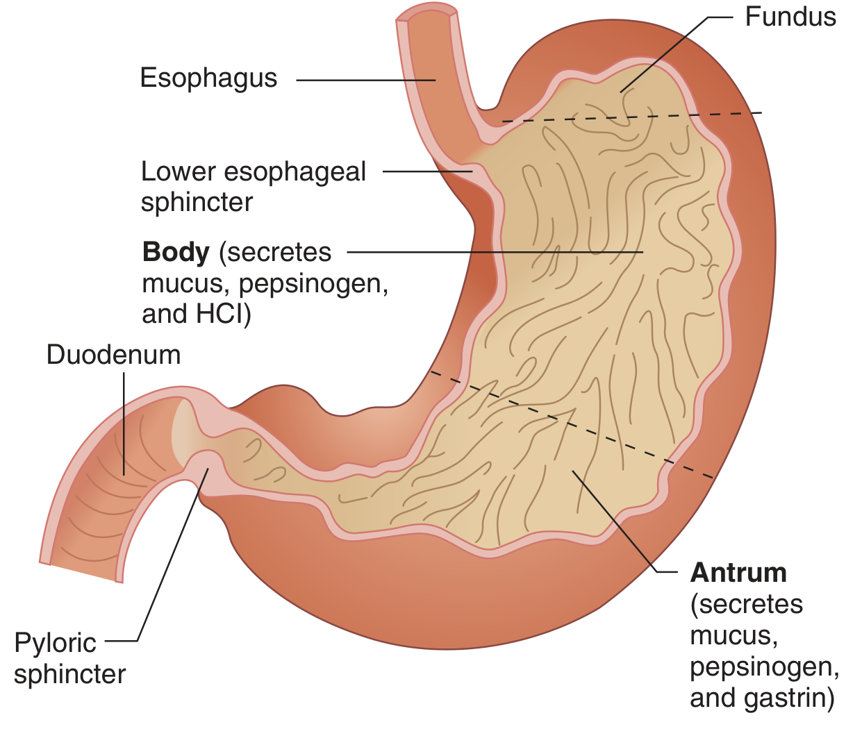
- Body/Fundus (proximal ~80%) - contains oxyntic (gastric) glands that secrete HCl, pepsinogen, intrinsic factor, and mucus
- Antrum/Pylorus (distal ~20%) - contains pyloric glands that secrete mucus and the hormone gastrin
(Guyton & Hall Medical Physiology)
Cell Types and Their Secretory Products
| Cell Type | Location | Secretory Product |
|---|---|---|
| Parietal (Oxyntic) cells | Oxyntic glands (body/fundus) | HCl + Intrinsic factor |
| Chief (Peptic) cells | Oxyntic glands (body/fundus) | Pepsinogen (+ gastric lipase) |
| Mucous neck cells | Oxyntic gland necks + surface | Mucus, HCO3-, pepsinogen |
| G cells | Pyloric glands (antrum) | Gastrin (into circulation) |
| ECL cells | Oxyntic glands | Histamine (paracrine) |
| D cells | Antrum and fundus | Somatostatin (paracrine) |
| Surface epithelial cells | Entire mucosa surface | Mucus, HCO3-, trefoil peptides |
(Ganong's Review of Medical Physiology, 26th ed.; Costanzo Physiology, 7th ed.)
Composition of Gastric Juice
Normal fasting gastric juice contains:
| Component | Details |
|---|---|
| HCl | ~160 mmol/L; pH ~0.8-2.0 |
| Pepsinogens | Inactive proteolytic precursors |
| Intrinsic factor | Glycoprotein essential for vitamin B12 absorption in the ileum |
| Gastric lipase | Begins fat digestion |
| Mucus | Viscoelastic gel; protective barrier |
| Cations | Na+, K+, Mg2+, H+ |
| Anions | Cl-, HPO42-, SO42- |
The only physiologically essential component is intrinsic factor - absence causes pernicious anemia. All others can be compensated for partially. (Costanzo Physiology, 7th ed.)
Cellular Mechanism of HCl Secretion
The parietal cell is the most metabolically active secretory cell in the body, packed with mitochondria.
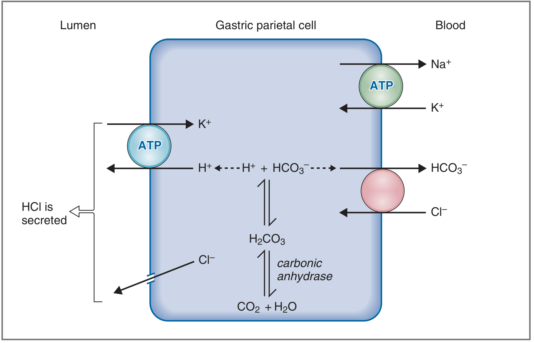
The steps are:
- CO2 + H2O → H2CO3 (catalyzed by carbonic anhydrase inside the parietal cell)
- H2CO3 dissociates → H+ + HCO3-
- H+ is actively pumped into the lumen via the apical H+/K+-ATPase (proton pump), exchanging H+ for K+
- K+ recycles back via apical K+ channels; Cl- enters from blood via a basolateral Cl-/HCO3- exchanger and exits into the lumen via apical Cl- channels
- HCO3- exits into the blood (causing "alkaline tide" in gastric venous blood during acid secretion)
- Final canalicular secretion: HCl ~150-160 mEq/L + KCl ~15 mEq/L + trace NaCl
The H+ concentration in secreted acid is ~3 million times that of arterial blood, requiring >1500 calories of energy per liter of gastric juice. (Guyton & Hall, Costanzo)
At rest, proton pumps are stored in intracellular tubulovesicles. When stimulated, these fuse with apical canalicular membranes, dramatically increasing the secretory surface area. (Ganong's)
Pepsinogen Secretion and Activation
- Chief cells secrete pepsinogen (MW ~42,500), an inactive zymogen
- At luminal pH < 5, HCl cleaves pepsinogen to active pepsin (MW ~35,000); autocatalytic at pH < 3.5
- Pepsin is a protease active at pH 1.8-3.5; it begins protein digestion
- Stimuli: ACh (most potent direct stimulus), gastrin, secretin (minor)
- At pH > 5, pepsin is irreversibly inactivated (Guyton & Hall)
Mucus and the Gastric Mucosal Barrier
- A viscid gel of mucopolysaccharides (glycoproteins) forms a 0.5-1 mm layer over the epithelium
- HCO3- secreted by surface cells is trapped in mucus, creating a pH gradient (pH ~7 at cell surface, pH ~2 in lumen)
- Trefoil peptides stabilize the mucus layer
- Prostaglandin E2 maintains the barrier, stimulates HCO3- secretion, and promotes mucosal blood flow
- Barrier is damaged by aspirin, NSAIDs, alcohol, and H. pylori (Bailey & Love's Surgery, 28th ed.; Costanzo)
Regulation of Gastric Secretion
Three Primary Stimulants
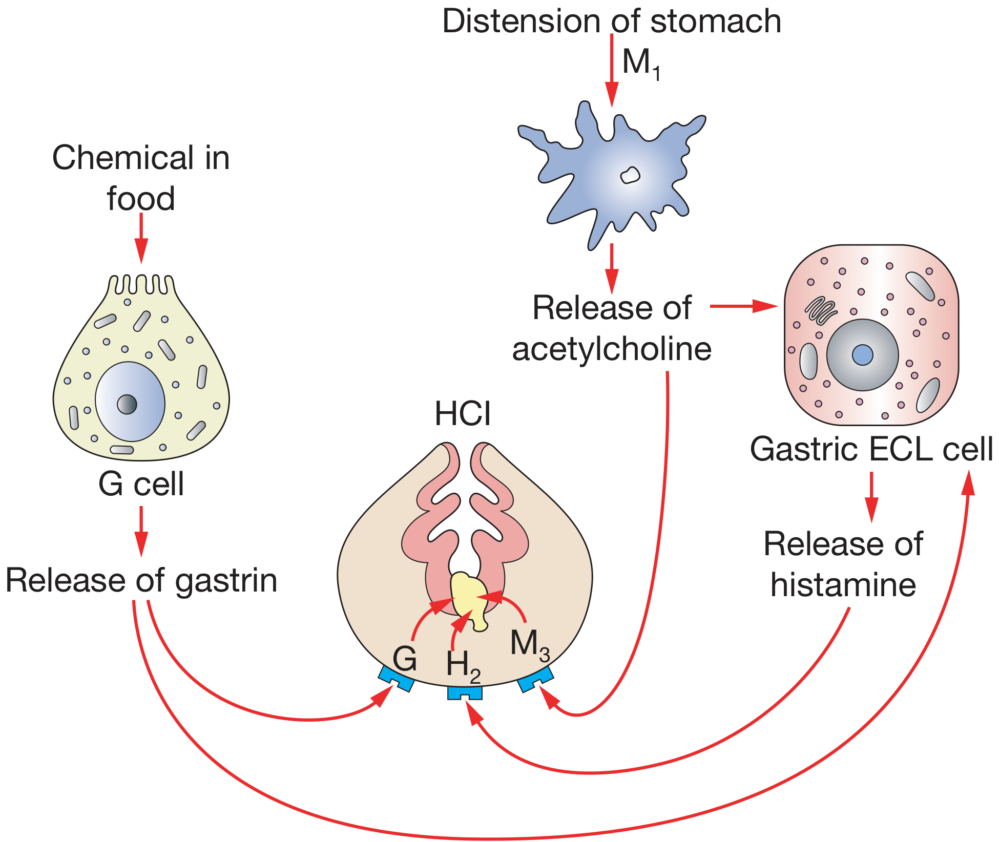
The parietal cell has three distinct receptor systems, each activating a separate second-messenger pathway:
| Stimulant | Receptor | Second Messenger | Source |
|---|---|---|---|
| Histamine | H2 receptor | cAMP ↑ (Gs → adenylyl cyclase) | ECL cells (paracrine) |
| Gastrin | CCK-B/gastrin receptor | Ca2+ ↑ (Gq → IP3/DAG) | G cells (endocrine) |
| Acetylcholine | M3 muscarinic receptor | Ca2+ ↑ (Gq → IP3/DAG) | Vagal nerve endings (neurocrine) |
All three pathways converge to activate the H+/K+-ATPase. Histamine is the dominant paracrine amplifier - gastrin and ACh act partly by stimulating ECL cells to release histamine, explaining why H2-blockers are so effective. (Ganong's, Guyton & Hall, Bailey & Love's)
Three Phases of Gastric Secretion
1. Cephalic Phase (~30% of total acid output)
- Triggered by sight, smell, taste, and thought of food - conditioned reflex (first demonstrated by Pavlov)
- Mediated entirely by the vagus nerve (dorsal vagal complex)
- Vagal output releases:
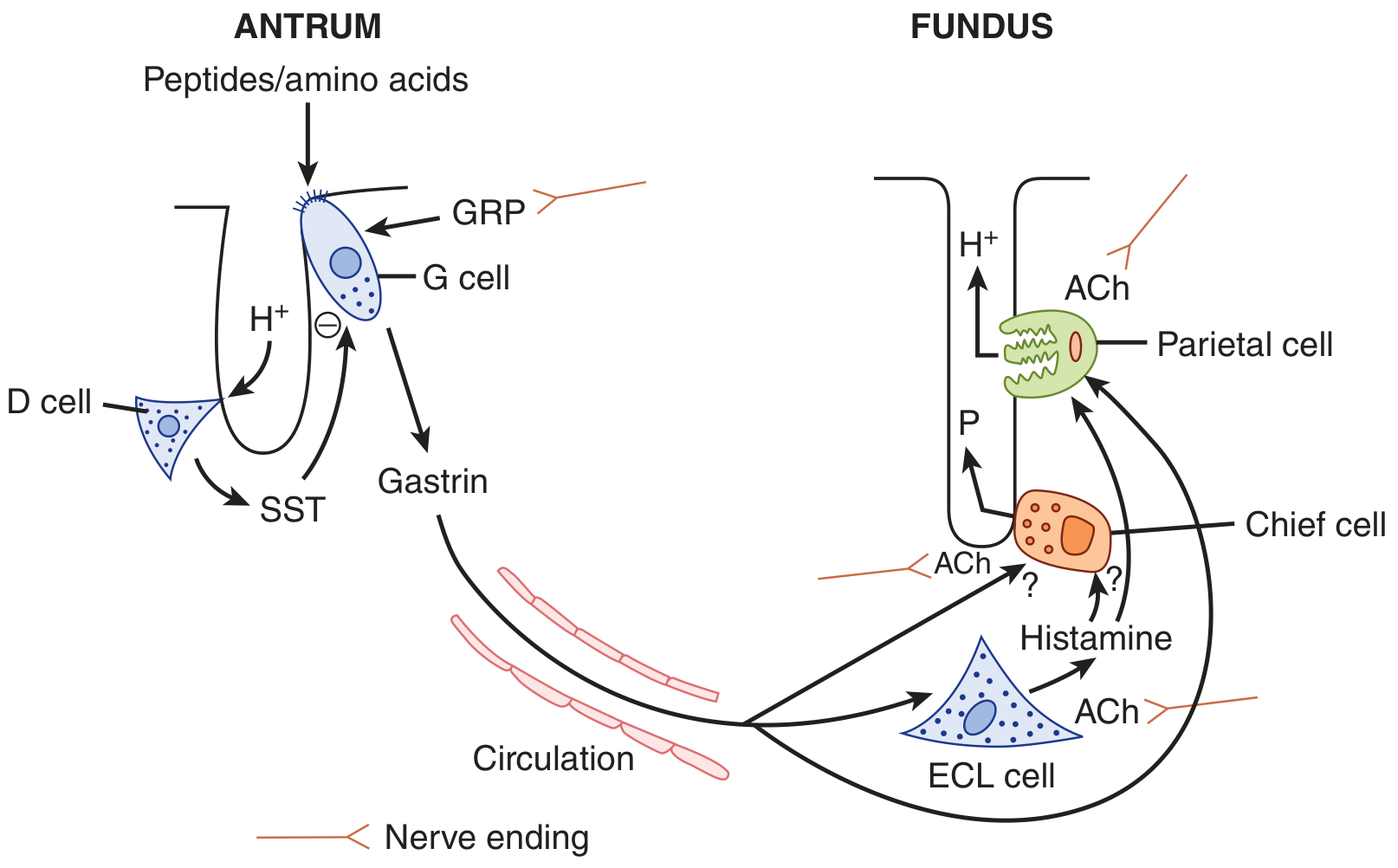
- GRP (gastrin-releasing peptide) → stimulates G cells → gastrin
- ACh → directly stimulates parietal cells (M3), chief cells, ECL cells
- Abolished by vagotomy
2. Gastric Phase (~60% of total acid output - quantitatively most important)
- Triggered by food entering the stomach
- Mechanisms:
- Gastric distension → local myenteric reflexes + vago-vagal reflexes → ACh release
- Protein breakdown products (oligopeptides, amino acids) → directly stimulate G cells → gastrin
- Meal also buffers luminal acidity, relieving somatostatin inhibition and allowing gastrin to rise
- Gastrin travels via the bloodstream to stimulate ECL cells and parietal cells
3. Intestinal Phase (~10% of total acid output)
- Stimulatory early component: amino acids absorbed in the small intestine → enteric G cells release a small amount of gastrin
- Inhibitory (dominant): chyme entering the duodenum triggers powerful brakes on gastric secretion
Inhibitory Mechanisms
1. Somatostatin (D cells) - Primary "Off Switch"
- Released by antral and fundal D cells in response to luminal acid (pH < 3)
- Acts paracrine on G cells (inhibits gastrin release), ECL cells (inhibits histamine release), and parietal cells directly
- This creates the key negative feedback loop: HCl → somatostatin → inhibits G/ECL cells → less gastrin/histamine → less HCl
- When a meal is present, buffering of luminal pH releases this inhibition, allowing gastrin to rise (Ganong's, Costanzo)
2. Enterogastric Reflexes (Intestinal Phase)
Triggered when chyme enters the duodenum:
- Distension of the duodenum
- Acid in the duodenum
- Protein breakdown products
- Hyperosmolar or hypo-osmolar fluids → Reverse enterogastric reflex via myenteric plexus + sympathetic nerves → inhibits gastric secretion and motility (Guyton & Hall)
3. Intestinal Hormones
| Hormone | Source | Inhibitory Action |
|---|---|---|
| Secretin | S cells, duodenum (acid) | Inhibits gastrin; also stimulates HCO3- |
| GIP (glucose-dependent insulinotropic peptide) | K cells, duodenum (fat/glucose) | Weak inhibitor |
| VIP (vasoactive intestinal peptide) | Enteric nerves | Weak inhibitor |
| Somatostatin | Intestinal D cells | Inhibits parietal cells |
| Prostaglandin E2 | Mucosal cells | Inhibits adenylyl cyclase (Gi pathway); opposes histamine |
(Guyton & Hall; Costanzo)
Interdigestive (Basal) Secretion
- Only a few mL/hour during fasting - predominantly mucus, minimal pepsinogen, almost no acid
- Emotional stimuli can raise this to >50 mL/hour (highly acidic and peptic) - contributing to stress ulcers (Guyton & Hall)
Intrinsic Factor
- Secreted by parietal cells in parallel with HCl
- A glycoprotein that binds vitamin B12 in the stomach and protects it from digestion
- The B12-IF complex is absorbed by specific receptors in the terminal ileum
- After total gastrectomy or parietal cell destruction (autoimmune gastritis), intrinsic factor loss leads to pernicious anemia
Clinical Correlations
| Condition | Acid Output | Gastrin | Mechanism |
|---|---|---|---|
| Gastric ulcer | ↓ | ↑ (secondary) | Mucosal barrier defect; reduced acid causes compensatory gastrin rise |
| Duodenal ulcer | ↑ | ↑ (exaggerated meal response) | Increased parietal cell mass; H. pylori-driven |
| Zollinger-Ellison syndrome | ↑↑ | ↑↑ | Pancreatic gastrinoma; unregulated gastrin secretion; massive acid output |
PPIs (e.g., omeprazole) block the final common pathway (H+/K+-ATPase) and thus abolish acid secretion regardless of the stimulus. H2-receptor antagonists effectively reduce histamine-driven acid but are not the final pathway, so their effect is surmountable. (Bailey & Love's Surgery, 28th ed.; Costanzo)
Sources:
- Guyton and Hall Textbook of Medical Physiology
- Ganong's Review of Medical Physiology, 26th Edition
- Costanzo Physiology, 7th Edition
- Bailey and Love's Short Practice of Surgery, 28th Edition
This is a shared conversation. Sign in to Orris to start your own chat.