
New Chat Recent Subscription Options OPD Orthopedic Drugs India Tamilarasan P Common opd drugs in ortho based on Indian standards
common OPD orthopedic drugs India formulary 2024 NSAID muscle relaxant
Common OPD Orthopedic Drugs - India
1. Analgesics & NSAIDs
| Drug | Dose (Oral) | Selectivity | Indian Brand Examples | Notes |
|---|---|---|---|---|
| Paracetamol | 500-1000 mg TDS | - | Crocin, Dolo 650 | First-line mild pain; safest GI profile |
| Aceclofenac | 100 mg BD | Relative COX-2 | Zerodol, Hifenac | Most commonly prescribed in India; good GI tolerability |
| Diclofenac Sodium | 50 mg BD/TDS (SR 100 mg OD) | Non-selective | Voveran, Diclomol | Strong anti-inflammatory; preferred by Indian orthopedic surgeons for OA synovitis |
| Ibuprofen | 400-800 mg TDS | Non-selective | Brufen, Combiflam | OTC availability; useful for acute MSK pain |
| Naproxen | 250-500 mg BD | Non-selective | Naprosyn | Longer half-life; twice daily dosing |
| Meloxicam | 7.5-15 mg OD | Relative COX-2 | Mobic, Melonex | Preferred in elderly; better GI safety |
| Nimesulide | 100 mg BD | Relative COX-2 | Nimulid, Nise | Widely used in India for acute pain; hepatotoxicity risk - limit to ≤15 days |
| Etoricoxib | 60-120 mg OD | Highly COX-2 | Nucoxia, Arcoxia | Best GI safety; avoid in CVD; useful for gout flares |
| Celecoxib | 200 mg OD/BD | Highly COX-2 | Celebrex | Preferred in high GI-risk patients |
| Ketorolac | 10 mg TDS (oral), 30 mg IM | Non-selective | Ketanov, Toradol | Short-term acute pain only (≤5 days); post-procedure |
| Indomethacin | 25-50 mg BD/TDS | Non-selective | Indocap | Gout flares; heterotopic ossification prophylaxis |
Indian practice note: A 2024 survey of Indian orthopedic surgeons published in IJSS confirmed diclofenac SR as the most frequently chosen NSAID for OA, especially for its high synovial fluid penetration and once-daily SR formulation.
2. GI Protectants (Mandatory Co-Prescription with NSAIDs)
| Drug | Dose | Brand | Indication |
|---|---|---|---|
| Pantoprazole | 40 mg OD (before food) | Pan, Pantodac | Routine co-prescription with non-selective NSAIDs |
| Rabeprazole | 20 mg OD | Razo, Rablet | Alternative PPI |
| Omeprazole | 20 mg OD | Omez | Budget option |
| Misoprostol | 200 mcg BD-QID | Cytotec | Mechanistically ideal (PGE1 analogue); rarely used due to GI side effects |
3. Muscle Relaxants
| Drug | Dose | Brand | Mechanism/Notes |
|---|---|---|---|
| Thiocolchicoside | 4-8 mg BD | Myoril, Mobik | Most popular in India; spinal & peripheral muscle relaxation |
| Tizanidine | 2-4 mg BD/TDS | Sirdalud | Central alpha-2 agonist; good for spasticity & back pain |
| Chlorzoxazone | 250-500 mg TDS | Parafon | Often combined with paracetamol (e.g., Flexon) |
| Eperisone | 50 mg TDS | Myonal, Epry | Peripheral relaxant; minimal sedation; popular in India |
| Baclofen | 5-10 mg TDS | Lioresal, Liofen | GABA-B agonist; for spasticity (cerebral/spinal origin) |
| Cyclobenzaprine | 5-10 mg TDS | Flexeril | Central relaxant; causes sedation |
| Tolperisone | 150 mg TDS | Mydocalm | Peripheral; minimal sedation; used in cervical/lumbar spondylosis |
- Aceclofenac 100 mg + Thiocolchicoside 4 mg (Zerodol-TH, Hifenac-TH)
- Diclofenac 50 mg + Thiocolchicoside 4 mg
- Paracetamol 325 mg + Chlorzoxazone 250 mg (Flexon)
4. Neuropathic Pain Agents
| Drug | Dose | Brand | Notes |
|---|---|---|---|
| Pregabalin | 75-150 mg BD | Lyrica, Pregeb | First-line neuropathic pain; also for fibromyalgia |
| Gabapentin | 300 mg OD titrating up | Gabapin, Neurontin | Cheaper alternative to pregabalin; start low in elderly |
| Duloxetine | 30-60 mg OD | Cymbalta, Duvanta | Useful in chronic musculoskeletal pain + depression overlay |
| Amitriptyline | 10-25 mg at night | Amitone, Sarotena | Low-dose for chronic pain; sedating |
5. Bone & Joint Supplements
| Drug | Dose | Notes |
|---|---|---|
| Calcium + Vitamin D3 | 500-1000 mg Ca + 400-800 IU D3 OD/BD | Routine in osteoporosis, fracture recovery, elderly patients |
| Vitamin D3 | 60,000 IU once weekly x 8 wks (loading) | Most Indians are Vitamin D deficient; very commonly prescribed |
| Glucosamine + Chondroitin | Glucosamine 500 mg TDS or 1500 mg OD | LEGS trial showed ~45% reduction in joint space narrowing in knee OA (Rheumatology, 2022, Elsevier) |
| Diacerein | 50 mg BD | IL-1 inhibitor; symptomatic slow-acting drug for OA (SYSADOA); popular in India |
| Avocado Soybean Unsaponifiables (ASU) | 300 mg OD | SYSADOA; structure-modifying in hip/knee OA |
| Omega-3 / Fish Oil | 1-2 g OD | Adjunct anti-inflammatory for RA & OA |
6. Anti-Osteoporosis Drugs
| Drug | Dose | Notes |
|---|---|---|
| Alendronate | 70 mg once weekly | First-line bisphosphonate; reduces vertebral + hip fracture risk |
| Risedronate | 35 mg once weekly / 150 mg once monthly | Alternative bisphosphonate |
| Zoledronic acid | 5 mg IV once yearly | For severe osteoporosis or bisphosphonate-intolerant patients |
| Calcitonin (Nasal) | 200 IU OD | Analgesic for acute vertebral compression fractures |
| Raloxifene | 60 mg OD | SERM; for postmenopausal osteoporosis |
| Strontium ranelate | 2 g OD | Dual action (anti-resorptive + bone forming); still used in India |
7. Drugs for Gout & Hyperuricemia
| Drug | Dose | Notes |
|---|---|---|
| Colchicine | 0.5-1 mg OD/BD for flare | First-line acute gout flare; also prophylaxis |
| Indomethacin | 50 mg TDS for flare | NSAID of choice for acute gout |
| Etoricoxib | 120 mg OD x 5-8 days | COX-2 option for acute gout |
| Allopurinol | 100-300 mg OD | First-line urate-lowering therapy (ULT); start 2-4 wks after flare resolves |
| Febuxostat | 40-80 mg OD | XO inhibitor; preferred in renal impairment; available in India (Buxstat, Zurig) |
8. DMARDs (for Inflammatory Arthritis - RA, PsA, AS)
| Drug | Dose | Notes |
|---|---|---|
| Methotrexate (MTX) | 7.5-25 mg once weekly | Anchor drug for RA; requires folic acid 5 mg supplementation |
| Hydroxychloroquine | 200-400 mg OD | Used in mild RA, lupus arthropathy; requires eye screening |
| Sulfasalazine | 500 mg to 1 g BD | Used in RA + spondyloarthropathies |
| Leflunomide | 10-20 mg OD | Alternative to MTX; teratogenic |
9. Corticosteroids
| Drug | Route/Dose | Use in Ortho OPD |
|---|---|---|
| Methylprednisolone acetate | Intra-articular 40-80 mg | Knee/shoulder OA flares, RA joint injections |
| Triamcinolone acetonide | Intra-articular 10-40 mg | Trigger finger, de Quervain's, plantar fasciitis, tennis elbow |
| Betamethasone | Intra-articular 3-6 mg | Bursa injections |
| Prednisolone | 5-10 mg OD (oral) | Bridge therapy in active RA; taper as DMARDs kick in |
| Dexamethasone | 4-8 mg IM/oral | Acute post-operative / inflammatory flares |
10. Topical Agents
| Drug | Form | Notes |
|---|---|---|
| Diclofenac gel 1% | Gel | Knee OA, soft tissue injuries |
| Ketoprofen gel | Gel | Topical NSAID |
| Capsaicin cream | Cream | Chronic joint pain; desensitizes substance P |
| Methyl salicylate + Menthol | Ointment (Moov, Volini) | OTC counter-irritant; very popular in India |
Key Prescribing Tips for Indian OPD Practice
- Always add a PPI whenever prescribing non-selective NSAIDs for >7 days.
- Nimesulide - limit to ≤15 days per course due to hepatotoxicity (CDSCO advisory).
- Thiocolchicoside is considered a controlled substance in some states - be aware of quantity restrictions.
- Vitamin D deficiency is extremely common across India; check 25-OH Vitamin D before supplementing long-term.
- Diacerein has a unique place in Indian OPD for knee OA - it is a symptomatic slow-acting drug (SYSADOA) and may reduce NSAID dependence over time.
- Pregabalin + Aceclofenac + Thiocolchicoside is one of the most frequently seen triple combinations prescribed for lumbar radiculopathy in Indian practice.
- For gout, always wait 2-4 weeks after flare resolution before starting allopurinol or febuxostat to avoid precipitating a flare.
Ivdp with lumbar radiculopathy
lumbar disc herniation prolapse MRI radiculopathy nerve compression
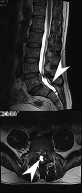
T2-weighted Magnetic Resonance Imaging (MRI) of the lumbar spine presented in two views. The top panel shows a sagittal section illustrating the lumbar vertebrae and intervertebral discs. A prominent disc herniation at the L5-S1 level (white arrowhead) is visible, protruding posteriorly into the spinal canal and causing compression of the thecal sac and cauda equina nerve roots. The bottom panel displays an axial cross-section at the L5-S1 level, further detailing the herniation. The arrow points to a significant left-sided focal protrusion that impinges upon the spinal canal, narrowing the space for neural elements. The high-intensity signal of the cerebrospinal fluid (CSF) in the thecal sac helps delineate the extent of mechanical compression. This diagnostic imaging is characteristic of lumbar radiculopathy secondary to disc prolapse, demonstrating the relationship between degenerative spinal structures and the central neural canal.
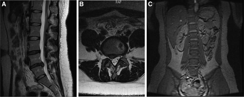
This diagnostic image composite features three MRI views of the human lumbar spine, demonstrating multilevel degenerative changes and disc pathology. Panel A is a T2-weighted sagittal MRI showing a loss of disc height and reduced signal intensity at the L4/L5 and L5/S1 levels, consistent with disc dehydration and degeneration. A posterior disc bulge is visible at L5/S1, indenting the thecal sac. Panel B provides a T2-weighted axial cross-section at the L5/S1 level, highlighting a right-sided posterior disc prolapse that causes narrowing of the lateral recess and potential nerve root compression. Panel C presents a coronal view of the abdomen and lumbar spine, illustrating a mild left-sided lateral curvature (scoliosis). Anatomical landmarks such as the kidneys and psoas muscles are visible, with the latter showing slight asymmetry corresponding to the spinal curvature. The study is highly relevant for musculoskeletal radiology and neurosurgery, illustrating common findings in chronic radiculopathy and lumbar disc herniation.
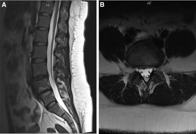
Diagnostic MRI scans of the lumbar spine showcasing degenerative disc disease and associated pathology. Figure A is a T2-weighted sagittal view of the lumbar spine. It demonstrates the vertebral bodies, intervertebral discs, and the spinal canal. Key findings include degenerative bulging at the L4/L5 level and a significant prolapse at the L5/S1 level with loss of disc height. There is visible signal intensity change at the adjacent vertebral endplates, consistent with terminal inflammation or Modic changes. Figure B is a T2-weighted axial view at a lumbar level, likely L5/S1, showing the relationship between the intervertebral disc and the neural structures. The disc demonstrates a posterior protrusion/prolapse that encroaches upon the spinal canal, potentially causing nerve root compression. The CSF-filled thecal sac appears as a bright (hyperintense) triangular region behind the disc, while the surrounding paraspinal musculature and posterior elements are clearly visible. These images serve as educational examples of lumbar disc herniation, discogenic pain, and the use of MRI in assessing mechanical back pain and radiculopathy.
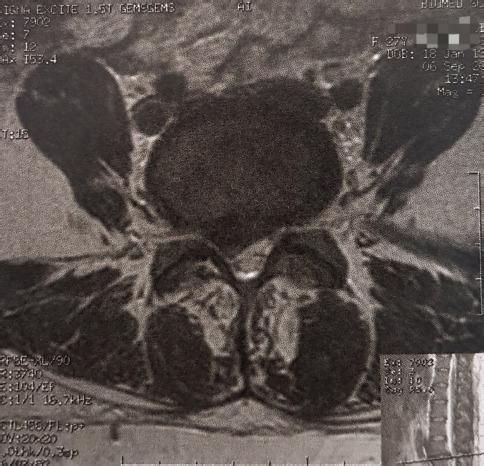
An axial T2-weighted Magnetic Resonance Imaging (MRI) scan of the lumbar spine, specifically at the L4-L5 level. The image provides a clear view of the vertebral body anteriorly and the spinal canal centrally. Pathological findings demonstrate a significant posterolateral disc herniation on the right side. The herniated disc material is seen protruding into the spinal canal, causing a mass effect and resulting in the compression of the right L5 nerve root. This disruption of the normal anatomical symmetry is evidenced by the narrowing of the lateral recess on the right compared to the relatively patent left side. Visible posterior elements include the laminae and paraspinal musculature. This diagnostic image serves as an educational example of degenerative disc disease and acute radiculopathy secondary to mechanical neural compression.
dermatome map lumbar L4 L5 S1 nerve root distribution leg
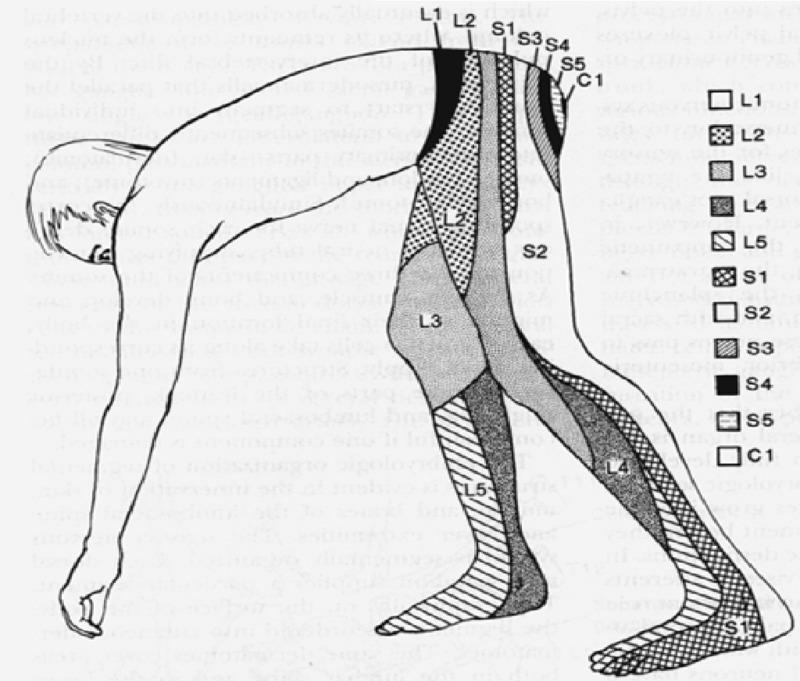
Anatomical diagram depicting a human dermatome map of the lumbosacral and coccygeal regions, shown from a lateral, bent-over perspective to highlight the sensory distribution along the lower extremities and posterior trunk. The illustration uses distinct shading patterns and alphanumeric labels to delineate specific cutaneous areas innervated by individual spinal nerve roots. Key segments include the lumbar dermatomes L1 through L5, sacral dermatomes S1 through S5, and the coccygeal dermatome C1. Visually, L1 covers the inguinal and hip region, L2-L3 transition down the anterior and lateral thigh, and L4-L5 extend down the lower leg to the dorsal foot. Sacral segments S1 and S2 cover the posterior leg and heel, while S3-S5 and C1 are concentrated in the perineal and gluteal regions. This medical illustration is a standard educational tool for neurological examinations, aiding in the localization of spinal nerve root compression or radiculopathy based on sensory loss patterns.
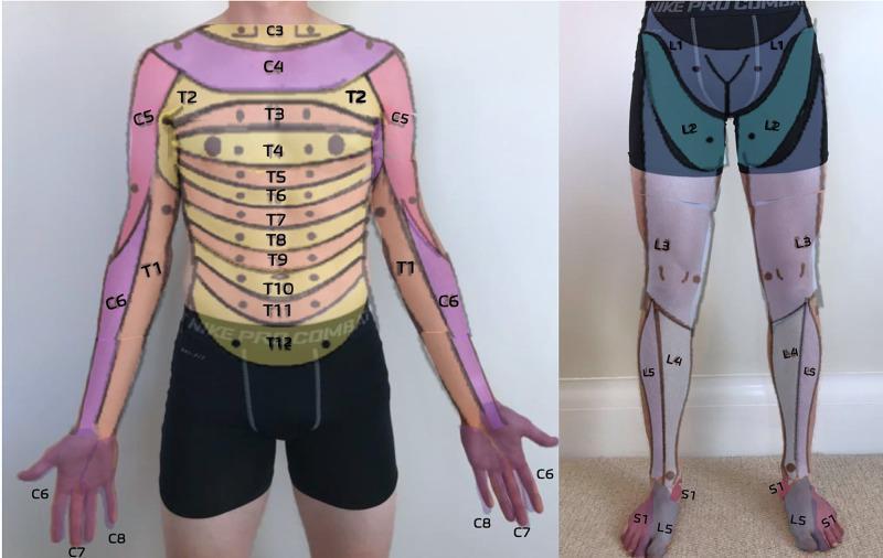
This clinical dermatome map, presented as an anatomical diagram overlaid on a human subject, illustrates the cutaneous sensory distribution of spinal nerves. The anterior trunk displays a sequential horizontal banding pattern representing thoracic dermatomes T2 through T12, with T4 roughly at the nipple line and T10 at the umbilicus. The upper extremities show cervical and upper thoracic distribution: C5 (lateral upper arm), C6 (lateral forearm and thumb), C7 (middle finger), C8 (medial hand and little finger), and T1 (medial forearm and arm). The lower extremity map details the lumbar and sacral levels: L1 (inguinal region), L2 (anterior mid-thigh), L3 (distal thigh and knee), L4 (medial lower leg), L5 (lateral lower leg and dorsal foot), and S1 (lateral foot and small toe). This resource is designed for primary care musculoskeletal medicine to assist in the neurological examination of sensory deficits, radiculopathy, or spinal cord injury level localization.
IVDP with Lumbar Radiculopathy - Complete Clinical Guide
What is IVDP?
- L5-S1 (most common)
- L4-L5
- L3-L4
- L2-L3 (uncommon)
Anatomy of Nerve Root Compression
MRI Appearances
Clinical Features by Root Level
| Disc Level | Root Compressed | Pain Distribution | Motor Weakness | Reflex Lost |
|---|---|---|---|---|
| L3-L4 | L4 | Anterolateral thigh → medial leg | Anterior tibialis (partial foot drop) | Knee jerk (diminished) |
| L4-L5 | L5 | Posterolateral gluteal → lateral thigh → anterolateral leg → dorsal foot → great toe | Extensor hallucis longus (EHL), extensor digitorum; foot drop | No classic reflex lost (posterior tibial) |
| L5-S1 | S1 | Midgluteal → posterior thigh → posterior calf → lateral foot → heel → lateral toes | Plantar flexors, hamstrings; difficulty walking on toes | Ankle jerk absent/diminished |
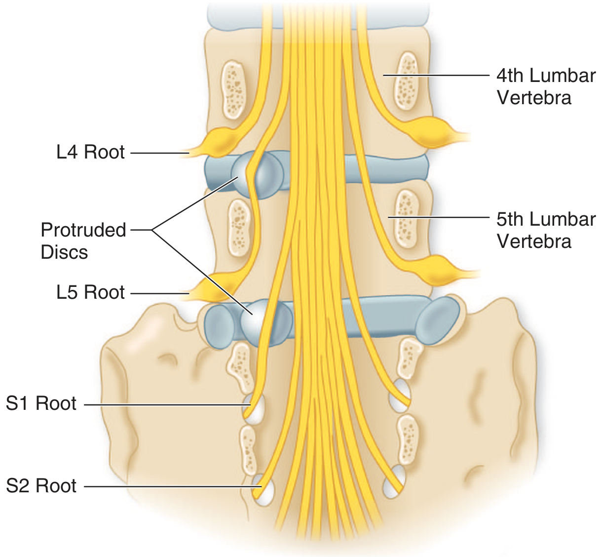
Key point: L4-L5 disc prolapse compresses L5 root (not L4), and L5-S1 disc prolapse compresses S1 root. This is because each lumbar nerve root exits below its numbered pedicle and the herniation typically catches the descending root.
Dermatome Reference

Clinical Diagnosis
History
- Sudden onset after flexion injury, lifting, or trivial trigger (sneeze, lurch)
- Low back pain radiating down one leg (sciatica) below the knee
- Worse on coughing, sneezing, straining (increased intradiscal pressure)
- Comfortable in lateral decubitus with knees flexed
Key Clinical Signs
| Test | Technique | Significance |
|---|---|---|
| Straight Leg Raise (SLR) / Lasegue's sign | Passive hip flexion with knee extended - pain reproduced at 30-70° | Most sensitive test for L4-L5, L5-S1 root compression |
| Crossed SLR | Pain in affected leg when contralateral leg is raised | Highly specific for central/paracentral disc herniation |
| Femoral stretch test | Prone - passive knee flexion - pain in front of thigh | L2, L3, L4 root lesions |
| Power testing | EHL weakness (L5), plantar flexion weakness (S1) | Quantify neurological deficit |
| Reflexes | Ankle jerk (S1), Knee jerk (L4) | Key objective signs |
Red Flags - Urgent Investigation / Referral
- Bilateral leg symptoms
- Saddle anaesthesia (perineal numbness)
- Bladder/bowel dysfunction
- Foot drop progressing acutely
- Fever + back pain (spinal infection)
- History of cancer (metastasis)
- Age >50, first episode, no trauma
Cauda Equina Syndrome = large central prolapse compressing multiple roots. Medical emergency - urgent MRI and surgical decompression.
Investigations
| Investigation | Details |
|---|---|
| X-ray lumbar spine (AP + Lateral) | Rule out fracture, spondylolisthesis, bony disease. May show disc space narrowing |
| MRI Lumbar Spine | Gold standard - shows disc herniation, nerve root compression, canal stenosis |
| CT Lumbar Spine | Alternative if MRI contraindicated |
| Nerve Conduction Study (NCS) / EMG | Confirm radiculopathy, differentiate from peripheral neuropathy |
| ESR, CRP, CBC | Rule out infective / inflammatory causes |
Management
Phase 1: Conservative (First 6-12 weeks - Majority resolve)
A. Analgesics & Anti-inflammatory Drugs
| Drug | Dose | Notes |
|---|---|---|
| Paracetamol | 500-1000 mg TDS | First line mild-moderate pain |
| Aceclofenac | 100 mg BD | Most prescribed NSAID in Indian ortho OPD |
| Diclofenac SR | 100 mg OD | Preferred for acute flares; high synovial penetration |
| Etoricoxib | 60-90 mg OD | COX-2; better GI safety; useful in elderly |
| Ibuprofen | 400-800 mg TDS | Budget-friendly option |
| + PPI (Pantoprazole 40 mg OD) | Always co-prescribe | Gastric protection |
B. Muscle Relaxants
| Drug | Dose | Notes |
|---|---|---|
| Thiocolchicoside | 4-8 mg BD | Most popular in India for lumbar spasm |
| Tizanidine | 2-4 mg BD-TDS | Central alpha-2 agonist; good for spasm |
| Eperisone | 50 mg TDS | Peripheral relaxant; minimal sedation |
| Tolperisone | 150 mg TDS | Good for lumbar/cervical spondylosis |
C. Neuropathic Pain Agents (for Radicular/Burning/Electric Pain)
| Drug | Dose | Notes |
|---|---|---|
| Pregabalin | 75 mg BD (can titrate to 150 mg BD) | First line for radiculopathy / sciatica |
| Gabapentin | 300 mg OD titrating to 300 mg TDS | Cheaper alternative; more sedating |
| Duloxetine | 30-60 mg OD | For chronic radicular pain with depressive overlay |
| Amitriptyline | 10-25 mg at night | Low-dose adjunct; helps sleep |
Note from Tintinalli's: Gabapentin/pregabalin are commonly used but their evidence for back pain specifically is modest; NSAIDs remain better supported for acute radiculopathy.
D. Short Course Oral Steroids
| Drug | Regimen | Notes |
|---|---|---|
| Methylprednisolone / Prednisolone | 4-6 day tapering pack (e.g., Medrol Dosepak) | For severe acute radiculopathy; reduces inflammatory edema around nerve root |
| Dexamethasone | 4-8 mg OD x 3-5 days | Acute severe flares |
E. Physical / Non-Pharmacological Measures
- Rest: 2-3 days only - prolonged bed rest is harmful
- Hot fomentation / TENS - symptomatic relief
- Lumbar corset/belt - short-term use only (max 2-3 weeks)
- Physiotherapy: McKenzie exercises, core strengthening - start after acute phase subsides
- Posture advice: Avoid forward bending, heavy lifting, prolonged sitting
Phase 2: Interventional (6-12 weeks if conservative fails)
Epidural Steroid Injection (ESI)
- Ideal candidate: Acute radicular pain unresponsive to analgesics and rest, significant functional impairment (Campbell's Operative Orthopaedics, 15th Ed.)
- Types:
- Transforaminal ESI - preferred; targets specific root, more ventral placement
- Interlaminar ESI - less specific
- Caudal ESI
- Drugs used: Methylprednisolone 40-80 mg + Bupivacaine 0.25%
- Benefit: Short-term leg pain relief (modest); no long-term functional benefit; does NOT reduce need for surgery (Firestein & Kelley's Textbook of Rheumatology)
- Complications: Dural puncture (5%), epidural hematoma, transient paralysis, chemical meningitis (rare)
- FDA note (2014): Rare but serious neurologic events (stroke, vision loss, death) - label warning issued
Phase 3: Surgical Indications
- Absolute (Emergency): Cauda equina syndrome - bilateral leg weakness, bladder/bowel involvement, saddle anesthesia
- Progressive neurological deficit (foot drop worsening)
- Failure of conservative management for >6-12 weeks with significant pain/disability
- Recurrent disc herniation with ongoing symptoms
- Single level disc herniation
- Neurologic deficit <6 weeks duration
- Monoradiculopathy
- Age <65 years
Surgical Options
| Procedure | Details |
|---|---|
| Microdiscectomy | Gold standard; minimally invasive; 85-95% success for leg pain |
| Open discectomy | Standard open approach |
| Transforaminal endoscopic discectomy | Newer; day surgery; growing use in India |
| PLIF / TLIF (fusion) | Only if disc excision alone is insufficient; recurrent herniation + instability |
Campbell's (2026): Radiographic findings alone are NEVER an indication for surgery. Correlation of imaging with symptoms is essential. Surgery for radiculopathy should not be fused unless there is a separate indication for instability.
Typical OPD Prescription (India) - IVDP with Radiculopathy
Rx:
1. Tab. Aceclofenac 100 mg + Thiocolchicoside 4 mg (Zerodol-TH / Hifenac-TH) - BD x 5 days
2. Tab. Pregabalin 75 mg - BD x 2 weeks (titrate as needed)
3. Tab. Pantoprazole 40 mg - OD (before breakfast)
4. Tab. Methylprednisolone 8 mg - OD x 3 days (for severe acute flares)

Advice:
- Bed rest 2-3 days only
- Hot fomentation to lumbar region
- Lumbar belt (soft) during activity
- Avoid heavy lifting, forward bending
- Physiotherapy referral after acute phase
- MRI Lumbar Spine (if not done)
- Review after 2 weeks
- Return immediately if: weakness in legs, bladder/bowel symptoms