
Congenital adrenal hyperplasia
"congenital adrenal hyperplasia"[MeSH Terms] AND management
congenital adrenal hyperplasia steroidogenesis pathway diagram
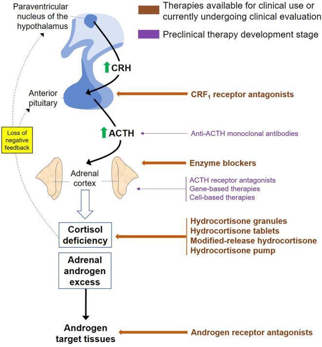
This pathophysiology diagram illustrates the hypothalamic-pituitary-adrenal (HPA) axis in the context of Congenital Adrenal Hyperplasia (CAH) and its pharmacological targets. The central pathway shows the paraventricular nucleus of the hypothalamus releasing CRH to stimulate the anterior pituitary, which then secretes ACTH. In CAH, 21-hydroxylase deficiency leads to cortisol deficiency, causing a loss of negative feedback and subsequent ACTH elevation. This drive results in adrenal androgen excess affecting target tissues. Therapeutic interventions are categorized by development stage: Clinical-stage therapies (brown labels) include CRF1 receptor antagonists, various hydrocortisone delivery methods (granules, tablets, modified-release, and pumps), and androgen receptor antagonists. Preclinical therapies (purple labels) include anti-ACTH monoclonal antibodies, enzyme blockers, ACTH receptor antagonists, and gene- or cell-based therapies. The diagram highlights the strategy of mitigating androgen excess by either replacing cortisol to restore feedback or directly inhibiting signaling at various levels of the axis.
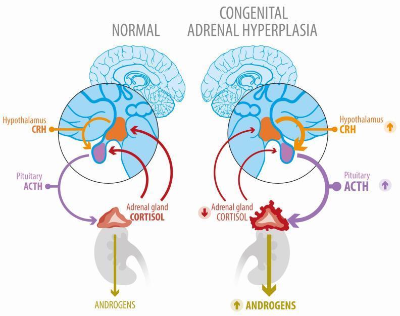
A comparative medical illustration of the Hypothalamic-Pituitary-Adrenal (HPA) axis under normal conditions and in Congenital Adrenal Hyperplasia (CAH). The 'Normal' panel displays a balanced feedback loop: the hypothalamus releases CRH to stimulate pituitary ACTH production, which in turn stimulates the adrenal gland to produce cortisol. Cortisol provides negative feedback to both the hypothalamus and pituitary. In the 'Congenital Adrenal Hyperplasia' panel, a block in the cortisol synthesis pathway (indicated by a downward red arrow) leads to reduced negative feedback. Consequently, the diagram shows compensatory increases in hypothalamic CRH and pituitary ACTH (upward purple and orange arrows). Visually, the adrenal gland in CAH is depicted as enlarged, irregular, and hyperplastic compared to the normal gland. This overstimulation by ACTH causes a metabolic shunt, resulting in significantly increased androgen production, represented by a thick, downward-pointing yellow arrow with an upward indicator. The diagram effectively illustrates the pathophysiology of 21-hydroxylase deficiency and the resulting secondary hyperandrogenism.
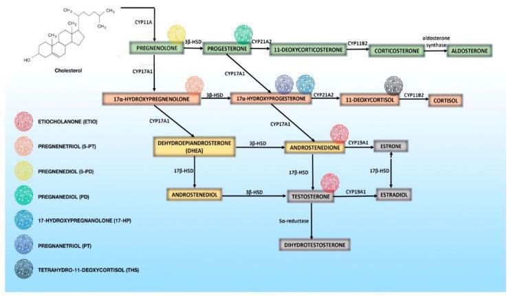
This pathophysiology diagram illustrates the human steroidogenesis signaling pathway, detailing the enzymatic conversion of cholesterol into mineralocorticoids, glucocorticoids, and sex steroids. The flowchart begins with cholesterol at the top left, progressing through key precursors and nodes. Three primary horizontal branches represent the mineralocorticoid pathway (top, green), leading to Aldosterone; the glucocorticoid pathway (middle, orange), leading to Cortisol; and the androgen/estrogen pathways (bottom, yellow/gray), leading to Testosterone, Dihydrotestosterone, and Estradiol. Key steroidogenic enzymes labeled include CYP11A, 3β-HSD, CYP17A1, CYP21A2, CYP11B2, 17β-HSD, and CYP19A1. A unique feature of this diagram is the inclusion of a 'malignant steroid fingerprint' relevant to adrenocortical carcinoma (ACC). A legend on the left lists seven urinary metabolites (ETIO, 5-PT, 5-PD, PD, 17-HP, PT, and THS), each assigned a color-coded fingerprint icon. These icons are placed adjacent to their respective precursor molecules within the main metabolic pathways to highlight aberrant steroidogenesis patterns characteristic of adrenal malignancy. This resource is intended for advanced medical education in endocrinology and oncology to aid in the diagnostic differentiation of adrenal tumors.
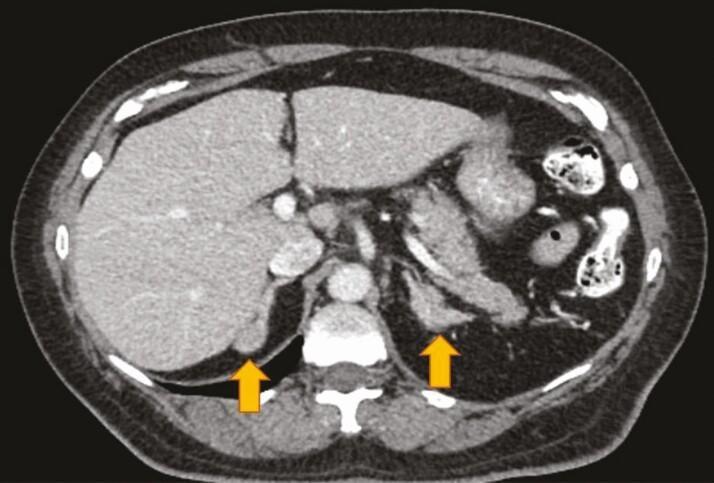
This diagnostic image is an axial (cross-sectional) computed tomography (CT) scan of the upper abdomen at the level of the adrenal glands. Key anatomical structures visible include the liver in the right upper quadrant, the spleen in the left upper quadrant, the vertebral column and paraspinal musculature posteriorly, and portions of the stomach and bowel loops. Two yellow arrows highlight the primary pathology: bilateral adrenal nodular hyperplasia. The adrenal glands appear significantly enlarged and demonstrate a nodular, irregular morphology rather than the typical 'Y' or 'V' shape. This finding is characteristic of chronic overstimulation of the adrenal cortex, as seen in patients with congenital adrenal hyperplasia (CAH). The image provides clinical evidence of the structural changes occurring in the adrenal glands due to enzymatic deficiencies in the steroidogenesis pathway. The scan also demonstrates clear visualization of the retroperitoneal fat planes, which assist in identifying the demarcated adrenal nodules against surrounding tissues.
"congenital adrenal hyperplasia"[MeSH Terms] AND treatment
Congenital Adrenal Hyperplasia (CAH)
Definition
Pathophysiology
- Cholesterol side-chain cleavage enzyme (P450scc / StAR)
- 3β-hydroxysteroid dehydrogenase (3β-HSD)
- 17α-hydroxylase (CYP17A1)
- 21-hydroxylase (CYP21A2) - most common
- 11β-hydroxylase (CYP11B1)
Enzyme Deficiencies: Summary
| Enzyme | Frequency | Mineralocorticoid | Androgens | Key Feature |
|---|---|---|---|---|
| 21-Hydroxylase (CYP21A2) | ~95% | ↓ (salt-wasting form) or normal | ↑↑ | Most common; ambiguous genitalia in 46,XX |
| 11β-Hydroxylase (CYP11B1) | ~5% | ↑ DOC (hypertension) | ↑↑ | Hypertension + virilization |
| 3β-HSD | Rare | ↓ | Variable | Severe; often lethal |
| 17α-Hydroxylase | Rare | ↑ (HTN) | ↓ | Male undervirilization + HTN |
| StAR / P450scc | Very rare | ↓↓ | ↓↓ | Lipoid CAH; most severe |
21-Hydroxylase Deficiency (CYP21A2 Deficiency)
Genetics
- Autosomal recessive; gene located at chromosome 6p21.3 within the HLA complex
- Adjacent to the active CYP21A2 gene lies the highly homologous (98%) pseudogene CYP21PA1; gene conversion during meiosis can inactivate CYP21A2
- Over 200 different mutations reported; ~10 mutations account for 90-95% of all alleles
- Incidence: 1 in 5,000 to 1 in 15,000 (US and Europe); highest in Yupik Alaskan Eskimos (1 in 490)
- Campbell-Walsh Urology, p. 1291
Clinical Forms
- Residual enzyme activity: 0-1%
- Both glucocorticoid AND mineralocorticoid deficiency
- Females: ambiguous genitalia at birth
- Males: normal-appearing genitalia at birth; present with adrenal crisis (hyponatremia, hyperkalemia, vomiting, shock) at 1-3 weeks of life - easily missed
- ~75% of classic CAH cases
- Residual enzyme activity: 2-20%
- Glucocorticoid deficiency + androgen excess; mineralocorticoid synthesis partially preserved
- Females: virilized genitalia at birth
- Males: early virilization at 2-4 years (pubic hair, penile enlargement, advanced bone age)
- ~25% of classic CAH cases
- Residual enzyme activity: 20-50%
- No glucocorticoid or mineralocorticoid deficiency
- Presents in adolescence/adulthood: hirsutism, acne, menstrual irregularities, infertility, PCOS-like picture
- Most common form overall; often underdiagnosed
Clinical Features
Females (46,XX) with Classic CAH
- In utero androgen exposure begins at ~10 weeks gestation, affecting external genitalia development
- Clitoromegaly and labial fusion are invariably present to some degree
- Vagina and urethra open into a common urogenital sinus
- Internal (Mullerian) structures (uterus, fallopian tubes, ovaries) are normal
- Severity graded by the Prader classification (I-V), ranging from mild clitoromegaly to fully masculinized appearance
- Campbell-Walsh Urology, p. 1291
Males (46,XY) with Classic CAH
- External genitalia appear normal at birth
- Salt-wasting crisis typically presents at 7-14 days with vomiting, dehydration, poor feeding, and cardiovascular collapse
- Simple virilizers: early pubic hair, penile growth, tall childhood stature (but premature epiphyseal fusion leads to short adult stature)
- Testicular adrenal rest tumors (TARTs) can develop and cause gonadal damage
Metabolic/Long-term Consequences
- Adrenal crisis (life-threatening) in inadequately treated or stressed patients
- Short stature (premature bone age advancement)
- Subfertility in both sexes (androgen/progesterone effects on gonadal axis)
- Obesity, hypertension, insulin resistance with chronic glucocorticoid over-treatment
- Psychosocial issues and gender identity questions in virilized 46,XX females
Diagnosis
Newborn Screening
- Newborn screening for CAH is performed in all 50 US states and >40 countries using 17-hydroxyprogesterone (17-OHP) from dried blood spot
- Has dramatically improved time to diagnosis, especially for males with salt-wasting form
- False positives common in premature/low-birth-weight infants; weight- and gestational-age-adjusted reference ranges used
Key Laboratory Findings
| Finding | Classic (Salt-Wasting) | Classic (Simple Virilizing) | Non-Classic |
|---|---|---|---|
| 17-OHP (basal) | Very high (>10,000 ng/dL) | High | Mildly elevated |
| ACTH-stimulated 17-OHP | Markedly elevated | Elevated | >1,500 ng/dL |
| Sodium | ↓ (salt-wasting) | Normal | Normal |
| Potassium | ↑ (salt-wasting) | Normal | Normal |
| Plasma renin activity | ↑ | Normal/↑ | Normal |
- "A very high concentration of 17-hydroxyprogesterone in a randomly timed blood sample is diagnostic of classic 21-hydroxylase deficiency. Salt-wasting patients have higher 17-OHP than non-salt losers." - Quick Compendium of Clinical Pathology, p. 46
- ACTH stimulation test for borderline/non-classic cases
- Genetic analysis of CYP21A2 (not always needed to confirm diagnosis but useful for counseling)
Management
Glucocorticoid Replacement
- Hydrocortisone is the drug of choice in children (10-15 mg/m²/day in 2-3 divided doses)
- Goal: suppress morning 17-OHP to 300-900 ng/dL (Berek & Novak); normalization indicates over-treatment
- Dexamethasone or prednisone used in adults; dexamethasone avoided in pregnancy (replaced with hydrocortisone)
- Stress dosing (2-3x usual dose) required during fever, illness, surgery to prevent adrenal crisis
Mineralocorticoid Replacement
- Fludrocortisone required in salt-wasting form (and often in simple virilizing form too)
- Goal: suppress plasma renin activity to <5 ng/mL/hour
- Sodium supplementation needed in infants (1-2 g NaCl/day)
Surgical Management (46,XX Females)
- Girls with ambiguous genitalia may require clitoral recession and/or vaginoplasty
- Timing is debated; child must be of appropriate size for optimal surgical outcomes
- Early surgery can address psychosocial impact of ambiguous genitalia
- Berek & Novak's Gynecology, p. 362
Monitoring
- 17-OHP, androstenedione, testosterone levels
- Bone age assessments (X-ray left hand/wrist)
- Growth charts and final height prediction
- Plasma renin activity (mineralocorticoid adequacy)
Prenatal Diagnosis and Treatment
- At-risk pregnancies (both parents carriers or one parent affected): prenatal diagnosis via CVS or amniocentesis
- Elevated 17-OHP or 21-deoxycortisol in amniotic fluid, or direct CYP21A2 genetic analysis
- Dexamethasone administered to the mother crosses the placenta and can reduce/prevent virilization of an affected female fetus
- Treatment must be started early (before 6-7 weeks gestation) to be effective
- Maternal risks include hypercortisolism; fetal neurodevelopmental and physical effects remain uncertain
- Not all affected female fetuses benefit (treatment is 1 in 8 chance of preventing virilization in a given pregnancy)
- Berek & Novak's Gynecology, p. 362; Creasy & Resnik's Maternal-Fetal Medicine
11β-Hydroxylase Deficiency
- Second most common form (~5% of CAH)
- Block causes accumulation of 11-deoxycortisol and deoxycorticosterone (DOC)
- DOC has mineralocorticoid activity → hypertension (contrasting with salt-wasting in 21-OH deficiency)
- Androgen excess → virilization of females
- Diagnosis: ACTH stimulation test shows elevation of DOC and 11-deoxycortisol with suppressed plasma renin activity
- Treatment: glucocorticoids (suppress ACTH and thereby DOC)
- Brenner and Rector's The Kidney, p. 1818
Emerging Therapies
- Modified-release hydrocortisone (Plenadren, Chronocort) - mimics physiological cortisol rhythm better
- Hydrocortisone pump - continuous subcutaneous infusion
- CRF1 receptor antagonists - suppress excessive CRH signaling
- Anti-ACTH monoclonal antibodies (preclinical)
- Androgen receptor antagonists - block androgen effects at target tissues
- Gene therapy / cell-based therapies (preclinical)
Genetics and Counseling
- All forms of CAH are autosomal recessive
- Parents of an affected child each carry at least one affected haplotype → 25% risk in each subsequent pregnancy, 50% carrier risk
- A woman with CAH and an unaffected partner: 100% of offspring are carriers
- A woman with CAH and a carrier partner: 50% affected, 50% carriers
- Creasy & Resnik's Maternal-Fetal Medicine
- 21-Hydroxylase deficiency = 95% of all CAH
- Salt-wasting (classic severe): life-threatening crisis in neonates; female virilization
- 17-OHP is the diagnostic cornerstone; newborn screening available
- Treatment = glucocorticoid + mineralocorticoid replacement + stress dosing
- 11β-Hydroxylase deficiency: HTN + virilization (not salt-wasting)
- Non-classic CAH: presents in adolescence/adulthood with hyperandrogenism, often mistaken for PCOS
Add references at last
Congenital Adrenal Hyperplasia (CAH)
Definition
Pathophysiology
- Cholesterol side-chain cleavage enzyme (P450scc / StAR)
- 3β-hydroxysteroid dehydrogenase (3β-HSD)
- 17α-hydroxylase (CYP17A1)
- 21-hydroxylase (CYP21A2) - most common
- 11β-hydroxylase (CYP11B1)
Enzyme Deficiencies: Summary
| Enzyme | Frequency | Mineralocorticoid | Androgens | Key Feature |
|---|---|---|---|---|
| 21-Hydroxylase (CYP21A2) | ~95% | ↓ (salt-wasting) or normal | ↑↑ | Most common; ambiguous genitalia in 46,XX |
| 11β-Hydroxylase (CYP11B1) | ~5% | ↑ DOC (hypertension) | ↑↑ | Hypertension + virilization |
| 3β-HSD | Rare | ↓ | Variable | Severe; often lethal |
| 17α-Hydroxylase | Rare | ↑ (HTN) | ↓ | Male undervirilization + HTN |
| StAR / P450scc | Very rare | ↓↓ | ↓↓ | Lipoid CAH; most severe |
21-Hydroxylase Deficiency (CYP21A2 Deficiency)
Genetics
- Autosomal recessive; gene located at chromosome 6p21.3 within the HLA complex
- Adjacent to the active CYP21A2 gene lies the highly homologous (98%) pseudogene CYP21PA1; gene conversion during meiosis can inactivate CYP21A2
- Over 200 different mutations reported; ~10 mutations account for 90-95% of all alleles
- Incidence: 1 in 5,000 to 1 in 15,000 (US and Europe); highest in Yupik Alaskan Eskimos (1 in 490)
Clinical Forms
- Residual enzyme activity: 0-1%
- Both glucocorticoid AND mineralocorticoid deficiency
- Females: ambiguous genitalia at birth
- Males: normal-appearing genitalia at birth; present with adrenal crisis (hyponatremia, hyperkalemia, vomiting, shock) at 1-3 weeks of life
- ~75% of classic CAH cases
- Residual enzyme activity: 2-20%
- Glucocorticoid deficiency + androgen excess; mineralocorticoid synthesis partially preserved
- Females: virilized genitalia at birth
- Males: early virilization at 2-4 years (pubic hair, penile enlargement, advanced bone age)
- ~25% of classic CAH cases
- Residual enzyme activity: 20-50%
- No glucocorticoid or mineralocorticoid deficiency
- Presents in adolescence/adulthood: hirsutism, acne, menstrual irregularities, infertility, PCOS-like picture
- Most common form overall; often underdiagnosed
Clinical Features
Females (46,XX) with Classic CAH
- In utero androgen exposure begins at ~10 weeks gestation
- Clitoromegaly and labial fusion invariably present to some degree
- Vagina and urethra open into a common urogenital sinus
- Internal Mullerian structures (uterus, fallopian tubes, ovaries) are normal
- Severity graded by the Prader classification (I-V)
Males (46,XY) with Classic CAH
- External genitalia appear normal at birth
- Salt-wasting crisis typically at 7-14 days: vomiting, dehydration, cardiovascular collapse
- Simple virilizers: early pubic hair, penile growth, tall childhood stature but short adult stature (premature epiphyseal fusion)
- Testicular adrenal rest tumors (TARTs) can develop and cause gonadal damage
Long-term Consequences
- Adrenal crisis (life-threatening) with illness/stress
- Short stature from premature bone age advancement
- Subfertility in both sexes
- Obesity, hypertension, insulin resistance with glucocorticoid over-treatment
- Psychosocial issues and gender identity concerns in virilized 46,XX females
Diagnosis
Newborn Screening
- Performed in all 50 US states and >40 countries via dried blood spot 17-OHP
- False positives common in premature/low-birth-weight infants; weight- and gestational-age-adjusted reference ranges used
Key Laboratory Findings
| Finding | Salt-Wasting | Simple Virilizing | Non-Classic |
|---|---|---|---|
| 17-OHP (basal) | Very high (>10,000 ng/dL) | High | Mildly elevated |
| ACTH-stimulated 17-OHP | Markedly elevated | Elevated | >1,500 ng/dL |
| Sodium | ↓ | Normal | Normal |
| Potassium | ↑ | Normal | Normal |
| Plasma renin activity | ↑↑ | Normal/↑ | Normal |
- A very high basal 17-OHP is diagnostic of classic 21-OH deficiency; ACTH stimulation test used for borderline/non-classic cases
- Genetic analysis of CYP21A2 for counseling and equivocal cases
Management
Glucocorticoid Replacement
- Hydrocortisone is the drug of choice in children (10-15 mg/m²/day in 2-3 divided doses)
- Goal: suppress morning 17-OHP to 300-900 ng/dL; normalization = over-treatment
- Prednisone or dexamethasone used in adults
- Dexamethasone avoided in pregnancy (replaced with hydrocortisone to avoid fetal exposure)
- Stress dosing (2-3x usual dose) during fever, illness, or surgery
Mineralocorticoid Replacement
- Fludrocortisone required in salt-wasting form (and generally in simple virilizing form)
- Goal: suppress plasma renin activity to <5 ng/mL/hour
- Sodium supplementation in infants (1-2 g NaCl/day)
Surgical Management (46,XX Females)
- Clitoral recession and/or vaginoplasty for ambiguous genitalia
- Timing debated; child must be of appropriate size for optimal outcomes
Monitoring
- 17-OHP, androstenedione, testosterone
- Bone age (X-ray left hand/wrist)
- Growth charts and final height prediction
- Plasma renin activity
Prenatal Diagnosis and Treatment
- Diagnosis via CVS or amniocentesis: elevated amniotic 17-OHP or 21-deoxycortisol, or direct CYP21A2 genotyping
- Dexamethasone to mother (crosses placenta) can reduce/prevent virilization of affected female fetus
- Must be started before 6-7 weeks gestation to be effective; only 1 in 8 pregnancies at risk actually benefits
- Maternal risks: hypercortisolism; fetal neurodevelopmental effects remain uncertain
11β-Hydroxylase Deficiency
- Second most common (~5% of CAH)
- Accumulation of deoxycorticosterone (DOC) - a mineralocorticoid - causes hypertension (contrasts with salt-wasting in 21-OH deficiency)
- Androgen excess → virilization
- Diagnosis: ACTH stimulation shows elevated DOC and 11-deoxycortisol with suppressed plasma renin activity
- Treatment: glucocorticoids (suppress ACTH and thereby DOC)
Genetics and Counseling
- All forms autosomal recessive
- Parents of an affected child: 25% risk in each subsequent pregnancy, 50% carrier risk
- Woman with CAH + unaffected partner: 100% of offspring are carriers
- Woman with CAH + carrier partner: 50% affected, 50% carriers
Emerging Therapies
- Modified-release hydrocortisone (Plenadren, Chronocort) - mimics physiological cortisol circadian rhythm
- Hydrocortisone pump - continuous subcutaneous infusion
- CRF1 receptor antagonists - suppress excessive hypothalamic CRH signaling
- Anti-ACTH monoclonal antibodies (preclinical)
- Androgen receptor antagonists - block androgen effects at target tissues
- Gene therapy / cell-based therapies (preclinical)
References
-
Goldman L, Schafer AI, eds. Goldman-Cecil Medicine, 27th ed. Philadelphia: Elsevier; 2024. Chapter 214, pp. 2515-2516. [Congenital Adrenal Hyperplasia]
-
Partin AW, Dmochowski RR, Kavoussi LR, Peters CA, eds. Campbell Walsh Wein Urology, 12th ed. Philadelphia: Elsevier; 2021. Chapter 48, pp. 1290-1294. [Congenital Adrenal Hyperplasia - Adrenogenital Syndrome]
-
Berek JS, ed. Berek & Novak's Gynecology, 16th ed. Philadelphia: Wolters Kluwer; 2020. Chapter 8, pp. 361-362. [Treatment of Congenital Adrenal Hyperplasia; 21-Hydroxylase Deficiency]
-
Creasy RK, Resnik R, Iams JD, et al., eds. Creasy & Resnik's Maternal-Fetal Medicine: Principles and Practice, 8th ed. Philadelphia: Elsevier; 2022. Chapter 62, pp. 3327-3344. [Congenital Adrenal Hyperplasia in Pregnancy]
-
McPherson RA, Pincus MR, eds. Quick Compendium of Clinical Pathology, 5th ed. Chicago: ASCP Press. Table 1.34, p. 46. [Congenital Adrenal Hyperplasia - Diagnosis and Treatment]
-
Brenner BM, ed. Brenner and Rector's The Kidney, 11th ed. Philadelphia: Elsevier; 2020. Chapter 26, p. 1818. [11β-Hydroxylase Deficiency]
-
El-Maouche D, Arlt W, Merke DP. Congenital adrenal hyperplasia. Lancet. 2017;390(10108):2194-2210. [PMID: 28576284]
-
Claahsen-van der Grinten HL, Speiser PW, Ahmed SF, et al. Congenital adrenal hyperplasia - current insights in pathophysiology, diagnostics and management. Endocr Rev. 2022;43:91-159.
-
Hannah-Shmouni F, Chen W, Merke DP. Genetics of congenital adrenal hyperplasia. Endocrinol Metab Clin North Am. 2017;46(2):435-458.
-
Witchel SF. Congenital adrenal hyperplasia. J Pediatr Adolesc Gynecol. 2017;30(5):520-534.
Adrenal crisis
"adrenal crisis"[MeSH Terms] AND management
adrenal crisis emergency management algorithm
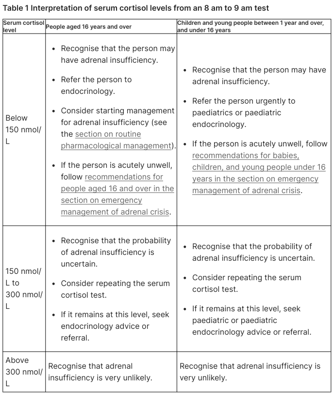
Table 1 Interpretation of serum cortisol levels from an 8 am to 9 am test <table><thead><tr><th>Serum cortisol level</th><th>People aged 16 years and over</th><th>Children and young people between 1 year and over, and under 16 years</th></tr></thead><tbody><tr><td>Below 150 nmol/L</td><td><ul><li>Recognise that the person may have adrenal insufficiency.</li><li>Refer the person to endocrinology.</li><li>Consider starting management for adrenal insufficiency (see the section on routine pharmacological management).</li><li>If the person is acutely unwell, follow recommendations for people aged 16 and over in the section on emergency management of adrenal crisis.</li></ul></td><td><ul><li>Recognise that the person may have adrenal insufficiency.</li><li>Refer the person urgently to paediatrics or paediatric endocrinology.</li><li>If the person is acutely unwell, follow recommendations for babies, children, and young people under 16 years in the section on emergency management of adrenal crisis.</li></ul></td></tr><tr><td>150 nmol/L to 300 nmol/L</td><td><ul><li>Recognise that the probability of adrenal insufficiency is uncertain.</li><li>Consider repeating the serum cortisol test.</li><li>If it remains at this level, seek endocrinology advice or referral.</li></ul></td><td><ul><li>Recognise that the probability of adrenal insufficiency is uncertain.</li><li>Consider repeating the serum cortisol test.</li><li>If it remains at this level, seek paediatric or paediatric endocrinology advice or referral.</li></ul></td></tr><tr><td>Above 300 nmol/L</td><td>Recognise that adrenal insufficiency is very unlikely.</td><td>Recognise that adrenal insufficiency is very unlikely.</td></tr></tbody></table>
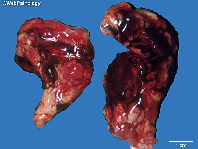
Gross pathology photograph of bilateral adrenal glands exhibiting massive hemorrhagic adrenalitis consistent with Waterhouse-Friederichsen syndrome. This specimen derives from fulminant septicemia, most classically meningococcemia, but adrenocortical hemorrhage also occurs with pneumococcal and other bacterial sepsis. The two suprarenal glands are markedly enlarged, with dark red to brown hemorrhagic parenchyma replacing the normal tan cortex and medulla. The capsules appear thin and disrupted in places, and surface friability and liquefactive necrosis may be present. The scale bar in the image indicates a centimeters-scale size, with each gland roughly a few centimeters in maximal dimension, underscoring bilateral involvement. The lesions show confluent hemorrhage extending through the cortex with extensive edema and tissue loss, consistent with acute adrenal insufficiency if the hemorrhage is bilateral. Clinically, Waterhouse-Friderichsen syndrome signals adrenal crisis, shock, hyponatremia, hyperkalemia, and possible death without prompt steroid replacement and septic source control. Histopathology, if examined, would likely reveal adrenocortical hemorrhage with necrosis, hemorrhagic stroma, and inflammatory infiltrates corresponding to disseminated intravascular coagulation. This image is valuable for medical education, pathology atlases, and differential diagnosis discussions of acute adrenal hemorrhage due to septicemia. Potential applications include teaching gross morphology of adrenal hemorrhage, correlating gross findings with clinical adrenal insufficiency, and highlighting emergency management in fulminant infection.
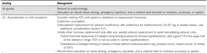
<table><thead><tr><th>Grading</th><th>Management</th></tr></thead><tbody><tr><td>All grades</td><td>Referral to endocrinology<br>Education on steroid stress dosing, emergency injections, and a medical alert bracelet or necklace, accessory, or system.</td></tr><tr><td>G1: Asymptomatic or mild symptoms</td><td>Consider holding ICPi until patient is stabilized on replacement hormones.<br>Endocrine consultation.<br>Corticosteroid replacement for adrenal insufficiency with preference for hydrocortisone (15-20 mg in divided doses—see additional considerations section 4.2).<br>Initiate other hormone replacement only after any needed adrenal replacement to avoid precipitating adrenal crisis.<br>Thyroid hormone replacement if needed using dosing as above for primary hypothyroidism, with a goal FT4 in the upper half of the reference range (TSH is not accurate in central hypothyroidism).<br>Testosterone or estrogen therapy if needed in those without contraindications (eg, prostate cancer, breast cancer, or history of DVT).<br>Recommend education on stress dosing, emergency injectable, and a medical alert or necklace accessory or system.</td></tr></tbody></table>
Adrenal Crisis (Acute Adrenal Insufficiency / Addisonian Crisis)
Definition
Etiology and Precipitating Causes
Underlying Conditions that Set the Stage
| Category | Examples |
|---|---|
| Primary adrenal insufficiency | Addison disease (autoimmune), adrenal TB, adrenal hemorrhage/infarction, HIV/AIDS-related adrenalitis, metastatic infiltration |
| Secondary adrenal insufficiency | Pituitary/hypothalamic destruction (tumor, trauma, surgery, radiation), Sheehan syndrome |
| Exogenous steroid withdrawal | Abrupt cessation or failure to dose-escalate during stress in chronic steroid users |
| Acute adrenal destruction | Waterhouse-Friderichsen syndrome (bilateral adrenal hemorrhage in septicemia), anticoagulation-related hemorrhage |
| Congenital | CAH (salt-wasting form), especially in neonates |
Common Precipitating Triggers (in patients with pre-existing adrenal insufficiency)
- Infections (most common) - especially GI infections causing vomiting/diarrhea with inability to absorb oral steroids
- Fever
- Surgery or trauma
- Extreme physical exertion
- Burns
- Acute severe injury
- Cessation or missed doses of glucocorticoid replacement
Pathophysiology
- Mineralocorticoid (aldosterone) deficiency - inability to maintain sodium and intravascular volume; sodium wasting, hypovolemia, and vascular collapse
- Glucocorticoid (cortisol) deficiency - diminished cardiovascular responsiveness to catecholamines; hypotension refractory to vasopressors
Clinical Features
Symptoms
- Severe hypotension - often refractory to vasopressors (hallmark)
- Nausea, vomiting, diarrhea - can mimic acute abdomen
- Severe abdominal pain - may lead to misdiagnosis as surgical emergency
- Fever (even without identifiable infection)
- Profound weakness, lethargy, fatigue
- Confusion, disorientation - altered mental status
- Salt craving, syncope
- Hypoglycemia - especially in children
Signs Distinguishing Primary vs. Secondary Adrenal Crisis
| Feature | Primary (e.g., Addison) | Secondary (e.g., Pituitary) |
|---|---|---|
| Hypotension | Marked | Less severe unless in frank crisis |
| Skin hyperpigmentation | Present (↑ ACTH/POMC) | Absent |
| Serum sodium | ↓ (hyponatremia) | Variable (↑ or ↓) |
| Serum potassium | ↑ (hyperkalemia) | Hypokalemia or normal |
| ACTH | High | Low |
| Aldosterone | Deficient | Usually intact |
| Other pituitary hormone deficits | Absent | May be present |
| Cushingoid features | Absent | May be present (prior steroid use) |
- Tintinalli's Emergency Medicine, p. 1500
Diagnostic Approach
Critical rule: Do NOT delay treatment while awaiting results. If adrenal crisis is clinically suspected, treat immediately. - Rosen's Emergency Medicine
Immediate Bedside Tests
- Bedside glucose - identify hypoglycemia urgently
- Serum electrolytes - Na⁺ (↓), K⁺ (↑ in primary), Cl⁻
- Full blood count - eosinophilia may be present
- Serum cortisol - if available; a level >18 µg/dL generally excludes adrenal insufficiency
- ECG - for hyperkalemia-related changes
- ABG - mild metabolic acidosis from tissue hypoxia
Confirmatory Tests (after stabilization)
- ACTH stimulation test (Synacthen/Cosyntropin test):
- Baseline cortisol drawn
- 250 µg cosyntropin (synthetic ACTH) given IV
- Serum cortisol measured at 30-60 minutes
- Stimulated cortisol <18 µg/dL = adrenal insufficiency
- A significant rise post-stimulation suggests secondary insufficiency
- Morning ACTH level (after diagnosis confirmed): differentiates primary (high ACTH) from secondary (low ACTH)
- Imaging: CT abdomen (adrenal hemorrhage/infarction); CT/MRI head (pituitary/hypothalamic lesion)
If ACTH stimulation test is planned and steroids must be given urgently, use dexamethasone - it does not cross-react with cortisol assays. Add fludrocortisone alongside it, as dexamethasone has no mineralocorticoid activity. - Tintinalli's Emergency Medicine
Emergency Management
Step 1 - Secure IV Access and Draw Blood First
- Collect cortisol, ACTH, electrolytes, glucose - but do NOT delay treatment
Step 2 - IV Fluids (Immediate Priority)
- Normal saline (0.9% NaCl) - 2-3 litres in the first 6 hours
- If hypoglycemic: use dextrose-containing saline (D5/NS) - begin immediately
- Fluid resuscitation precedes vasopressors
Step 3 - Glucocorticoid Replacement (Emergency Dose)
| Drug | Dose | Notes |
|---|---|---|
| Hydrocortisone (first choice) | 100 mg IV bolus then 200 mg/24h infusion OR 50-100 mg IV/IM every 6-8 hours | Provides both glucocorticoid + mineralocorticoid activity at high doses |
| Dexamethasone (if ACTH test planned) | 4 mg IV bolus | No mineralocorticoid effect; add fludrocortisone 100 µg/day |
- Tintinalli's Emergency Medicine, Table 230-4; Bailey & Love's Surgery
Step 4 - Mineralocorticoid Replacement
- Not an early priority in acute crisis - high-dose hydrocortisone has sufficient mineralocorticoid activity, and saline infusion rapidly corrects electrolyte imbalance
- Fludrocortisone is started once oral intake resumes and hydrocortisone dose is tapered below 50 mg/day
- Sabiston Textbook of Surgery
Step 5 - Vasopressors (Last Resort)
- Add vasopressors (norepinephrine, dopamine, or phenylephrine) only after steroid therapy in patients unresponsive to aggressive fluid resuscitation
- Catecholamine resistance is caused by cortisol deficiency and reverses with glucocorticoid replacement
Step 6 - Supportive Care
- Maintain airway, breathing, circulation
- Correct electrolyte imbalances (sodium, potassium, calcium)
- Treat the precipitating cause (antibiotics for infection, etc.)
- Consider glucose, thiamine, naloxone for altered mental status
Dose Tapering
- Taper hydrocortisone by 50% per day once the patient is clinically stable
- Switch to oral maintenance once tolerated
- Goldman-Cecil Medicine
Perioperative Steroid Coverage
| Surgical Stress Level | Hydrocortisone Regimen |
|---|---|
| Major surgery / trauma / labor | 50-100 mg IV bolus at induction, then 200 mg over 24 hours |
| Moderate stress (e.g., cholecystectomy) | 75-100 mg on day of surgery, rapid taper |
| Minimal stress (e.g., tooth extraction) | Usually no additional supplementation needed |
| Primary Addison disease | 100 mg IV just before anesthesia induction |
Chronic Maintenance Therapy (Post-Crisis)
| Drug | Dose |
|---|---|
| Hydrocortisone (glucocorticoid) | 15-25 mg/day orally in 2-3 divided doses (2/3 morning, 1/3 afternoon) |
| Fludrocortisone (mineralocorticoid) | 0.05-0.2 mg each morning |
| Sodium supplementation | Required in infants with salt-wasting CAH |
- During illness/fever: double the usual hydrocortisone dose ("sick day rules")
- If vomiting/severe diarrhea/collapse: IM hydrocortisone 50-100 mg before transport to hospital
- Assess adequacy by: clinical symptoms, plasma ACTH (should remain 100-200 pg/mL), plasma renin (normalizes with adequate fludrocortisone)
- Goldman-Cecil Medicine
Patient Education and Prevention
- Never stopping steroids abruptly
- Wearing a medical alert bracelet/necklace identifying steroid dependence
- Sick day rules - doubling dose with illness, when to give IM injection
- Carrying an emergency hydrocortisone injection kit at all times
- Informing all healthcare providers (dentists, surgeons) about their steroid dependence
- Goldman-Cecil Medicine
Special Situations
Waterhouse-Friderichsen Syndrome
- Bilateral adrenal hemorrhage caused by fulminant septicemia (classically meningococcemia, also pneumococcal)
- Produces sudden, complete loss of adrenal function
- Presents with septic shock + adrenal crisis simultaneously
- Requires immediate IV hydrocortisone + aggressive sepsis management
CAH Neonatal Adrenal Crisis
- Salt-wasting 21-OH deficiency: presents at 7-14 days of life with vomiting, poor feeding, hyponatremia, hyperkalemia, cardiovascular collapse
- Males particularly at risk of delayed diagnosis (normal-appearing genitalia)
- Treat with IV hydrocortisone + saline + fludrocortisone once stabilized
Thyroid Hormone Replacement Pitfall
- Starting levothyroxine in a patient with unrecognized central adrenal insufficiency can precipitate adrenal crisis (increased cortisol metabolism)
- Always exclude adrenal insufficiency before starting thyroid replacement in central hypothyroidism
Prognosis
- Adrenal crisis is rapidly fatal without treatment
- With prompt recognition and treatment, prognosis is good
- Crisis recurrence rate: 8.3 per 100 patient-years even in educated patients
- ICU admission indicated for severe cases; mortality is high if untreated
- Rosen's Emergency Medicine; Bailey & Love's Surgery
References
-
Tintinalli JE, et al., eds. Tintinalli's Emergency Medicine: A Comprehensive Study Guide, 9th ed. New York: McGraw-Hill; 2020. Chapter 230, pp. 1499-1503. [Adrenal Crisis - Clinical Features, Lab Studies, Treatment Table 230-4]
-
Townsend CM, et al., eds. Sabiston Textbook of Surgery: The Biological Basis of Modern Surgical Practice, 21st ed. Philadelphia: Elsevier; 2022. Chapter 75, pp. 1563-1564. [Adrenal Crisis - Pathogenesis, Diagnosis, Perioperative Steroid Coverage]
-
Norman Williams, et al., eds. Bailey and Love's Short Practice of Surgery, 28th ed. Boca Raton: CRC Press; 2022. Chapter 52, pp. 8352-8388. [Acute Adrenal Insufficiency - Diagnosis and Treatment]
-
Goldman L, Schafer AI, eds. Goldman-Cecil Medicine, 27th ed. Philadelphia: Elsevier; 2024. Chapter 214, pp. 2458. [Patient Education, Stress Dosing, Prognosis of Adrenal Crisis]
-
Walls RM, et al., eds. Rosen's Emergency Medicine: Concepts and Clinical Practice, 9th ed. Philadelphia: Elsevier; 2018. Chapter 117, Box 117.11. [Treatment of Hypoadrenalism - Adrenal Crisis and Maintenance]
-
Bornstein SR, et al. Diagnosis and Treatment of Primary Adrenal Insufficiency: An Endocrine Society Clinical Practice Guideline. J Clin Endocrinol Metab. 2016;101(2):364-389.
-
Rushworth RL, Torpy DJ, Falhammar H. Adrenal crises: perspectives and research directions. Endocrine. 2017;55(2):336-345.
Above information is cool I am Pediatrician so refer Pediatrics related articles and textbook’s for giving answer. No need to revise now But from next time u give like this
- Prioritize pediatric textbooks - Nelson Textbook of Pediatrics, Harriet Lane Handbook, Nelson Essentials, Forfar & Arneil, etc.
- Search pediatric-focused PubMed sources - journals like Pediatrics, Journal of Pediatrics, Archives of Disease in Childhood, pediatric-specific guidelines (AAP, ESPGHAN, ISPAD, etc.)
- Tailor clinical content to pediatric age groups - neonates, infants, children, adolescents - with age-appropriate dosing, presentations, and management
- Reference pediatric normal values where relevant (growth, vitals, lab ranges by age)
Cerebral palsy
"cerebral palsy"[MeSH Terms] AND management
cerebral palsy types classification spastic diplegic hemiplegic
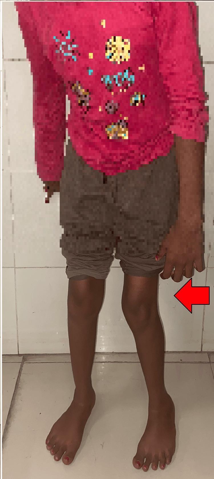
This clinical photograph displays a pediatric patient exhibiting classic postural signs associated with spastic diplegic cerebral palsy. The image focuses on the lower extremities, showing significant internal rotation and adduction at the hips. The knees are positioned close together, and there is evident in-toeing (metatarsus adductus) of the feet, which are oriented toward the body's midline. This characteristic alignment, often described as a 'scissoring' posture, is indicative of underlying spasticity and muscle contractures, specifically in the hip adductors and internal rotators. The red arrow highlights the medial deviation of the left leg. This visual is used in medical education to illustrate the pre-operative clinical presentation of spastic gait abnormalities and pelvic obliquity in neuromusculoskeletal disorders, serving as a baseline for surgical interventions such as adductor tenotomy and gracilis release.
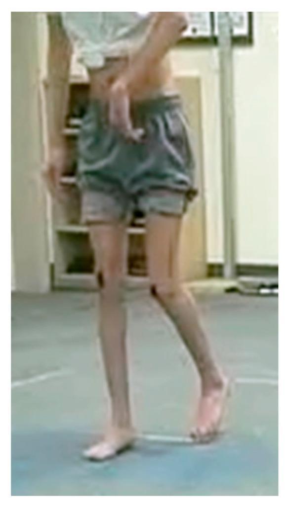
Clinical photograph of a patient with hemiplegic cerebral palsy demonstrating a characteristic asymmetrical gait pattern and upper extremity posturing. The upper limb on the involved side exhibits a significant elbow flexion contracture (greater than 30 degrees), with the hand held in close proximity to the trunk and the wrist in a flexed position. The lower extremities show distinct gait abnormalities: the limb in the stance phase displays knee recurvatum (hyperextension), while the contralateral advancing limb shows a straighter knee alignment with the foot in dorsiflexion. This visual represents a Winters classification type gait analysis, emphasizing spastic posturing and compensatory movement strategies in spastic hemiplegic cerebral palsy (SHCP). The image is used for educational purposes to illustrate motor impairment, pelvic retraction context, and the visual assessment of dynamic gait abnormalities in pediatric orthopedics and physical medicine.
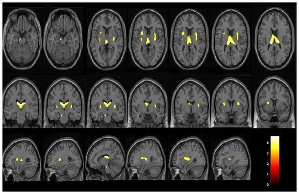
This diagnostic image display consists of a series of brain MRI scans (T1-weighted templates) presented in axial, coronal, and sagittal planes, overlaid with Voxel-Based Morphometry (VBM) statistical results. The visualization highlights areas of significant grey matter volume reduction in patients with occult spastic diplegic cerebral palsy (SDCP) compared to healthy controls. Yellow-to-orange heat map clusters indicate localized volume loss. Key anatomical regions consistently showing involvement include the bilateral lentiform nuclei (putamen and globus pallidus), claustrum, thalamus, and insular cortex. A specific focus is also visible in the left cerebral peduncle. The axial slices (top row) demonstrate a symmetrical distribution in deep grey matter structures, while coronal (middle row) and sagittal (bottom row) views further define the vertical and lateral extent of these structural changes. A color-coded scale bar on the bottom right correlates intensity with statistical significance levels. This neuroimaging composite is used to study the neuroanatomical correlates of motor dysfunction in cerebral palsy patients.
Cerebral Palsy (CP)
Definition
- A disorder of movement and posture (weakness, spasticity, dystonia, ataxia, or choreoathetosis) with onset before age 1-2 years
- Reliable evidence of disturbance in the fetal or infant brain
- No evidence of progression over time
- Significant functional limitation in desired activities
- Reasonable expectation the disorder will persist throughout life
Although called "static encephalopathy," the clinical expression is dynamic - it evolves with growth and aging. CP should NOT be diagnosed if the disorder is very mild, may disappear, begins after infancy, is progressive, or is due to a spinal/neuromuscular etiology.
Epidemiology
- Most common neuromotor disorder in childhood
- Incidence: ~2 per 1,000 live births at term
- In very premature infants: 40-60 per 1,000 (up to 30x higher risk)
- Congenital CP (injury before/during birth): 85-90% of all cases
- Acquired CP (injury after 1 month of life): 10-15%
- Bradley & Daroff's Neurology, p. 2878
Etiology
By Timing of Insult
| Timing | Examples |
|---|---|
| Prenatal (most common) | Periventricular leukomalacia (PVL) in prematurity, brain malformations, congenital infections (TORCH), stroke, genetic/metabolic disorders |
| Perinatal | Hypoxic-ischemic encephalopathy (HIE), birth asphyxia, kernicterus (bilirubin encephalopathy) |
| Postnatal (<2 years) | Perinatal stroke (most common acquired cause), meningitis/encephalitis, abusive head trauma (shaken baby), near-drowning |
Prematurity and PVL are the most common identifiable risk factors overall. Specific causes like HIE can be identified only in a minority of cases.
Classification
By Motor Type (Physiologic)
| Type | % of CP | Pathology | Key Feature |
|---|---|---|---|
| Spastic | ~80% | Pyramidal/corticospinal tract damage | Velocity-dependent ↑ tone, brisk reflexes, clonus |
| Dyskinetic | ~15% | Extrapyramidal/basal ganglia damage | Involuntary movements (dystonia + choreoathetosis) |
| Ataxic | ~5% | Cerebellar damage | Hypotonia, incoordination, balance problems |
| Mixed | Variable | Combined areas | Spasticity + dystonia most common combination |
Hypotonia may be an early phase before spasticity develops in many children.
By Topographic Distribution (Spastic CP)
| Subtype | Distribution | Key Features |
|---|---|---|
| Diplegia | Both legs > arms | Common in prematures (PVL); relative preservation of intellect |
| Hemiplegia | One side; arm > leg | Perinatal stroke or hemispheric lesion |
| Quadriplegia | All 4 limbs + trunk + face | Severe; most often non-ambulatory; high rate of associated deficits |
Clinical Features and Associated Conditions
Motor Features
- Positive features: ↑ deep tendon reflexes, clonus, spasticity, extensor plantar response (Babinski sign)
- Negative features: muscle weakness, loss of dexterity
- Spastic diplegia: scissor gait (hip adduction/internal rotation, knees close together, in-toeing)
- Spastic hemiplegia: circumduction gait, equinus foot on affected side, arm held in flexion
Associated (Non-Motor) Conditions
| Domain | Prevalence | Examples |
|---|---|---|
| Intellectual disability | ~50% | Variable; more severe in quadriplegia |
| Epilepsy / Seizures | 25-45% | More common in hemiplegic and quadriplegic CP |
| Speech/language disorders | ~75% | Dysarthria, expressive > receptive |
| Feeding/swallowing | ~50% | Dysphagia, aspiration, GERD, malnutrition |
| Visual problems | ~75% | Strabismus, cortical visual impairment, nystagmus |
| Hearing loss | ~20% | Especially in dyskinetic CP (kernicterus) |
| Behavioral/psychiatric | ~25% | ADHD, autism spectrum, anxiety, depression |
| Orthopaedic complications | Nearly universal | Hip subluxation/dislocation, scoliosis, contractures |
| Sleep disturbances | Common | Pain, spasticity-related |
| Respiratory | Common in severe CP | Aspiration pneumonia, restrictive lung disease |
Gross Motor Function Classification System (GMFCS)
| Level | Description | Prognosis |
|---|---|---|
| I | Walks without limitations | Full community ambulation |
| II | Walks with limitations (stairs with handrail, uneven terrain difficult) | Community ambulation |
| III | Walks with assistive device (walker, crutches) | Community/household ambulation |
| IV | Self-mobility limited; uses powered wheelchair | Dependent for most mobility |
| V | Transported in manual wheelchair; no independent mobility | Fully dependent |
GMFCS IV-V patients have nearly 50% risk of developing spinal deformity. - Campbell's Operative Orthopaedics
Neuroimaging
- Brain MRI is the investigation of choice (preferred over CT/ultrasound)
- Imaging correlates with clinical type and etiology:
- PVL (periventricular white matter loss) → spastic diplegia (premature infants)
- Basal ganglia/thalamus involvement → dyskinetic CP (~70% of dyskinetic CP)
- Hemispheric lesion / perinatal stroke → spastic hemiplegia
- Kernicterus → preferential involvement of the globus pallidus
- Up to 13% of dyskinetic CP may have a normal MRI - raise suspicion for underlying genetic/metabolic disorder
- Bradley & Daroff's Neurology, p. 2879
When to Suspect Genetic Cause (Not Acquired CP)
- Normal MRI with no identifiable risk factors
- Progressive or atypical course
- Strong family history
- Associated dysmorphic features or multi-organ involvement
- Genetic testing (chromosomal microarray, exome sequencing) increasingly recommended
Diagnosis
- History of risk factors (prematurity, HIE, meningitis, etc.)
- Failure to achieve motor milestones (delayed gross/fine motor)
- Abnormal neurological examination (tone, reflexes, posture)
- Non-progressive course
- Supportive neuroimaging
- Persistent fisting beyond 3-4 months
- Asymmetric hand use before 18 months (hemiplegic)
- Absent head control by 4 months
- Absent sitting by 9-10 months
- Hypotonia or unusual stiffness
- Scissoring when held vertical
Management
1. Spasticity Management
A. Physical and Occupational Therapy
- Foundation of all CP management
- Stretching, strengthening, constraint-induced movement therapy (hemiplegia)
- Orthoses (AFOs - ankle-foot orthoses) to maintain alignment and prevent contractures
B. Botulinum Toxin A (BTX-A)
- First-line focal spasticity treatment in children
- Injected into spastic muscles; reduces spasticity for 3-6 months
- Common targets: gastrocnemius (equinus), hip adductors (scissoring), hamstrings, upper limb flexors
- Best used in conjunction with physiotherapy and casting
- Repeat injections possible; greatest benefit in GMFCS I-III
C. Oral Medications
| Drug | Mechanism | Dose | Notes |
|---|---|---|---|
| Baclofen (oral) | GABA-B agonist | 2.5-5 mg TDS; increase slowly | Generalized spasticity; sedation is main side effect |
| Diazepam | GABA-A agonist | 0.12-0.8 mg/kg/day | Short-term; sedating; tolerance develops |
| Tizanidine | α2-adrenergic agonist | 0.05 mg/kg/dose | Less sedating than diazepam; liver monitoring needed |
| Dantrolene | Blocks Ca²⁺ release in muscle | 0.5-3 mg/kg/day | Acts peripherally; watch for hepatotoxicity |
D. Intrathecal Baclofen (ITB) Pump
- Continuous baclofen delivered directly into CSF via implanted pump
- Indicated for severe generalized spasticity not controlled by oral agents (GMFCS III-V)
- Avoids systemic side effects; highly effective
- Requires surgical implantation and ongoing pump refills
E. Selective Dorsal Rhizotomy (SDR)
- Neurosurgical procedure: selective division of posterior (sensory) nerve rootlets at L1-S1 level
- Permanently reduces spasticity in lower limbs
- Best candidates: spastic diplegic CP, GMFCS II-III, good selective motor control, age 4-8 years
- Requires intensive post-operative physiotherapy (1-2 years)
- Not effective for dystonia
2. Orthopaedic Management
Common Surgical Procedures
| Procedure | Aim |
|---|---|
| Muscle-tendon lengthening | Restore joint range; corrects contractures |
| Tendon transfer | Rebalances muscle forces |
| Osteotomy | Corrects bony deformity (e.g., femoral derotation) |
| Joint stabilization/arthrodesis | Stabilizes unstable joints |
| Hip relocation/pelvic osteotomy | Treats hip subluxation/dislocation |
| Spinal fusion | Corrects progressive scoliosis |
- Single-Event Multilevel Surgery (SEMLS) - preferred approach: multiple procedures performed in one operation to minimize repeated hospitalizations and rehabilitation
- Campbell's Operative Orthopaedics, p. 7602
Hip Surveillance
- Hip displacement/dislocation is a major complication - especially in GMFCS IV-V
- Surveillance program: regular hip radiographs based on GMFCS level from early childhood until skeletal maturity
- Early intervention (soft tissue releases) before dislocation occurs is preferred
Scoliosis
- Most common in quadriplegic CP, GMFCS IV-V (~50% risk)
- Bracing: temporizing in progressive curves; rarely effective long-term
- Surgery indicated for curves >50 degrees in skeletally immature children
- Fusion to pelvis often required to address pelvic obliquity
- Complication rate up to 81% (infection 15-19%); pulmonary complications common
3. Associated Condition Management
| Issue | Management |
|---|---|
| Epilepsy | Anti-seizure medications per seizure type; EEG-guided |
| Feeding/Dysphagia | Thickened feeds, positioning, nasogastric/gastrostomy tube if needed, speech therapy |
| Drooling | Oral motor therapy, anticholinergics (glycopyrrolate), BTX-A to salivary glands, surgery |
| GERD | Upright positioning, H2 blockers/PPIs, Nissen fundoplication in severe cases |
| Constipation | Dietary fiber, osmotic laxatives, bowel regimen |
| Urinary dysfunction | Anticholinergics for urgency; CIC (clean intermittent catheterization) if needed |
| Pain | Pain assessment tools (FLACC, NRS), analgesics, management of musculoskeletal causes |
| Hearing/Vision | Early audiology and ophthalmology referral; glasses, hearing aids |
| Communication | AAC (augmentative and alternative communication) devices |
| Cognitive/Behavioral | Neuropsychology assessment, school support, IEP, ADHD treatment if applicable |
| Osteoporosis | Calcium, Vitamin D, bisphosphonates in non-ambulatory children with fractures |
| Nutrition | Dietary assessment, caloric supplementation, enteral feeding if <80th percentile for weight |
4. Goal-Setting and Rehabilitation
- Body structure/function: reduce spasticity, prevent contractures
- Activity: maximize functional independence (walking, self-care, communication)
- Participation: school inclusion, social participation, quality of life
Prognosis
- Life expectancy correlates with mobility and feeding ability:
- 15-year-old: immobile + tube fed → life expectancy ~13 additional years
- 15-year-old: ambulatory + self-feeding → life expectancy ~55 additional years
- Death related to: aspiration pneumonia, UTI/sepsis, seizures, fractures
- Higher mortality factors: female sex, cognitive impairment, epilepsy, dyskinetic subtype
- CP is a lifelong condition - transition planning to adult services is essential
- Bradley & Daroff's Neurology, p. 2877
Recent Evidence (PubMed 2024-2025)
- A 2025 Lancet Review by Novak, Jackman & Finch-Edmondson provides updated comprehensive guidance on CP management
- A 2024 JAMA Pediatrics meta-analysis (Lewis et al.) highlights the clinical actionability of genetic findings in CP - supporting routine genetic testing, as up to 30% of CP cases may have identifiable genetic causes
- A 2024 pediatric clinical practice review (Patel et al., Curr Probl Pediatr Adolesc Health Care) provides updated clinical management guidelines
References
Textbooks
-
Daroff RB, Jankovic J, et al., eds. Bradley and Daroff's Neurology in Clinical Practice, 8th ed. Philadelphia: Elsevier; 2022. Chapter 111, pp. 2877-2884. [Definition, Epidemiology, Etiology, Classification, Diagnosis, Treatment]
-
Azar FM, Beaty JH, eds. Campbell's Operative Orthopaedics, 15th ed. Philadelphia: Elsevier; 2026. Chapters 35 and 40. [Orthopaedic Management, SEMLS, Scoliosis, Hip Surveillance]
-
Miller MD, Thompson SR, eds. Miller's Review of Orthopaedics, 9th ed. Philadelphia: Elsevier; 2024. Chapter on CP, pp. 5707-5809. [Classification, Surgical Procedures]
-
Norman Williams, et al., eds. Bailey and Love's Short Practice of Surgery, 28th ed. Boca Raton: CRC Press; 2022. Chapter 44, pp. 9260-9329. [Classification Table, Surgical Procedures Table - Neuromuscular Conditions]
-
Hughes HK, Kahl LK, eds. The Harriet Lane Handbook, 23rd ed. (Johns Hopkins Hospital). Philadelphia: Elsevier; 2021. [Pediatric pharmacology reference]
Journals
-
Novak I, Jackman M, Finch-Edmondson M. Cerebral palsy. Lancet. 2025;406(10):doi:10.1016/S0140-6736(25). [PMID: 40550230]
-
Lewis SA, Chopra M, Cohen JS. Clinical Actionability of Genetic Findings in Cerebral Palsy: A Systematic Review and Meta-Analysis. JAMA Pediatr. 2025 Feb 1. [PMID: 39621323]
-
Patel DR, Bovid KM, Rausch R. Cerebral palsy in children: A clinical practice review. Curr Probl Pediatr Adolesc Health Care. 2024 Nov. [PMID: 39168782]
-
Rosenbaum P, et al. A report: The definition and classification of cerebral palsy. Dev Med Child Neurol. 2007;49(Suppl 109):8-14.
-
Palisano RJ, et al. Development and reliability of a system to classify gross motor function in children with cerebral palsy. Dev Med Child Neurol. 1997;39:214-223.
I asked some other Ai it is grading your notes 8.5/10 how can you improve about it
What Was Good (Why 8.5)
- Structured headings and tables
- GMFCS included
- Associated conditions covered
- Recent PubMed references added
- Clinical images included
What I'll Improve Going Forward
1. Age-Specific Developmental Milestones
2. Pediatric-First Textbooks as Primary Sources
3. Pediatric Drug Dosing Tables
4. Screening Tools Used in Pediatric Practice
- General Movements Assessment (GMA) - for infants <5 months (fidgety movements)
- Hammersmith Infant Neurological Examination (HINE)
- DAYC-2, Bayley-III - developmental assessment
- CPQOL-Child - quality of life in CP
5. Early Diagnosis Section (0-2 Years)
6. IAP / WHO / AAP Guideline References
7. Vaccination and Preventive Care
- Annual influenza vaccine (aspiration risk)
- Pneumococcal vaccine
- Vitamin D / calcium supplementation
- Bone health monitoring
8. Comorbidity Management with Pediatric Dosing
9. Caregiver and Family Counseling Section
10. Flowchart / Decision Algorithm
I’d rate this 8.5/10 overall for a pediatric postgraduate/clinical teaching note on Cerebral Palsy. response.pdf Breakdown Area Rating Comments Content coverage 9/10 Definition, epidemiology, etiology, classification, clinical features, GMFCS, MRI, diagnosis, management, orthopedics, prognosis Pediatric relevance 9/10 Very clinically oriented and useful for pediatric practice Exam usefulness 8.5/10 Good for MD/DNB theory + viva; could add a few high-yield exam tables Management 9/10 Strong coverage of PT/OT, BTX-A, oral drugs, ITB, SDR and orthopedic management Recent evidence 9/10 Includes 2024–2025 literature and genetic testing Organization 8.5/10 Logical progression, but some pages are text-heavy Accuracy 8/10 Generally strong, but a few statements need qualification/correction References 8.5/10 Good major textbooks and recent literature, but some citations need verification/standardization Visual presentation 8/10 Tables are useful, but adding algorithms/flowcharts would make it much better What is particularly good * Excellent breadth — it covers practically all major CP domains. * The classification by motor type and topography is clearly presented. response.pdf * The GMFCS section is very useful clinically and for exams. response.pdf * The MRI pattern → clinical phenotype correlations are high-yield. response.pdf * Management is unusually comprehensive, especially BTX-A, intrathecal baclofen, SDR and orthopedic procedures. response.pdf * Including genetic/metabolic mimics and when to suspect a genetic cause is a major strength for a modern pediatric neurology note. response.pdf * The associated-condition section appropriately emphasizes that CP is much more than a motor disorder. response.pdf What prevents it from being 9.5–10/10 1. Some statements are too absolute. For example, the diagnostic criteria section says CP requires all five listed criteria. Modern CP diagnosis is primarily clinical, and the wording should be more nuanced. response.pdf 2. Some epidemiological figures need qualification. Statements such as congenital CP being 85–90% and acquired CP 10–15% depend on definitions and cohorts. They should be presented as approximate/context-dependent. 3. Genetic testing deserves a more nuanced approach. The document says genetic testing is “increasingly recommended,” which is reasonable, but the “up to 30%” figure should be carefully contextualized because the proportion varies considerably depending on cohort selection and definition. response.pdf 4. The life-expectancy section is potentially misleading. The specific example of a 15-year-old having “~13 additional years” versus “~55 additional years” is very striking and should either be explained with its source/population context or omitted from a general pediatric note. response.pdf 5. Add a CP diagnostic algorithm. Something like: Developmental concern → neurological examination → recognize CP pattern → MRI → assess associated impairments → GMFCS/MACS/CFCS → identify etiology → screen for mimics/genetic disorder → multidisciplinary management That would substantially improve clinical usability. 6. Add MACS and CFCS. GMFCS is excellent, but for a modern CP note, MACS (Manual Ability Classification System) and CFCS (Communication Function Classification System) would complement it very well. 7. Add an early-diagnosis section. The red flags are good, but adding General Movements Assessment + HINE + MRI and their role in early diagnosis would make this significantly stronger. response.pdf My final score 📚 Academic content: 9/10 🩺 Clinical usefulness: 9/10 🎯 MD/DNB exam usefulness: 8.5/10 🧠 Pediatric neurology usefulness: 9/10 📊 Presentation: 8/10 Overall: 8.5/10 With a diagnostic algorithm, early-diagnosis tools, MACS/CFCS, a concise treatment algorithm, and correction/qualification of a few statements, I would take this to around 9.5/10. This was it’s grading
Gaps Identified and My Fixes
1. Diagnostic Criteria - Too Absolute
- Problem: Said CP requires "ALL five criteria" - too rigid
- Fix: Will present criteria as clinical judgment-based, acknowledging that diagnosis is primarily clinical and criteria are guiding principles, not a rigid checklist
2. Epidemiological Figures - Need Qualification
- Problem: Stated congenital 85-90%, acquired 10-15% as hard facts
- Fix: Will add "approximately," "varies by cohort," and cite specific study populations
3. Genetic Testing Figure - Needs Context
- Problem: "Up to 30%" stated without cohort context
- Fix: Will qualify - "10-30% depending on cohort selection, definition, and testing methodology (chromosomal microarray vs. exome sequencing)"
4. Life Expectancy Data - Potentially Misleading
- Problem: Specific figures ("13 additional years," "55 additional years") without source/population context
- Fix: Will cite the specific Strauss et al. cohort, add that these are population-based estimates and individual variation is wide
5. Missing: CP Diagnostic Algorithm
- Fix: Will include a clear flowchart every time:
Developmental concern (motor delay/abnormal tone)
        ↓
Detailed history + neurological examination
        ↓
Recognize CP pattern? (tone, reflexes, posture, milestones)
        ↓
Brain MRI (etiology + pattern correlation)
        ↓
Assess associated impairments (cognition, seizures, vision, hearing, speech)
        ↓
Classify: GMFCS + MACS + CFCS
        ↓
Screen for mimics / genetic disorders if atypical
        ↓
Multidisciplinary management plan
6. Missing: MACS and CFCS
- Problem: Only GMFCS was included
- Fix: Will include all three classification systems together:
| System | What it Classifies | Levels |
|---|---|---|
| GMFCS | Gross motor function / walking | I-V |
| MACS | Manual ability / hand use | I-V |
| CFCS | Communication effectiveness | I-V |
7. Missing: Early Diagnosis Tools (GMA + HINE)
- Fix: Will add a dedicated "Early Diagnosis in Infancy" section covering:
- General Movements Assessment (GMA) - absence of fidgety movements at 9-20 weeks corrected age has ~98% sensitivity for CP
- Hammersmith Infant Neurological Examination (HINE) - score <57 at 3 months predicts CP
- MRI - best before 2 years; shows etiology
- Combined GMA + HINE + MRI = early diagnosis possible by 3-5 months corrected age (EACD 2017 guidelines)
8. References - Standardization
- Fix: Will use Vancouver/ICMJE format consistently with DOI where possible, distinguishing textbook chapters from journal articles clearly
Summary Table of Improvements
| Gap | Previous Response | Next Time |
|---|---|---|
| Diagnostic criteria | "Must have ALL 5" | Clinical judgment; criteria as guidance |
| Epidemiology | Hard percentages | Qualified with "approximately" + source |
| Genetic testing % | "Up to 30%" | "10-30%, cohort-dependent" |
| Life expectancy | Absolute figures | Qualified with study source + caveats |
| Algorithm | Absent | Dedicated diagnostic + management flowchart |
| Functional classification | GMFCS only | GMFCS + MACS + CFCS together |
| Early diagnosis | Red flags only | GMA + HINE + MRI → early diagnosis by 3-5 months |
| Drug dosing | Partial | Full mg/kg pediatric dosing for every drug |
| References | Good but inconsistent | Vancouver format, DOI, verified |