
Ankylosis spondylitis ortho clinical a notes detailed
Ankylosing Spondylitis (AS) - Detailed Orthopaedic Clinical Notes
1. DEFINITION & OVERVIEW
- Part of the seronegative spondyloarthritides group (Moll & Wright, 1970s) - includes AS, psoriatic arthritis, reactive arthritis, IBD-associated arthritis
- Also termed r-axSpA (radiographic axial spondyloarthritis) in modern classification
- Campbell's Operative Orthopaedics 15th Ed, p. 2420
2. EPIDEMIOLOGY
| Feature | Detail |
|---|---|
| Age of onset | 20-40 years (average 23 years) |
| Sex ratio | Male : Female = 3:1 (r-axSpA); 1:1 for nr-axSpA |
| Delay to diagnosis | 8.5-11.4 years from symptom onset |
| HLA-B27 | 88-96% of AS patients are HLA-B27 positive |
| Risk if HLA-B27 positive | Only 5% of HLA-B27-positive individuals develop AS |
| Risk in HLA-B27+ relatives of AS patients | 20% risk |
| Population prevalence | 0.5-1.4% (US); varies with HLA-B27 frequency by region |
- Campbell's Operative Orthopaedics, p. 2420; Harrison's 22E, p. 2926
3. PATHOLOGY & PATHOGENESIS
Key Mechanism: Enthesitis
Progression:
- Inflammation at enthesis → periarticular osteopenia
- Inflammatory bone resorption → "squaring off" of vertebral bodies (Romanus lesions - sclerotic shiny corners)
- Ossification of annulus fibrosus (Sharpey fibres) → syndesmophytes (vertical, thin, marginal)
- Sparing of anterior longitudinal ligament initially
- Fusion of posterior elements - facet joints, interspinous/supraspinous ligaments, ligamentum flavum
- Final result: Bamboo spine appearance
- Progression: caudal to cephalad (sacroiliac joints → lumbar → thoracic → cervical)
Pathological features:
-
Sacroiliitis (bilateral, symmetrical) - synovial portion of SIJ first
-
Costovertebral joint involvement → restricted chest expansion
-
Hip involvement ("root joint") - diffuse joint space loss, entheseal new bone at femoral head
-
Andersson lesion - inflammatory pseudarthrosis at mobile unfused segments (can mimic infection)
-
Grainger & Allison's Diagnostic Radiology, p. 1119-1121; Campbell's p. 2420
4. CLINICAL FEATURES
4a. Spinal Symptoms
- Inflammatory back pain (cardinal symptom): insidious onset, worse in morning/after rest, improves with activity
- Morning stiffness - lasting >30 min, improves with exercise
- Buttock pain (sacroiliac joint origin) - may alternate sides
- Progressive loss of lumbar lordosis → thoracic and cervical kyphosis
- Inability to look above the horizon ("stooped" posture)
- Inability to lie flat in bed
4b. Peripheral Manifestations
- Hip joint - most common peripheral joint (second most after SIJ overall); diffuse joint space loss
- Shoulder, knee (asymmetric, oligoarticular, lower limb predominant)
- Heel pain (calcaneal enthesopathy, Achilles enthesitis)
4c. Extra-articular (Extra-spinal) Features - "3 A's"
| System | Manifestation |
|---|---|
| Eyes | Acute anterior uveitis (iritis) - most common extra-articular feature; unilateral, recurrent |
| Cardiovascular | Aortic insufficiency, conduction defects (heart block) |
| Pulmonary | Upper lobe fibrosis/cavitation; restricted chest expansion from costochondral/costovertebral fusion |
| Renal | Amyloid deposition → renal failure (secondary amyloidosis) |
| Neurological | Atlantoaxial instability (25-90% of AS patients); cauda equina syndrome (late) |
4d. Clinical Examination Signs
| Test | Method | Positive Result |
|---|---|---|
| Schober Test (Modified) | Mark 5 cm below and 10 cm above L5-S1 landmark. Patient bends forward fully. | Distance increases <5 cm (normal >5 cm increase, i.e., <15 cm total from 10 cm) |
| Chest expansion | Measure at 4th ICS/nipple line | <2.5 cm = restricted (normal >5 cm) |
| Occiput-to-wall distance | Patient stands with heels/back against wall | >0 cm = abnormal (cervical kyphosis) |
| Finger-to-floor distance | Forward flexion, fingertip to floor | Increased distance |
| FABER test | Flexion, ABduction, External Rotation of hip | SI joint pain |
| Gaenslen's test | Hyperextension of hip | SI joint tenderness |
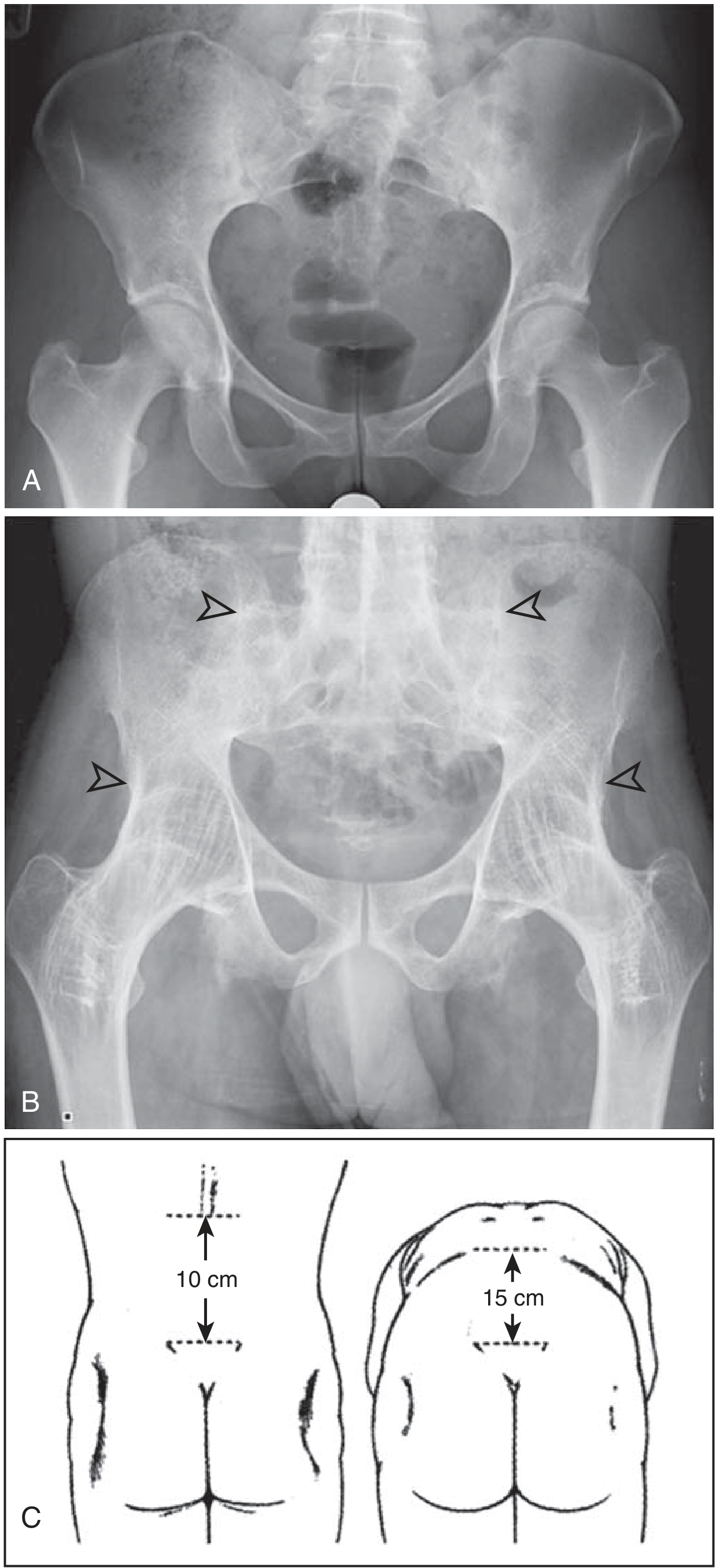
5. DIAGNOSTIC CRITERIA
Modified New York Criteria (1984) - Gold Standard for AS
- Low back pain for ≥3 months, improved by exercise, not relieved by rest
- Limitation of lumbar spine motion in sagittal AND frontal planes
- Chest expansion reduced relative to normal for age and sex
- Sacroiliitis grade ≥2 bilaterally, OR grade 3-4 unilaterally
Sacroiliitis Grading (Modified New York Criteria):
| Grade | Description |
|---|---|
| 0 | Normal |
| 1 | Suspicious - slight blurring of cortical margins |
| 2 | Minimal - small localized erosions/sclerosis; joint width normal |
| 3 | Moderate - severe erosions, sclerosis, widening/narrowing; partial ankylosis |
| 4 | Total ankylosis (complete fusion) |
- Harrison's Principles 22E, p. 2926-2927; Firestein & Kelley's Rheumatology
ASAS Criteria (2009) - for Axial SpA (broader, includes nr-axSpA):
- Imaging arm: Sacroiliitis on MRI or X-ray + ≥1 SpA feature
- Clinical arm: HLA-B27 positive + ≥2 SpA features
6. INVESTIGATIONS
| Investigation | Finding |
|---|---|
| X-ray pelvis (AP) | Sacroiliitis (bilateral, symmetric); bamboo spine; syndesmophytes |
| MRI SIJ | Best for early disease - subchondral bone marrow edema (STIR/T2 fat-sat); detects sacroiliitis before X-ray changes |
| CT SIJ | Best for cortical erosions, ankylosis; not for early disease (radiation) |
| ESR/CRP | Elevated in active disease |
| HLA-B27 | Positive in 88-96% |
| RF / ANA | Negative (seronegative) |
| CBC | Normocytic anemia of chronic disease |
| Bone scan | Increased uptake in SIJ (less specific; superseded by MRI) |
MRI Findings (Grainger & Allison's Radiology):
- T1W: Erosions, joint space loss (damage)
- T2 fat-suppressed / STIR: Subchondral edema (active inflammation)
- Romanus lesion MRI equivalent = corner bone edema of vertebral bodies
- Costovertebral joint edema on STIR sagittal
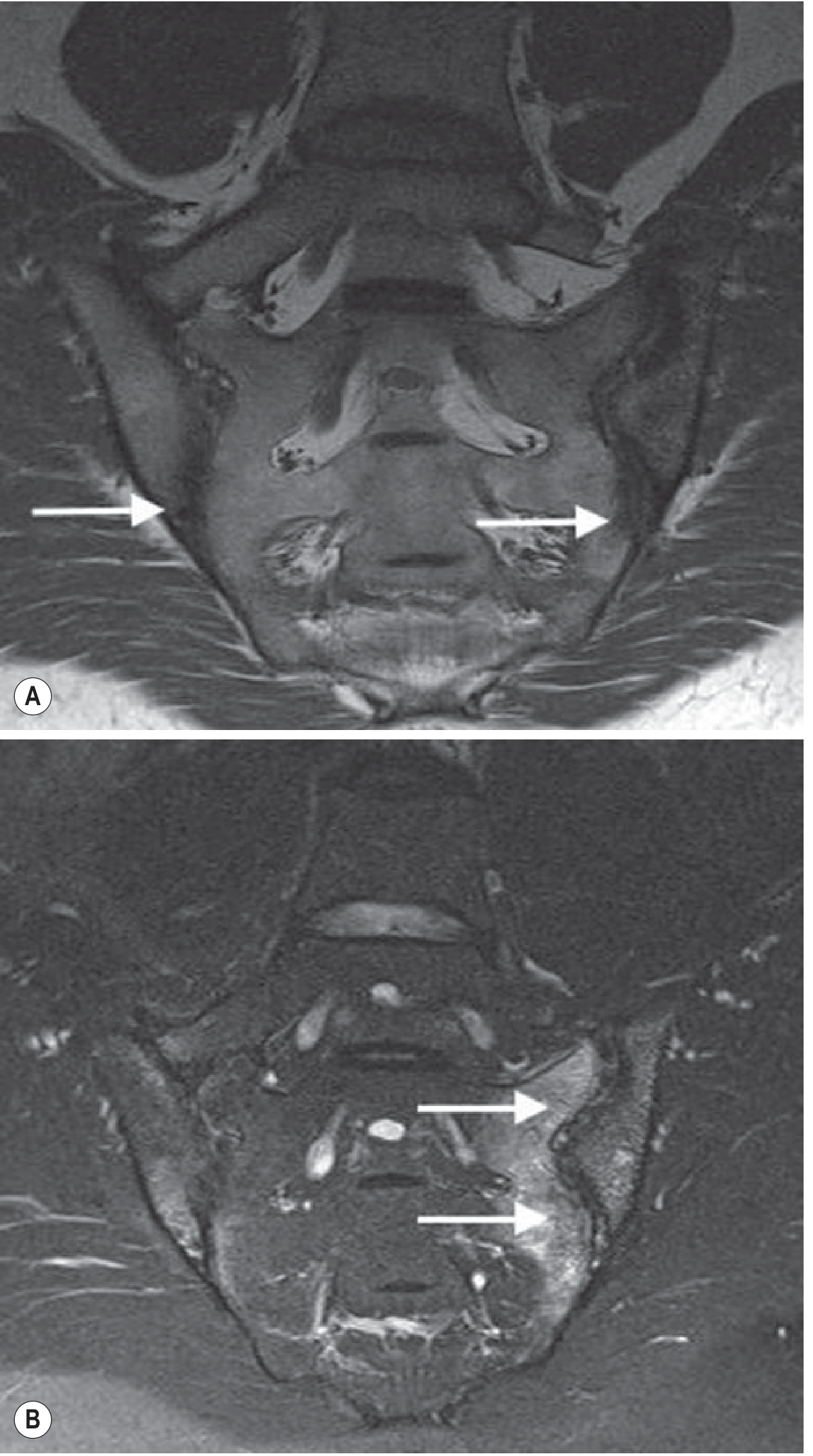
7. RADIOLOGY - KEY FEATURES
Plain X-ray Progression:
- Periarticular osteopenia
- Loss of SIJ definition / blurring of cortical margins
- Romanus lesion ("shiny corners") - sclerosis at anterior vertebral corners
- Squaring of vertebral bodies (erosion + reactive sclerosis)
- Syndesmophytes - thin, vertical, marginal bone outgrowths (annular ossification)
- Facet joint fusion
- SI joint erosions + sclerosis
- Bamboo spine - complete vertebral body fusion + posterior element fusion
- Complete bilateral SIJ ankylosis
- "Trolley track" sign (posterior element fusion on AP view)
- "Dagger sign" (fused interspinous ligament on AP view)
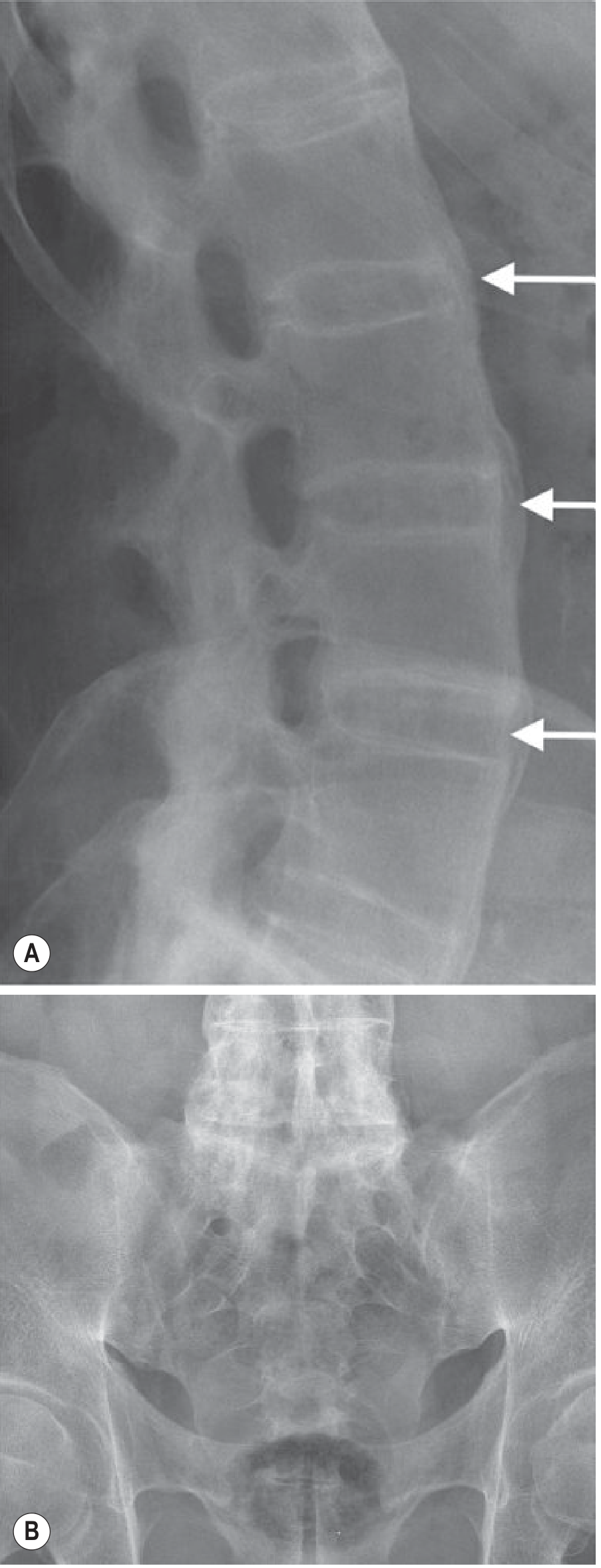
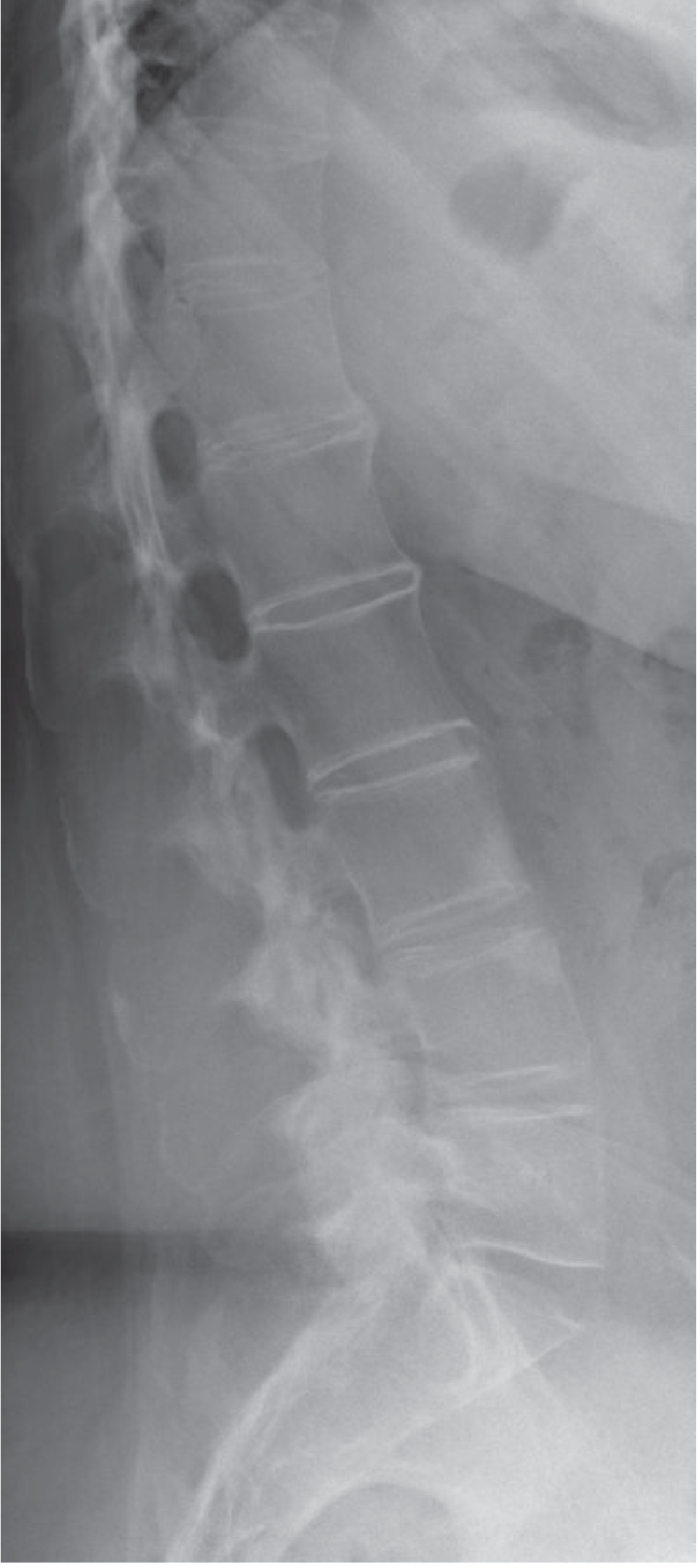
Distinguishing Syndesmophytes from Osteophytes:
| Feature | AS Syndesmophytes | DISH / Spondylosis Osteophytes | Psoriatic/Reactive ArthritisParavertebral Ossifications |
|---|---|---|---|
| Orientation | Vertical (marginal) | Horizontal/curved | Bulky, non-marginal |
| Distribution | Multilevel, symmetrical | Skip levels | Asymmetric, skip levels |
| SIJ | Bilateral, symmetric | Normal | Unilateral or asymmetric |
8. DIFFERENTIAL DIAGNOSIS
| Condition | Key Differentiator |
|---|---|
| Diffuse idiopathic skeletal hyperostosis (DISH) | Flowing ossification along anterior vertebral bodies (≥4 levels), spares SIJ and facet joints, no erosions |
| Psoriatic arthritis | Bulky non-marginal syndesmophytes, asymmetric SIJ, DIP involvement, nail changes |
| Reactive arthritis (Reiter's) | Follows GI/urogenital infection; triad: urethritis, conjunctivitis, arthritis; asymmetric SIJ |
| Rheumatoid arthritis | Seropositive, symmetric small joint involvement, no bamboo spine |
| Lumbar spondylosis | Degenerative, older age, horizontal osteophytes, no SIJ disease |
| Brucellosis | Infectious sacroiliitis (unilateral); systemic features; blood cultures positive |
9. MANAGEMENT
9a. Non-operative Treatment
- Daily stretching exercises for hip flexors and hamstrings
- Spinal extension exercises to prevent kyphosis
- Deep breathing exercises to maintain chest expansion
- Swimming is ideal (reduces axial loading)
- Sleeping supine on firm mattress with one pillow to maintain sagittal alignment
- Avoid hip flexion contractures
| Drug Class | Agents | Role |
|---|---|---|
| NSAIDs (1st line) | Indomethacin (drug of choice), diclofenac, naproxen | Reduce inflammation and pain; continuous use may slow radiographic progression |
| DMARDs (2nd line, mainly peripheral) | Sulfasalazine, methotrexate, minocycline | Slow disease, no cure; less effective for axial disease |
| TNF-α inhibitors (3rd line/biologic) | Etanercept, infliximab, adalimumab, golimumab, certolizumab | Highly effective for active axial and peripheral disease; improve QoL, CRP, sleep; early continuous use inhibits radiographic progression |
| IL-17A inhibitors | Secukinumab, ixekizumab | FDA approved for AS; alternative to anti-TNF |
| JAK inhibitors | Tofacitinib, upadacitinib | Effective across SpA spectrum |
9b. Operative Management
| Surgery | Indication |
|---|---|
| Total Hip Arthroplasty (THA) | Most common; hip involvement with severe pain/loss of function; risk of heterotopic ossification postoperatively |
| Spinal osteotomy | Severe fixed kyphosis with sagittal imbalance |
| Fracture fixation | Long posterior constructs (anterior + posterior for cervical; long posterior for thoracolumbar) |
Types of Spinal Osteotomy:
| Type | Description | Correction |
|---|---|---|
| Smith-Petersen Osteotomy (SPO) | Posterior column resection (Chevron opening-wedge anteriorly); relies on disc mobility | ~10° per level |
| Pedicle Subtraction Osteotomy (PSO) | Posterior + posterior-middle column resection; posterior closing wedge; three-column osteotomy | 30-35° per level |
| Vertebral Column Resection (VCR) | All three columns removed; maximum correction | >45°; highest risk |
-
Type I: Apex lumbar
-
Type II: Apex thoracolumbar
-
Type III: Apex thoracic
-
Type IV: Apex cervical / cervicothoracic junction
-
Campbell's Operative Orthopaedics 15th Ed, p. 2420-2421
10. COMPLICATIONS & SPECIAL CONSIDERATIONS
Spinal Fractures in AS - HIGH RISK
- Osteopenic + fused spine = highly susceptible to fracture from minor trauma
- Most common site: lower cervical spine
- Often missed on plain X-rays (distorted anatomy from ossification)
- Key sign: Widened anterior disc space (hyperextension injury through disc)
- Always immobilize in position found - do NOT extend (risk of sudden neurological deficit)
- Image entire spine (associated fractures at multiple levels possible)
- CT/MRI/bone scan often needed
Atlantoaxial Instability
- 25-90% of AS patients have atlantoaxial instability
- Must be assessed pre-operatively
- Cervical spine deformity can cause disabling functional deficits
Cauda Equina Syndrome (late)
- Rare; due to arachnoid adhesions from chronic inflammation
Heterotopic Ossification after THA
- Increased risk in AS patients
- Prophylaxis: NSAIDs (indomethacin) or low-dose radiation
Andersson Lesion (Spondylodiscitis)
- Inflammatory pseudarthrosis at mobile segment in otherwise fused spine
- Mimics infection; distinguish by lack of systemic features and MRI pattern
11. ASSESSMENT TOOLS (Orthopaedic/Rheumatology)
| Tool | Purpose |
|---|---|
| BASDAI (Bath AS Disease Activity Index) | Disease activity (0-10 scale; ≥4 = active) |
| BASFI (Bath AS Functional Index) | Functional assessment |
| BASMI (Bath AS Metrology Index) | Spinal mobility (5 measurements) |
| ASDAS (AS Disease Activity Score) | Combines patient-reported + CRP |
| mSASSS (Modified Stoke AS Spinal Score) | Radiographic progression |
12. PROGNOSIS
- After ankylosis occurs: pain often improves (burnt-out phase)
- Progressive kyphosis → fixed sagittal imbalance is the main disabling outcome
- Hip involvement → adds significantly to functional loss
- Uveitis: requires regular ophthalmology follow-up to prevent vision changes
- Renal amyloidosis: serious late complication
- Spinal fractures: frequently life-threatening
SUMMARY TABLE
| Feature | Details |
|---|---|
| Etiology | Unknown; chronic inflammatory |
| Genetics | HLA-B27 (88-96%) |
| Age/Sex | 20-40 yr; M:F = 3:1 |
| Cardinal symptom | Inflammatory back pain + morning stiffness |
| Earliest joint | Sacroiliac joints (bilateral, symmetric) |
| Progression | Caudal → cephalad |
| X-ray hallmark | Bamboo spine; Romanus lesion; bilateral SIJ fusion |
| MRI early finding | Subchondral bone marrow edema (STIR) |
| Diagnostic criteria | Modified New York 1984 |
| 1st-line drug | NSAIDs (indomethacin) |
| Most common surgery | Total Hip Arthroplasty |
| Key surgical risk | Spinal fractures - often missed, often catastrophic |
- Campbell's Operative Orthopaedics 15th Ed (2026), pp. 2316, 2420-2421
- Grainger & Allison's Diagnostic Radiology, pp. 1119-1121
- Harrison's Principles of Internal Medicine 22E (2025), pp. 2926-2928
- Miller's Review of Orthopaedics 9th Ed, p. 63
- Firestein & Kelley's Textbook of Rheumatology (2-Volume Set)
I need physiotherapy managementdetailed and evidence based
"ankylosing spondylitis"[MeSH Terms] AND "exercise therapy"[MeSH Terms]
"spondyloarthritis" AND "physical therapy" AND "rehabilitation"
"axial spondyloarthritis" AND "exercise"
PMID: 36261247
PMID: 38851879
PMID: 31410786
PMID: 39798984
PMID: 33547227
"ankylosing spondylitis" AND "hydrotherapy OR aquatic OR yoga OR pilates OR breathing"
PMID: 39342350
PMID: 35633390
PMID: 31578051
"ankylosing spondylitis" AND "TENS OR electrotherapy OR ultrasound OR manual therapy OR TENS"
"ankylosing spondylitis" AND "ASAS EULAR recommendations" AND "management"
"axial spondyloarthritis" AND "group exercise" AND "home exercise"
PMID: 36270658
"ankylosing spondylitis" AND "aerobic exercise OR strength training" AND "disease activity"
PMID: 36369692
PMID: 35052296
Physiotherapy Management of Ankylosing Spondylitis (AS / axSpA)
Evidence-Based Clinical Notes
1. RATIONALE FOR PHYSIOTHERAPY IN AS
- Inflammation leads to progressive ankylosis - mobility must be actively maintained
- Fused spine can still develop kyphotic deformity; posture must be trained
- Drug therapy (even biologics) does not restore lost mobility - only exercise does
- Functional disability, fatigue, and quality of life are exercise-responsive outcomes
- Evidence confirms: exercise improves BASDAI, BASFI, BASMI, pain, and QoL
Key paradox (Perrotta et al., RMD Open 2021, PMID: 33547227): Biomechanical stress at entheses may theoretically promote inflammation, yet exercise programmes consistently demonstrate therapeutic benefit. The net benefit of structured exercise outweighs any theoretical entheseal risk. Exercise is recommended, not avoided.
2. PHYSIOTHERAPY GOALS (Outcome Domains)
| Goal | Measurement Tool |
|---|---|
| Reduce pain | VAS / NRS / BASDAI question 2 |
| Reduce morning stiffness | Duration of stiffness; BASDAI questions 5-6 |
| Improve spinal mobility | BASMI (5 measures) |
| Improve function | BASFI |
| Reduce disease activity | BASDAI, ASDAS |
| Improve chest expansion | Tape measure at 4th ICS |
| Improve posture / sagittal balance | Occiput-to-wall, tragus-to-wall distance |
| Improve aerobic fitness | 6-min walk test |
| Improve QoL | ASQoL, SF-36 |
| Reduce fatigue | FACIT-Fatigue, BASDAI question 1 |
3. EVIDENCE SUMMARY
Level 1 Evidence - Meta-Analyses & Systematic Reviews
| Study | Population | Finding |
|---|---|---|
| Boudjani et al., 2023 PMID: 36369692 - Disabil Rehabil | AS patients | Exercise moderately improves BASDAI (-0.60), BASFI (-0.63), BASMI (-0.52). Flexibility + strength combined = large effect on mobility. Aerobic addition = large effect on function. |
| Ortolan et al. (ASAS/EULAR SLR), 2023 PMID: 36261247 - Ann Rheum Dis | axSpA RCTs | Exercise ES on BASDAI: 0.14-1.43 (moderate to high). Education ES on BASDAI: 0.06-0.59 (small-moderate). Both confirmed efficacious. |
| Regnaux et al. (Cochrane), 2019 PMID: 31578051 | AS, 14 RCTs, n=1579 | Exercise vs. no intervention: pain reduction clinically meaningful (MD -2.1 on VAS); BASFI improvement moderate quality; group > home exercise. |
| Gravaldi et al., 2022 PMID: 35052296 - Healthcare | AS, 12 RCTs | Supervised physiotherapy > usual care for disease activity (SMD -0.37) and function (SMD -0.36). No significant difference between supervised vs. home exercise. |
| Wang et al. (mind-body), 2024 PMID: 39342350 - J Orthop Surg Res | axSpA, 15 RCTs | Tai Chi/Yoga/Pilates/Qigong: significant improvement in BASFI (MD -0.76), BASDAI (MD -0.76), VAS pain (MD -0.89), BASMI (MD -0.44), QoL (MD -2.14). Safe, no serious adverse events. |
| Medrado et al., 2022 PMID: 35633390 - Rheumatol Int | Inflammatory arthritis | Aquatic exercise superior to home exercise for pain; improved disease activity vs. land and control groups. |
RCT Level 1 Evidence
| Study | Finding |
|---|---|
| van Wissen et al., 2025 RCT PMID: 38851879 - Rheumatology | 1-year supervised aerobic + strength exercise (max 64 sessions): PSC1 favored intervention (MD -1.8, 95%CI -2.4 to -1.2). All physical secondary outcomes (BASFI, 6MWT, SF-36 PCS) significantly improved. Effect sizes 0.4-0.7. |
4. PHYSIOTHERAPY ASSESSMENT
4a. Subjective Assessment
- Pain: site, character, onset, duration, radiating, aggravating/relieving factors
- Morning stiffness: duration and severity
- Functional limitations: ADL, work, recreation
- Sleep quality (pain-related)
- Fatigue level
- Psychological status (depression/anxiety common in AS)
- Smoking status (reduces exercise tolerance)
- Medications (NSAIDs, biologics)
4b. Objective Assessment (BASMI - 5 components)
| Test | Method | Scoring |
|---|---|---|
| 1. Lumbar side flexion | Distance fingertip to floor, both sides; best measurement | cm |
| 2. Modified Schober | 5 cm below and 10 cm above PSIS; full flexion | Increase in cm (normal >5 cm) |
| 3. Occiput-to-wall distance | Heels and back against wall; chin neutral | cm (0 = normal) |
| 4. Tragus-to-wall distance | Same position, measure tragus | cm |
| 5. Cervical rotation | Passive maximum rotation, goniometer | degrees |
Additional Objective Tests
| Assessment | Details |
|---|---|
| Chest expansion | Maximal inspiration - expiration at 4th ICS; normal ≥5 cm; <2.5 cm = restricted |
| Thomas test | Hip flexor tightness |
| Hamstring length | Popliteal angle |
| Hip range of motion | All planes; compare bilateral |
| 6-minute walk test | Aerobic capacity / functional endurance |
| Posture analysis | Sagittal: loss of lumbar lordosis, increased thoracic/cervical kyphosis |
| Gait assessment | Reduced arm swing, trunk stiffness, antalgic gait |
5. PHYSIOTHERAPY INTERVENTIONS
5a. THERAPEUTIC EXERCISE (Core Intervention)
PRINCIPLE: Exercise is ALWAYS recommended. The type, setting, and intensity should be individualized.
A. STRETCHING & FLEXIBILITY EXERCISES
| Exercise | Muscle/Region Targeted | Notes |
|---|---|---|
| Thoracic extension over rolled towel/foam roller | Thoracic kyphosis correction | Daily; 5-10 min |
| Lumbar rotation stretch (knee to chest, lateral) | Lumbar mobility | Both sides; hold 20-30 sec |
| Hip flexor stretch (Thomas stretch position) | Iliopsoas | Prevent hip flexion contracture |
| Hamstring stretch | Posterior chain | Sitting or supine |
| Piriformis stretch | Hip external rotators | Important for SIJ mobility |
| Chin tuck + cervical extension | Cervical kyphosis correction | Key for occiput-to-wall distance |
| Doorway pectoral stretch | Pectoralis major/minor | Counters thoracic kyphosis |
| Lateral trunk bending | Lateral spinal mobility | BASMI component |
B. STRENGTHENING EXERCISES
| Muscle Group | Exercises | Evidence |
|---|---|---|
| Spinal extensors (core) | Prone extension ("Superman"), bird-dog, McKenzie extension exercises | Prevent/correct kyphosis |
| Deep cervical flexors | Chin tuck with resistance | Counter cervical kyphosis |
| Scapular stabilizers | Rows, prone Y-T-W, wall angels | Counter protracted posture |
| Hip extensors | Glute bridges, prone hip extension | Prevent hip flexion deformity |
| Core stabilizers | Dead bug, planks, Pallof press | Lumbar support |
| Lower limb | Squat, leg press, step-ups | Functional strength |
C. AEROBIC EXERCISE
| Mode | Notes |
|---|---|
| Swimming (preferred) | Non-impact; full body; improves breathing mechanics; excellent in AS |
| Cycling (stationary or outdoor) | Low impact; maintains hip and knee mobility |
| Walking | Low impact; accessible; improves fitness |
| Elliptical trainer | Low impact |
| Avoid high-impact running / contact sports | Risk of spinal fracture in fused spine |
D. BREATHING EXERCISES
| Exercise | Technique |
|---|---|
| Diaphragmatic breathing | Belly breathing; hand on abdomen; 10 min/day |
| Lateral costal expansion | Hands on lower ribs; breathe into hands laterally |
| Segmental breathing | Anterior/posterior/lateral chest targeting |
| Incentive spirometry | Useful for objective measurement of progress |
| Pursed-lip breathing | Optimizes air flow in restrictive pattern |
| Combination with trunk extension | Breathe in during extension; breathe out during flexion |
E. POSTURAL TRAINING
- Sleeping position: Supine on firm mattress, one flat pillow only (prevents cervical kyphosis)
- No prone sleeping initially if severe kyphosis is present
- Standing posture: Wall standing exercises - head, shoulders, buttocks touching wall
- Ergonomic advice for work (raise monitor height, avoid flexed posture)
- Avoid sitting in deep flexion for prolonged periods
- Brügger posture correction
- Mirror/wall feedback training
- Thoracic extension mobilizations before posture exercises
F. SPINAL MOBILITY / RANGE OF MOTION EXERCISES
| Direction | Exercise |
|---|---|
| Flexion | Standing wall slides; seated forward lean |
| Extension | Prone press-ups (McKenzie); thoracic extension over roller |
| Lateral flexion | Standing side lean; seated lateral reach |
| Rotation | Supine hook-lying rotation; seated trunk rotation |
| Cervical mobility | Chin tucks, extension, rotation, lateral flexion (all within pain-free range) |
5b. GROUP vs. HOME vs. SUPERVISED EXERCISE
| Setting | Evidence |
|---|---|
| Supervised group exercise | Cochrane 2019: Group superior to home exercise; peer support, adherence better |
| Supervised individual | Gravaldi 2022: Supervised > usual care; NOT significantly superior to home exercise |
| Home exercise | Non-inferior to supervised if compliance maintained; critical for long-term self-management |
| Spa/inpatient rehabilitation | Short-term intensive programmes show benefit; sustained with follow-up home programme |
| Long-term supervised (64 sessions/year) | van Wissen 2025 RCT: Significantly superior to usual care in severe functional limitations (ES 0.4-0.7) |
5c. MIND-BODY EXERCISE (Tai Chi, Yoga, Pilates, Qigong)
- BASFI improvement: MD -0.76 (P<0.00001)
- BASDAI improvement: MD -0.76 (P<0.00001)
- VAS pain: MD -0.89 (P<0.00001)
- BASMI: MD -0.44 (P=0.0006)
- QoL (ASQoL): MD -2.14 (P=0.003)
- No serious adverse events in any included study
- Tai Chi had the most pronounced pain-reduction effect vs. Qigong
5d. AQUATIC THERAPY / HYDROTHERAPY
- Aquatic exercise superior to home exercise for pain
- Improved disease activity vs. land-based and control groups
- Improved physical function
- Water buoyancy reduces joint loading
- Warm water (32-35°C) reduces muscle spasm and pain
- Water resistance provides gentle resistance training
- Facilitates movement in patients with severe stiffness or pain
5e. MANUAL THERAPY & ELECTROPHYSICAL AGENTS
| Modality | Role in AS | Evidence Level |
|---|---|---|
| Gentle joint mobilization | Peripheral joint involvement (hip, shoulder); avoid aggressive spinal manipulation in fused spine (fracture risk) | Limited; clinical consensus |
| Soft tissue massage | Para-spinal muscle spasm and tenderness; not curative but symptomatic relief | Expert opinion |
| Thermotherapy (heat) | Morning stiffness - warm shower/heat pack before exercise | Clinical practice standard |
| Cryotherapy | Post-exercise inflammation reduction | Adjunctive |
| TENS | Short-term pain relief; may improve compliance with exercise | Limited RCT evidence |
| Therapeutic ultrasound | Enthesitis; local sites only | Weak/inconclusive evidence |
| Transcutaneous electrical stimulation | Adjunctive analgesia | Insufficient RCT evidence in AS |
Important: Spinal mobilization/manipulation is contraindicated in severely fused spine due to risk of chalk-stick fracture. Only gentle, graded mobilization techniques should be used in early/inflammatory disease.
5f. PATIENT EDUCATION (Cornerstone - Equal Status with Exercise)
| Topic | Content |
|---|---|
| Disease understanding | Nature of AS, inflammatory vs. mechanical pain, progression |
| Exercise rationale | Why daily exercise is essential; "use it or lose it" principle |
| Activity pacing | Balance between activity and rest; avoid boom-bust cycles |
| Posture & ergonomics | Sleeping position, workstation setup, car seat adjustment |
| Self-monitoring | Use of BASDAI/BASFI; when to seek review |
| Flare management | Continue gentle movement; increase anti-inflammatory coverage |
| Smoking cessation | Smoking worsens disease progression and reduces lung function |
| Fall prevention | Fused spine = high fracture risk from minor trauma; must be emphasized |
| Sexual activity | Adaptive positioning if required |
| Fatigue management | Sleep hygiene, pacing, energy conservation strategies |
6. PHYSIOTHERAPY ACROSS DISEASE STAGES
Stage 1 - Early / Active Inflammatory Phase
- Gentle ROM exercises within pain tolerance
- Hydrotherapy (warm water reduces pain)
- Deep breathing exercises
- Postural education and sleeping position advice
- NSAID timing before exercise (take 30-60 min before morning exercises)
- Avoid aggressive stretching into pain; work in pain-free range
- 10-15 min of gentle daily activity initially; build up gradually
Stage 2 - Established / Progressive Phase
- Full structured exercise programme: flexibility + strength + aerobic
- Supervised group exercise or individual physiotherapy
- Intensive chest expansion programme
- Postural correction programme
- Spinal mobility exercises (all planes)
- Aquatic therapy if land-based exercise limited
Stage 3 - Advanced / Ankylosed Phase
- Focus shifts to maintaining what is left of mobility
- Peripheral joint exercises become more important
- Fall prevention and balance training
- Respiratory physiotherapy (diaphragmatic breathing, incentive spirometry)
- Gait training; walking aids if needed
- Pre/post-operative rehabilitation if osteotomy or THA planned
7. POST-OPERATIVE PHYSIOTHERAPY
After Total Hip Arthroplasty (THA)
- Pre-op: Assess and optimize hip ROM and strength; respiratory physiotherapy
- Day 1-2 post-op: Bed exercises, deep breathing, transfers
- Week 1-2: Gait training (walking frame → stick); hip precautions (no forced adduction/internal rotation)
- Heterotopic ossification (HO) prevention: NSAIDs (indomethacin 25mg TDS × 6 weeks) or low-dose radiation within 72 hrs
- 6-week programme: progressive ROM, strength, functional training
- HO risk is high in AS - monitor; aggressive physio of hip may worsen HO; balance needed
After Spinal Osteotomy
- Pre-op: Chest physiotherapy, breathing exercises, strengthen paravertebral muscles
- Post-op: Respiratory physiotherapy (vital capacity may be reduced postoperatively)
- Gradual mobilization in brace/orthosis
- Progressive core and postural re-education
- Gait retraining in corrected posture
8. SPECIAL CONSIDERATIONS
Flare Management
- Do NOT stop exercise during mild-moderate flare
- Reduce intensity; shift to gentle ROM, hydrotherapy
- Swimming / pool walking ideal during flares
- Use ice/heat for symptomatic relief
- Maintain breathing exercises even during severe flares
Fatigue
- Fatigue is the most disabling symptom for many patients
- ASAS/EULAR SLR 2023 confirms non-pharmacological interventions (exercise, cognitive-behavioural approaches) moderately effective for fatigue
- Pacing strategies; sleep hygiene; aerobic exercise shown to reduce fatigue
Kinesiophobia (Fear of Movement)
- Common in AS (Ertem, 2025 PMID: 39959836)
- Graded exposure to movement; reassurance that exercise is safe
- CBT-based pain neuroscience education effective
Osteoporosis
- Common in AS (paradox: despite osteoproliferation, systemic osteoporosis occurs)
- Weight-bearing exercise (recommended as per ASAS/EULAR)
- Co-manage with rheumatologist (DEXA scan, calcium/vitamin D)
Pregnancy
- Continue low-impact exercise (swimming, walking, pelvic floor)
- Avoid prone exercises after 1st trimester
- Aquatic therapy preferred
- NSAIDs avoided in 3rd trimester; exercise even more important
9. FREQUENCY / INTENSITY / DURATION - PRESCRIPTION SUMMARY
| Component | Frequency | Duration | Intensity |
|---|---|---|---|
| ROM / Flexibility | Daily | 15-20 min | Gentle, pain-free |
| Strengthening | 3×/week | 30-40 min | Moderate; progressive overload |
| Aerobic | 3-5×/week | 30-45 min | Moderate (50-70% HRmax); build gradually |
| Breathing | Daily | 10-15 min | Full breath; maximal expansion |
| Postural retraining | Daily | 10 min | Wall standing, mirror feedback |
| Aquatic therapy | 2-3×/week | 30-45 min | Warm water 32-35°C |
| Supervised physio review | Monthly (active) then 3-monthly (stable) | 45-60 min/session | Individual + group |
10. OUTCOME MONITORING
| Timepoint | Assessment |
|---|---|
| Baseline | Full BASMI; BASDAI; BASFI; chest expansion; VAS pain; 6MWT; posture |
| 6-8 weeks | BASMI, BASFI, VAS; adjust programme based on progress |
| 3 months | Full re-assessment; decide on continuation / change of programme |
| 6 months | Formal re-assessment; report to rheumatologist |
| Annual | Full BASMI + imaging for disease progression |
11. ASAS/EULAR 2022 - PHYSIOTHERAPY RECOMMENDATIONS (Key Points)
- Non-pharmacological management should be offered to all patients at all stages
- Education and exercise are consistently confirmed efficacious
- Both supervised and home exercise are effective; supervised preferred for technique acquisition
- Smoking cessation is recommended (smoking = worse outcome)
- Physical therapy should be personalised and ongoing - not time-limited
- Patient self-management (home exercise, lifestyle modification) is essential
SUMMARY TABLE - Evidence-Based Physiotherapy Hierarchy in AS
| Intervention | Evidence Level | Effect |
|---|---|---|
| Structured exercise (any type) | Level 1 (multiple meta-analyses) | Moderate on BASDAI, BASFI, BASMI |
| Flexibility + strength combined | Level 1 (Boudjani 2023) | Large on mobility (BASMI) |
| Aerobic + flexibility + strength | Level 1 (Boudjani 2023) | Large on function (BASFI) |
| Long-term supervised exercise | Level 1 RCT (van Wissen 2025) | ES 0.4-0.7 on all physical outcomes |
| Mind-body exercise | Level 1 (Wang 2024 meta-analysis) | Significant on BASDAI, BASFI, VAS, QoL |
| Group > home exercise | Level 1 (Cochrane 2019) | Adherence and outcomes |
| Aquatic therapy | Level 1 (Medrado 2022) | Superior to home for pain |
| Patient education | Level 1 (ASAS/EULAR SLR 2023) | Small-moderate ES |
| Breathing exercises | Clinical standard | Chest expansion maintenance |
| Manual therapy | Limited / Low | Adjunctive only |
| TENS / Electrotherapy | Insufficient evidence | Short-term analgesia only |
- ASAS-EULAR 2022 Recommendations - Ramiro et al., Ann Rheum Dis 2023
- ASAS/EULAR SLR 2022 - Non-pharmacological - Ortolan et al., Ann Rheum Dis 2023
- Cochrane Review: Exercise in AS - Regnaux et al., 2019
- Meta-analysis: Exercise types in AS - Boudjani et al., Disabil Rehabil 2023
- RCT: Long-term supervised exercise - van Wissen et al., Rheumatology 2025
- Meta-analysis: Mind-body exercise in axSpA - Wang et al., J Orthop Surg Res 2024
- Systematic review: Aquatic exercise in inflammatory arthritis - Medrado et al., Rheumatol Int 2022
- Physiotherapy effectiveness SR/MA in AS - Gravaldi et al., Healthcare 2022
- Paradoxical role of exercise review - Perrotta et al., RMD Open 2021
- Goldman-Cecil Medicine, Chapter on AS
Don't make it that detailed evidence based make a clear notes like a detailed notes one after the other. Give me only pt management
Physiotherapy Management of Ankylosing Spondylitis
Clinical Notes
GOALS OF PHYSIOTHERAPY
- Reduce pain and stiffness
- Maintain/improve spinal and peripheral joint mobility
- Prevent fixed deformity (kyphosis, hip flexion contracture)
- Strengthen postural muscles
- Maintain chest expansion and respiratory function
- Improve functional ability and quality of life
- Patient education and self-management
1. PATIENT EDUCATION
- Explain nature of disease - inflammatory, progressive, but manageable
- Stress that daily exercise is non-negotiable - "use it or lose it"
- Sleeping position: supine on firm mattress, one flat pillow - prevents cervical kyphosis and hip flexion contracture
- Avoid soft mattress, high pillows, and prolonged sitting in flexed posture
- Ergonomics: raise monitor height, avoid forward-bending posture at work
- Take NSAIDs 30-60 min before morning exercise session
- Smoking cessation - worsens disease and reduces lung function
- Warn about fracture risk - any trauma, even minor, needs medical attention
- Teach self-monitoring using BASDAI and BASFI
2. POSTURAL TRAINING
- Wall standing exercise daily - head, shoulders, buttocks touching wall; hold for 10 min
- Mirror feedback posture correction
- Thoracic extension drills before daily activities
- Train "chin tuck" habit to counter cervical kyphosis
- Brügger postural relief position (sit upright, chest open, shoulders back)
- Ergonomic advice for work, driving, and sleeping
3. SPINAL MOBILITY EXERCISES
- Forward flexion - seated reach or standing wall slide
- Extension - prone press-up (McKenzie)
- Lateral flexion - standing side lean, both sides
- Rotation - supine hook-lying knee roll, both sides
- Extension over foam roller / rolled towel placed across thoracic spine
- Seated thoracic rotation with arms crossed
- Thread-the-needle stretch
- Chin tuck + slow extension
- Lateral flexion - ear toward shoulder
- Rotation left and right
- All movements slow, within pain-free range
4. FLEXIBILITY / STRETCHING
- Hip flexors - Thomas position stretch (prevents hip flexion contracture)
- Hamstrings - supine straight leg raise or seated reach
- Piriformis - figure-4 stretch
- Pectorals - doorway stretch (counters thoracic kyphosis)
- Quadratus lumborum - lateral trunk stretch
- Calf / Achilles - standing wall stretch (enthesitis at heel)
- Spinal extensors - child's pose (gentle; not held long in flexion)
5. STRENGTHENING EXERCISES
- Prone "Superman" extension
- Bird-dog (opposite arm/leg)
- McKenzie extension in lying
- Dead bug
- Plank (progress from knees to toes)
- Side plank
- Pallof press
- Prone Y-T-W
- Seated rows / band rows
- Wall angels
- Glute bridges
- Prone hip extension
- Step-ups
- Chin tuck with light resistance band
6. BREATHING EXERCISES
- Diaphragmatic breathing - hand on belly; breathe in to expand belly, not chest
- Lateral costal expansion - hands on lower ribs; breathe into the hands sideways
- Anterior chest expansion - hands on upper chest; expand toward hands
- Maximal deep breaths - slow inhale to maximum, hold 3-5 sec, slow exhale
- Combine with trunk extension - inhale during extension, exhale during flexion
- Incentive spirometry - daily; objective tracking of progress
- Target: maintain chest expansion ≥ 2.5 cm
7. AEROBIC EXERCISE
- Swimming - preferred; non-impact, full body, improves breathing
- Cycling (stationary or outdoor) - low impact, maintains hip mobility
- Walking - accessible, safe, effective
- Elliptical trainer
- High-impact running or contact sports
- Any activity with high risk of fall or spinal impact (fused spine = fracture risk)
8. AQUATIC THERAPY / HYDROTHERAPY
- Pool temperature: 32-34°C
- 3 sessions per week, 30-45 min each
- Combine flexibility, ROM, gentle strengthening, and aerobic in water
- Best during flares or when land-based exercise is poorly tolerated
- Buoyancy reduces joint loading; warm water relieves morning stiffness
9. MIND-BODY EXERCISE
- Tai Chi - best evidence for pain reduction
- Yoga - flexibility + breathing + relaxation
- Pilates - core, posture, mobility
- Qigong - gentle movement + breathing
10. HEAT & PHYSICAL MODALITIES (Adjunctive)
- Warm shower or hot pack before morning exercises - reduces stiffness, aids warm-up
- Ice/cold pack post-exercise - reduces post-activity inflammation
- TENS - short-term pain relief; improves compliance with exercise
- Therapeutic ultrasound - adjunctive for local enthesitis sites
- Manual therapy: gentle peripheral joint mobilization (hip, shoulder) is acceptable; spinal manipulation is CONTRAINDICATED in fused spine - chalk-stick fracture risk
11. GROUP vs. HOME EXERCISE
| Setting | Role |
|---|---|
| Supervised group physio | Best for adherence; peer support; technique correction; preferred initially |
| Home programme | Essential for long-term maintenance; non-inferior to supervised if compliance is maintained |
| Inpatient/spa rehab | Beneficial for intensive short bursts; must be followed by home programme |
12. MANAGEMENT BY DISEASE STAGE
- Gentle ROM within pain-free range
- Hydrotherapy; diaphragmatic breathing
- Postural education; sleeping position
- Short sessions (10-15 min) daily; build gradually
- NSAIDs timed before exercise
- Full programme: flexibility + strength + aerobic + breathing
- Group exercise class
- Intensive chest expansion
- All planes of spinal mobility daily
- Maintain remaining peripheral joint mobility
- Respiratory physiotherapy - key to prevent complications
- Fall prevention and balance training
- Gait training; walking aids if needed
- Pre/post-operative rehabilitation (THA or spinal osteotomy)
13. FLARE MANAGEMENT
- Do NOT stop exercise during a flare
- Reduce intensity, not frequency
- Shift to hydrotherapy / pool walking
- Gentle ROM and breathing exercises maintained
- Apply heat before, ice after
- Return to full programme as flare settles
14. POST-OPERATIVE REHABILITATION
- Day 1-2: Bed exercises, breathing, transfers
- Week 1-2: Gait training (frame → stick); hip precautions (no forced adduction/IR)
- 6 weeks: Progressive hip ROM, strength, functional training
- Monitor for heterotopic ossification (HO) - if developing, avoid aggressive hip physio
- Pre-op: Chest physio + breathing exercises + core strengthening
- Post-op: Respiratory physio; mobilize in orthosis/brace
- Gradual core and postural re-education
- Gait retraining in corrected posture
15. OUTCOME MEASURES
| Tool | Measures |
|---|---|
| BASMI | Spinal mobility (5 components) |
| BASFI | Functional ability |
| BASDAI | Disease activity (patient-reported) |
| VAS / NRS | Pain |
| Chest expansion | Respiratory |
| Occiput-to-wall | Cervical kyphosis |
| 6-minute walk test | Aerobic capacity |
QUICK SUMMARY
| Component | Frequency | Duration |
|---|---|---|
| ROM + Stretching | Daily | 15-20 min |
| Strengthening | 3×/week | 30-40 min |
| Aerobic | 3-5×/week | 30-45 min |
| Breathing | Daily | 10-15 min |
| Postural training | Daily | 10 min |
| Aquatic therapy | 2-3×/week | 30-45 min |
Ye snow pt assessment
Physiotherapy Assessment of Ankylosing Spondylitis
Clinical Notes
A. SUBJECTIVE ASSESSMENT (History)
1. Patient Profile
- Name, age, sex, occupation, dominant hand
- Body weight and BMI
- Duration of symptoms and diagnosis
2. Chief Complaint
- Site of pain - low back, buttocks, neck, chest
- Character - dull ache, stiffness, burning
- Onset - insidious (typical of AS)
- Duration - chronic (>3 months)
- Radiation - buttocks, posterior thigh (referred from SIJ)
3. Pain Assessment
- VAS / NRS (0-10) at rest, activity, night
- Morning stiffness:
- Duration (>30 min = inflammatory)
- Improves with activity? (Yes = inflammatory back pain)
- Worsens with rest? (Yes = inflammatory)
- Night pain - wakes from sleep? (Inflammatory feature)
4. Inflammatory Back Pain Features (ASAS Criteria - ≥4/5 = positive)
- Age of onset < 40 years
- Insidious onset
- Improvement with exercise
- No improvement with rest
- Pain at night (improves on getting up)
5. Functional History
- ADL: dressing, bathing, bending, reaching overhead
- Work: desk/manual; prolonged sitting or standing
- Mobility: walking distance, stairs, transfers
- Sleep: position, pillows used, quality
- Driving ability
6. Extra-articular Features - Screen for:
- Eye: redness, pain, blurred vision (uveitis)
- Skin: rashes, psoriasis
- Bowel: diarrhoea, blood in stool (IBD)
- Heel pain (enthesitis)
- Chest tightness or shortness of breath (costovertebral fusion)
7. Past Medical & Treatment History
- When diagnosed; by whom
- Previous physiotherapy - what helped, what didn't
- Medications: NSAIDs, DMARDs, biologics (TNF-i, IL-17i)
- Surgical history - THA, osteotomy
- Imaging: latest X-ray, MRI, DEXA (osteoporosis)
- Comorbidities: cardiovascular, respiratory, osteoporosis
8. Social History
- Living situation (alone/family)
- Exercise habits before and after diagnosis
- Smoking status (worsens disease)
- Alcohol
- Recreational activities, sports
9. Patient's Goals
- What does the patient want to achieve from physiotherapy?
B. OBJECTIVE ASSESSMENT
1. OBSERVATION
- Gait - antalgic gait, reduced arm swing, trunk stiffness
- Transfers - getting up from chair or plinth (rigid en bloc movement)
- Undressing - difficulty reaching behind, putting on shoes
| Region | Normal | AS Finding |
|---|---|---|
| Cervical | Gentle lordosis | Forward head posture / kyphosis |
| Thoracic | Gentle kyphosis | Increased kyphosis |
| Lumbar | Lordosis | Loss of lordosis / flat back |
| Pelvis | Neutral tilt | Posterior pelvic tilt |
| Hips | Extended | Slight flexion deformity |
| Knees | Extended | Slight flexion compensation |
2. SPINAL MOBILITY ASSESSMENT
BASMI - Bath AS Metrology Index (5 components)
| Test | Method | Normal | AS Finding |
|---|---|---|---|
| Modified Schober | Mark 5 cm below and 10 cm above PSIS in standing. Measure distance on full forward flexion | Increase ≥5 cm (total ≥15 cm) | < 5 cm increase = restricted |
| Lateral lumbar flexion | Standing; slide hand down lateral thigh as far as possible; measure fingertip to floor; best of both sides | >10 cm from baseline | Reduced |
| Occiput-to-wall distance | Stand heels against wall; eyes horizontal; measure occiput to wall | 0 cm | >0 cm = cervical kyphosis |
| Tragus-to-wall distance | Same position; measure tragus of ear to wall | Minimal | Increased with cervical involvement |
| Cervical rotation | Measure passive maximum rotation, goniometer | ~80° | Reduced bilaterally |
3. CHEST EXPANSION
- Measured at 4th intercostal space / nipple level
- Maximum inspiration minus maximum expiration
- Normal: ≥5 cm
- < 2.5 cm = significantly restricted (costovertebral fusion)
4. PERIPHERAL JOINT ASSESSMENT
- Active and passive ROM: flexion, extension, abduction, adduction, IR, ER
- Thomas test - hip flexor tightness (angle of hip flexion contracture)
- FABER test (Flexion, ABduction, External Rotation) - SIJ provocation
- Trendelenburg test - gluteus medius weakness
- Pain at end range and groin pain
- FABER (Patrick's test) - hip flexion, abduction, external rotation; SI pain = positive
- Gaenslen's test - hyperextension of one hip; SI pain = positive
- Posterior pelvic pain provocation (P4 / thigh thrust) - axial load through femur
- Distraction test - ASIS pushed laterally; SIJ pain = positive
- Compression test - iliac crests compressed toward midline; SIJ pain = positive
- Sacral thrust test - prone; downward pressure on sacrum
- Active ROM: flexion, abduction, rotation
- Often restricted with upper thoracic involvement
- Knee and ankle ROM if symptomatic
- Heel: tenderness at Achilles insertion and plantar fascia (enthesitis sites)
5. NEUROLOGICAL ASSESSMENT
- Sensation: light touch and pinprick - lower limbs (check for cauda equina involvement)
- Reflexes: knee jerk (L3/4), ankle jerk (S1)
- Power: hip flexors, knee extensors/flexors, ankle dorsiflexion/plantarflexion
- Babinski sign (rule out myelopathy - especially with cervical involvement)
- Bladder/bowel function - cauda equina red flag
6. MUSCLE ASSESSMENT
| Muscle Group | Test |
|---|---|
| Paraspinals | Prone extension endurance (Sorensen test) |
| Hip flexors | Thomas test (tightness); manual muscle test (MMT) |
| Hip extensors | Glute bridge endurance |
| Hip abductors | Side-lying abduction; Trendelenburg |
| Core | Dead bug, plank hold time |
| Deep cervical flexors | Craniocervical flexion test |
| Pectorals | Tight in thoracic kyphosis - assess length |
7. RESPIRATORY ASSESSMENT
- Breathing pattern at rest: diaphragmatic vs. accessory muscle breathing
- Chest expansion (as above)
- Respiratory rate
- Auscultation if respiratory symptoms present
- SpO2 if clinically indicated
- Incentive spirometry reading (baseline FEV1/FVC if available)
8. FUNCTIONAL TESTS
| Test | Purpose |
|---|---|
| 6-Minute Walk Test (6MWT) | Aerobic capacity and functional endurance |
| Sit-to-stand test (5×) | Lower limb strength and functional mobility |
| Timed up and go (TUG) | Mobility, balance, fall risk |
| Finger-to-floor distance | Overall spinal flexibility |
| Wall squatting | Hip and knee ROM functional screen |
9. OUTCOME MEASURES (Baseline Score - Record and Reassess)
| Outcome Measure | What It Assesses | Score Range |
|---|---|---|
| BASDAI | Disease activity (6 questions) | 0-10 (≥4 = active disease) |
| BASFI | Functional ability (10 questions) | 0-10 (higher = worse) |
| BASMI | Spinal mobility (5 physical tests) | 0-10 (higher = worse) |
| VAS / NRS | Pain intensity | 0-10 |
| Chest expansion | Respiratory | cm |
| Occiput-to-wall | Cervical kyphosis | cm |
| 6MWT | Aerobic capacity | metres |
| ASQoL | Quality of life | 0-18 (higher = worse) |
10. RED FLAGS - Screen Before Starting Physiotherapy
| Red Flag | Significance |
|---|---|
| Severe worsening of deformity after minor trauma | Chalk-stick fracture - refer immediately |
| New neurological signs (weakness, numbness, bowel/bladder) | Cauda equina / cord compression - urgent referral |
| Fever + spine pain | Spondylodiscitis / infection |
| Chest pain + dyspnoea | Cardiac / pulmonary involvement |
| Sudden loss of prior kyphotic deformity | Fracture through ankylosed segment |
C. ASSESSMENT SUMMARY FORMAT
- Demographics + diagnosis duration
- Dominant symptoms: pain site + VAS score + stiffness duration
- Postural findings
- BASMI components (individual scores)
- Chest expansion measurement
- SIJ provocation test results
- Peripheral joint ROM (esp. hip)
- Neurological screen result
- Functional test results
- BASDAI and BASFI scores
- Outcome measure baseline
- Short-term and long-term goals
- Physiotherapy plan
QUICK ASSESSMENT CHECKLIST
- Inflammatory back pain features confirmed
- VAS / NRS recorded
- Morning stiffness duration noted
- Posture analyzed (lateral view)
- BASMI completed (all 5 tests)
- Chest expansion measured
- SIJ provocation tests done
- Hip ROM assessed (Thomas test)
- Neurological screen done
- 6MWT / functional test done
- BASDAI + BASFI scored
- Red flags screened
- Patient goals identified