
Ketoconazole help in ringworm infection
ringworm tinea corporis skin lesion
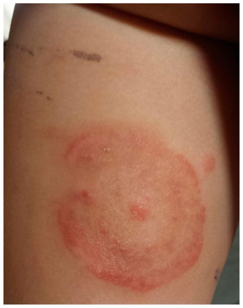
This clinical photograph displays a dermatological presentation of tinea corporis (ringworm) in a pediatric patient. The primary feature is an annular, erythematous plaque located on the trunk or proximal limb. The lesion is characterized by a well-demarcated, raised red border with evidence of mild scaling or vesiculation, indicating active peripheral growth. The center of the plaque shows partial clearing with a lighter pink hue, though it remains occupied by a few scattered inflammatory papules. Above the primary lesion, incidental linear hyperpigmented marks are visible, likely representing excoriations or minor superficial trauma. The image serves as a classic educational example of a fungal skin infection, specifically demonstrating the centrifugal spread and central clearing characteristic of Microsporum canis dermatophytosis.
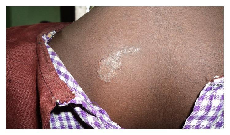
Clinical photograph of a skin lesion on the upper back of a pediatric patient with a dark skin tone, consistent with Tinea corporis (ringworm). The image demonstrates a hypopigmented, well-circumscribed patch with an irregular, slightly raised, and scaly border. Centrally, the lesion shows characteristic clearing but maintains a whitish, fine-textured scaling. A linear extension of the scaling is visible, trailing away from the primary circular focus. This presentation illustrates a common superficial fungal infection (dermatophytosis) of the glabrous skin. The anatomical location is the posterior trunk near the right scapular region. The visual features are diagnostic of a fungal etiology, highlighting the annular morphology and peripheral scale distribution typically seen in localized tinea infections.
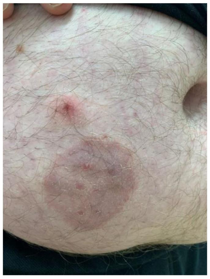
This clinical photograph displays a dermatological presentation of tinea corporis (zoonotic dermatophytosis) on the abdomen of an adult male. The primary feature is a well-circumscribed, annular, erythematous plaque approximately 4-5 cm in diameter. The lesion exhibits a classic 'ringworm' morphology with a slightly raised, scaly inflammatory border and a partially cleared central area containing subtle papular elements. Superior to the primary plaque is a secondary, smaller, raised erythematous papule with a central punctum or crust. The surrounding skin shows mild diffuse erythema and scattered smaller inflammatory macules. Hair follicles are visible throughout the affected and unaffected regions. This image serves as an educational example of fungal infection (likely Trichophyton mentagrophytes) transmitted from a pet (guinea pig) to a human host, emphasizing the visual diagnostic features of annular inflammatory skin lesions.
Ketoconazole for Ringworm (Dermatophytosis)
What is Ringworm?
- Tinea corporis - body/trunk
- Tinea cruris - groin
- Tinea pedis - feet (athlete's foot)
- Tinea capitis - scalp
- Tinea unguium - nails (onychomycosis)
How Ketoconazole Works
Topical Ketoconazole - The Primary Use for Ringworm
| Formulation | Concentration | Indication |
|---|---|---|
| Cream | 2% | Tinea corporis, cruris, pedis, cutaneous Candida, seborrheic dermatitis |
| Foam | 2% | Same as cream |
| Gel | 2% | Same as cream |
| Shampoo | 1% | Tinea capitis (adjuvant), tinea versicolor |
- Fitzpatrick's Dermatology, Table 160-11 (Topical Antifungal Agents)
Efficacy
- Tinea pedis, tinea corporis, tinea cruris
- Tinea versicolor (Malassezia)
- Cutaneous candidiasis
When Topical Ketoconazole Is NOT Enough
- Tinea capitis (scalp) - hair follicle involvement means topical penetration is insufficient; oral agents (griseofulvin, terbinafine, itraconazole, fluconazole) are mandatory. Ketoconazole 2% shampoo is used only as an adjuvant here.
- Tinea unguium (nails) - requires months of oral itraconazole or terbinafine.
- Extensive or recalcitrant tinea corporis/pedis - oral agents may be needed.
Comparison with Other Topical Antifungals
| Drug | Class | Notes |
|---|---|---|
| Ketoconazole 2% | Imidazole | Broad spectrum; also treats seborrheic dermatitis |
| Miconazole 2% | Imidazole | Similar efficacy; widely OTC available |
| Clotrimazole 1% | Imidazole | Comparable efficacy for tinea |
| Terbinafine 1% | Allylamine | Often preferred for tinea pedis; shorter course |
| Tolnaftate | Thiocarbamate | Narrower spectrum; no Candida activity |
Precautions for Topical Ketoconazole
- Can stain skin and clothing
- Avoid use on open wounds (risk of tattooing)
- Pregnancy category C - use with caution
- Avoid contact with eyes
- If no improvement in 4 weeks, reassess diagnosis and consider alternative therapy or oral treatment
Summary
I have ketoconazole soap its effective on ringworm infection
ketoconazole soap dermatophyte tinea efficacy
ketoconazole soap effectiveness ringworm tinea corporis evidence
Ketoconazole Soap vs. Cream - The Key Difference: Contact Time
The Problem with Soap
| Formulation | Contact Time | Skin Penetration |
|---|---|---|
| Cream/Gel 2% | Stays on skin continuously | High - sustained drug levels in keratin |
| Shampoo 2% | 5-10 minutes, then rinsed | Moderate - proven for tinea versicolor with this method |
| Soap | ~30-60 seconds, rinsed off | Very low - insufficient time for penetration |
What the Evidence Actually Shows
- Ketoconazole 2% shampoo - when applied and left on for 5-10 minutes before rinsing, has well-documented efficacy (78-84% mycological cure rates in clinical trials on tinea versicolor). The key is the contact time.
- Ketoconazole 2% cream - applied once or twice daily without rinsing, is the gold standard for ringworm and achieves 70-100% cure rates when used for 2-4 weeks. - Jawetz Medical Microbiology 28e
- Ketoconazole soap - there is essentially no quality clinical trial evidence supporting soap as a standalone treatment for tinea corporis (ringworm). The published literature does not list soap as a recognized treatment option in dermatophytosis management. - Fitzpatrick's Dermatology, Narr. Review on Tinea Corporis (PMC10718250)
Where Soap CAN Help
- Adjuvant hygiene - reduces fungal load on the skin surface during active treatment
- Tinea versicolor - Malassezia (a yeast, not a dermatophyte) lives superficially on skin and is somewhat more susceptible to brief antifungal contact than dermatophytes
- Prevention/recurrence reduction - regular use after successful treatment can help prevent relapse, especially in people prone to fungal infections
- Widespread body infection - used alongside the cream, not as a replacement
Practical Advice
- It will not reliably cure the infection on its own
- You need a ketoconazole 2% cream (or miconazole/clotrimazole cream if ketoconazole is unavailable) applied directly to the lesion and left on skin - once or twice daily for at least 2-4 weeks
- You can continue using the soap as a body wash in addition to the cream - it does no harm and may reduce spreading
- If the lesion is on the scalp, neither soap nor cream alone will work - oral antifungals are required