
Phantom limb
phantom limb pain mechanisms treatment 2025
"phantom limb" pain mechanisms treatment
phantom limb pain cortical reorganization somatosensory cortex diagram
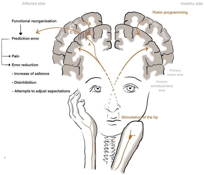
A pathophysiology diagram illustrating theoretical models of phantom limb pain (PLP) through cortical reorganization. The image displays a coronal cross-section of the brain, comparing a 'Healthy side' and an 'Affected side' (post-amputation). On the healthy side, stimulation of the lip correctly activates the face representation in the primary somatosensory area (S1), while motor programming activates the hand representation in the primary motor area. On the affected side, functional reorganization is shown where lip stimulation erroneously activates both the face and the amputated hand's S1 representation. This mismatch is labeled as a 'Prediction error,' leading to the perception of 'Pain.' Three theoretical pathways are identified: (A) Maladaptive plasticity, where neural activation directly involves nociceptive neurons; (B) Persistent pain representation, involving motor-to-S1 feedback; and (C) Predictive coding, where the system attempts 'Error reduction' through increased salience, disinhibition, and attempts to adjust expectations. This schematic serves as a medical educational tool for understanding neuroplasticity and the sensory-motor incongruency underlying neuropathic pain syndromes.
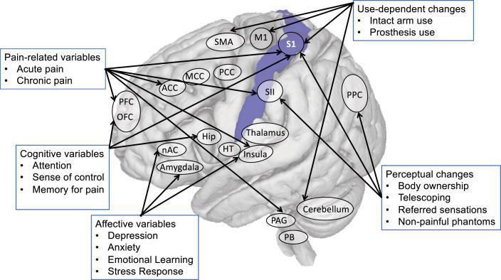
This medical illustration depicts a human brain highlighting the primary somatosensory cortex (S1) and its functional connections within the context of cortical remapping and phantom limb pain. The S1 area is highlighted in purple along the postcentral gyrus. The diagram uses arrows to illustrate the complex interplay between anatomical structures and psychological/behavioral drivers. Key cortical areas shown include the primary motor cortex (M1), supplementary motor area (SMA), secondary somatosensory cortex (SII), and posterior parietal cortex (PPC). Subcortical and limbic structures such as the Thalamus, Amygdala, Hippocampus (Hip), and Periaqueductal Grey (PAG) are also interconnected. Five categorized boxes detail variables influencing S1 organization: 1) Pain-related (acute and chronic); 2) Cognitive (attention, sense of control, memory); 3) Affective (depression, anxiety, stress response); 4) Use-dependent (intact arm or prosthesis use); and 5) Perceptual (body ownership, telescoping, referred sensations). This diagram serves as a conceptual model for neuroplasticity and the multi-factorial nature of chronic pain syndromes.
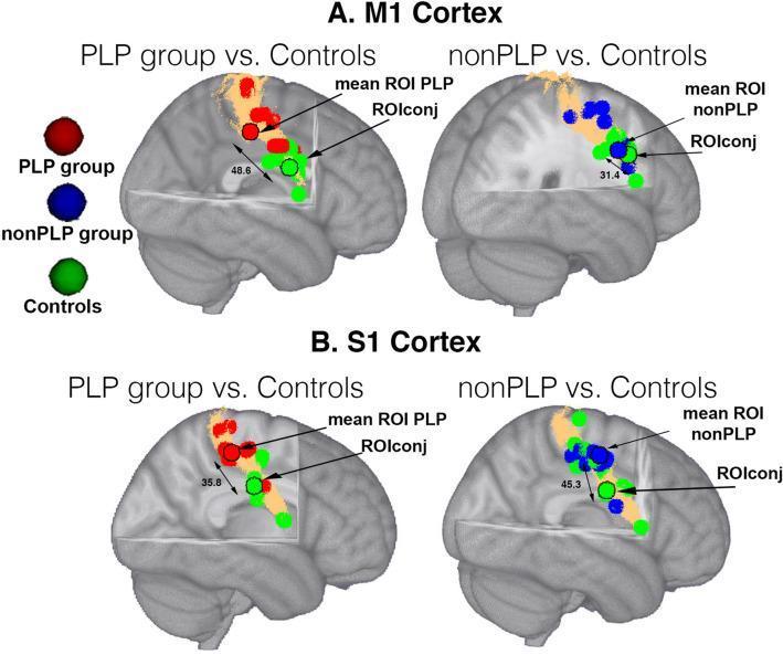
This diagnostic neuroimaging figure displays individual and group-level regions of interest (ROIs) mapped onto a 3D MNI152 brain template, focusing on the primary motor cortex (M1, Panel A) and primary somatosensory cortex (S1, Panel B). The visualization compares three experimental groups: Phantom Limb Pain (PLP, red), non-phantom limb pain (nonPLP, blue), and healthy controls (green). Copper-colored surface masks highlight cortical areas defined by the Juelich atlas. Small colored spheres represent peak coordinates for individual subjects, while spheres with black contours indicate the mean group ROI. A specific 'ROIconj' (conjunction ROI) is identified as the area where control activations overlap. Double-headed arrows with numerical values quantify cortical distances in millimeters (mm) between mean ROIs and the conjunction area. The image illustrates cortical reorganization and spatial shifts in neural activity within the sensorimotor cortex associated with amputation and chronic phantom limb pain, serving as a study of functional neuroanatomy and maladaptive plasticity.
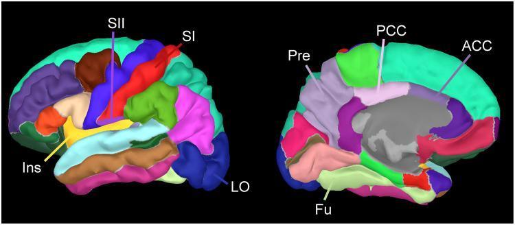
This diagnostic anatomical diagram displays a 3D cortical parcellation of the human brain based on the Desikan-Killiany atlas. The image features two views: a lateral view of the left hemisphere (left) and a medial view of the right hemisphere (right). Various cortical regions are color-coded to delineate functional areas involved in pain processing and connectivity analyses. In the lateral view, the primary somatosensory cortex (SI) is highlighted in red along the postcentral gyrus, the secondary somatosensory cortex (SII) is shown in blue-violet within the parietal operculum, and the insular cortex (Ins) is visible in yellow. The lateral occipital area (LO) is also indicated in deep blue. In the medial view, the anterior cingulate cortex (ACC) is depicted in teal, the posterior cingulate cortex (PCC) in light pink/green, the precuneus (Pre) in lavender, and the fusiform gyrus (Fu) in light green. This visualization serves as a neuroimaging template for mapping regional current density and connectivity in clinical research, specifically for studying the 'pain matrix' and its correlation with subjective pain scales like the Visual Analogue Scale (VAS).
Phantom Limb
Definition
- Phantom limb sensation - non-painful awareness of the missing limb (movement, position, temperature, tingling)
- Phantom limb pain (PLP) - actual pain perceived in the absent limb, affecting 50%-80% of amputees
- Ganong's Review of Medical Physiology, 26th Ed.
Epidemiology
- 50%-80% of amputees experience phantom sensations or pain
- Truly bothersome PLP with functional impact is present in fewer than 10% of amputees - Campbell's Operative Orthopaedics, 15th Ed. 2026
- As of 2005, ~1.6 million Americans lived with limb loss; projected to reach 3.6 million by 2050
- PLP is more frequent after proximal amputations (forequarter, hindquarter)
- Higher risk in women and upper-extremity amputees
- Approximately 30% of amputees experience telescoping (the phantom limb feels like it is retracting into the stump); this is associated with more PLP
Pathophysiology
Peripheral Mechanisms
- Severed nerve endings at the amputation site form neuromas (abnormal regenerative growths)
- Neuromas generate ectopic spontaneous discharge
- Abnormal peripheral input drives and amplifies central reorganization
Spinal Mechanisms
- Increased excitability of dorsal horn neurons (central sensitization)
- Reduction of inhibitory interneuron tone
- Structural changes at primary sensory neuron endings and projection neurons
Supraspinal / Cortical Mechanisms
- The ventral posterior thalamic nucleus remaps: in leg amputees, the region that once received input from the leg now responds to stump stimulation
- Somatosensory cortex reorganization (maladaptive plasticity): in arm/hand amputees, the mouth representation shifts into the hand area of primary somatosensory cortex (S1)
- Stroking the face of an arm amputee can produce sensation in the missing limb - this "referred sensation" is direct evidence of cortical remapping
- The mismatch between expected and actual sensory feedback generates a prediction error, which may be experienced as pain (predictive coding model)
- PET and MRI studies confirm activity in the cortical area of the amputated limb during PLP episodes
Psychological Factors
- Pain is exacerbated by emotional stress, anxiety, and depression
- Pre-existing chronic pain before amputation strongly predicts PLP
- Poor coping strategies and reduced social support correlate with greater PLP severity
Clinical Features
| Feature | Detail |
|---|---|
| Quality | Stabbing, throbbing, burning, cramping |
| Distribution | More intense distally (fingers, toes) |
| Triggers | Weather changes, pressure on stump, emotional stress, micturition |
| Pre-amputation link | Pain quality often mirrors the pain felt before amputation |
| Telescoping | ~30% of patients; phantom gradually shortens toward the stump |
| Sites beyond limbs | Breast, rectum, penis, testicle, eye, tongue, teeth |
- Bradley and Daroff's Neurology in Clinical Practice
Treatment
Pharmacological
| Agent | Evidence |
|---|---|
| Ketamine | Proven effective in controlled studies (NMDA antagonist - reduces central sensitization) |
| Opioids | Effective in controlled studies |
| Calcitonin | Effective in controlled studies |
| Tricyclic antidepressants | First-line for neuropathic pain broadly; no dedicated controlled trials for PLP |
| Anticonvulsants (gabapentin, pregabalin) | Commonly used; evidence modest |
| Sodium channel blockers | Used for neuropathic component |
Non-pharmacological
| Intervention | Notes |
|---|---|
| Mirror therapy | Uses visual feedback of the intact limb to "trick" the brain; a 2024 systematic review (PMID: 37967831) confirms benefit for lower limb amputees |
| TENS | Gate control mechanism; minor effect but non-invasive |
| Spinal cord stimulation | Electrode near spinal cord interferes with ascending nociceptive signals; patients feel tingling instead of pain |
| Deep brain stimulation (DBS) | Reported benefit in refractory cases |
| Virtual reality | Emerging; provides visuoproprioceptive feedback; may surpass mirror therapy for functional tasks - Frontiers in Pain Research, 2025 |
| Myoelectric prosthesis | Active use may alleviate cortical reorganization and reduce PLP |
| Epidural anesthesia pre-amputation | May reduce incidence by preventing acute pain sensitization |
| Botulinum toxin | Systematic review (PMID: 38517393) explored its role in both PLP and residual limb pain |
| Cognitive behavioral therapy | Addresses fear-avoidance and catastrophizing |
| Physical measures | Massage, ice, heat, ultrasound, biofeedback, relaxation, stump compression for edema |
General Supportive Measures
- Control stump edema
- Treat comorbid anxiety and depression
- Improve sleep hygiene
- Encourage smoking cessation
Key Points Summary
- Phantom limb sensation is normal post-amputation; educate patients proactively
- Disabling PLP affects fewer than 10% of amputees, but even mild PLP affects quality of life
- Cortical remapping (maladaptive plasticity) is the dominant current theory
- Pre-amputation pain is the strongest modifiable predictor - good perioperative analgesia may reduce incidence
- No single treatment is curative; combination therapy targeting multiple levels yields the best outcomes
- Emerging technologies (VR, mixed reality) show promise per a 2025 Frontiers editorial