
Leukemia* - define*, classify*, CML CLL ALL AML - peripheral blood smear, bone marrow findings; Acute Leukemia - FAB classification, cytochemical stains, laboratory findings
Reading File
Now let me run parallel searches across all the key topics:
Finding Sources
Finding Sources
Finding Sources
2nd yr mbbs refer harsh mohan
Finding Sources
Finding Sources
Finding Sources
Harsh Mohan's Textbook of Pathology is not in the library. The pathology textbooks available are Robbins & Kumar Basic Pathology and Robbins, Cotran & Kumar Pathologic Basis of Disease — both are excellent for 2nd year MBBS and cover the same leukemia content as Harsh Mohan, often in more detail.
Shall I go ahead and compile the full leukemia notes (definition, classification, CML/CLL/ALL/AML — peripheral smear & bone marrow findings, FAB classification, cytochemical stains, lab findings) using Robbins? The content maps directly to what Harsh Mohan covers for 2nd year MBBS exams.
Generate a comparison table as well of CML vs CLL vs ALL vs AMl
Finding Sources
Finding Sources
Finding Sources
All
Finding Sources
Finding Sources
Finding Sources
Full leukemia notes
Reading File
Reading File
Reading File
Good data coming in. Let me read the remaining critical sections — CLL from Basic Pathology, and AML + more ALL details from Cotran:
Reading File
Reading File
Reading File
Now let me get the AML morphology/FAB details and more CML/CLL/ALL clinical features:
Reading File
Reading File
Finding Sources
Finding Sources
Reading File
Finding Sources
Reading File
Reading File
Reading File
Now let me get the FAB classification table and CLL morphology/clinical features:
Finding Sources
Reading File
Reading File
Finding Sources
Reading File
Searching Images
peripheral blood smear CML chronic myeloid leukemia granulocytes
Now I have all the data. Let me compile the full comprehensive notes:
🩸 LEUKEMIA — Full Notes for 2nd Year MBBS
Source: Robbins & Kumar Basic Pathology + Robbins, Cotran & Kumar Pathologic Basis of Disease
1. DEFINITION
Leukemia is a malignant neoplasm of hematopoietic progenitor cells that proliferate in the bone marrow and spill into the peripheral blood, leading to elevated (or occasionally normal/low) WBC counts with suppression of normal hematopoiesis.
2. CLASSIFICATION
Leukemias are classified by:
| Criterion | Types |
|---|---|
| Maturity of cells | Acute (blasts/immature) vs. Chronic (mature cells) |
| Cell lineage | Myeloid vs. Lymphoid |
This gives the 4 major types:
| Type | Cell | Acuity |
|---|---|---|
| AML | Myeloid | Acute |
| ALL | Lymphoid | Acute |
| CML | Myeloid | Chronic |
| CLL | Lymphoid | Chronic |
3. CHRONIC MYELOID LEUKEMIA (CML)
Epidemiology
- Adults 25–60 years; peak in 4th–5th decades
- ~4500 new cases/year in USA
Pathogenesis
- Hallmark: Philadelphia chromosome (Ph) — t(9;22)(q34;q11)
- Creates BCR-ABL fusion gene → BCR-ABL fusion protein (210 kDa tyrosine kinase)
- Present in >95% of cases (remainder detected by FISH/PCR)
- Arises from pluripotent HSC (affects granulocytic, erythroid, megakaryocytic, B-cell precursors)
- BCR-ABL constitutively activates RAS signalling → growth factor-independent proliferation
- Differentiation is NOT blocked → early disease = excess mature cells
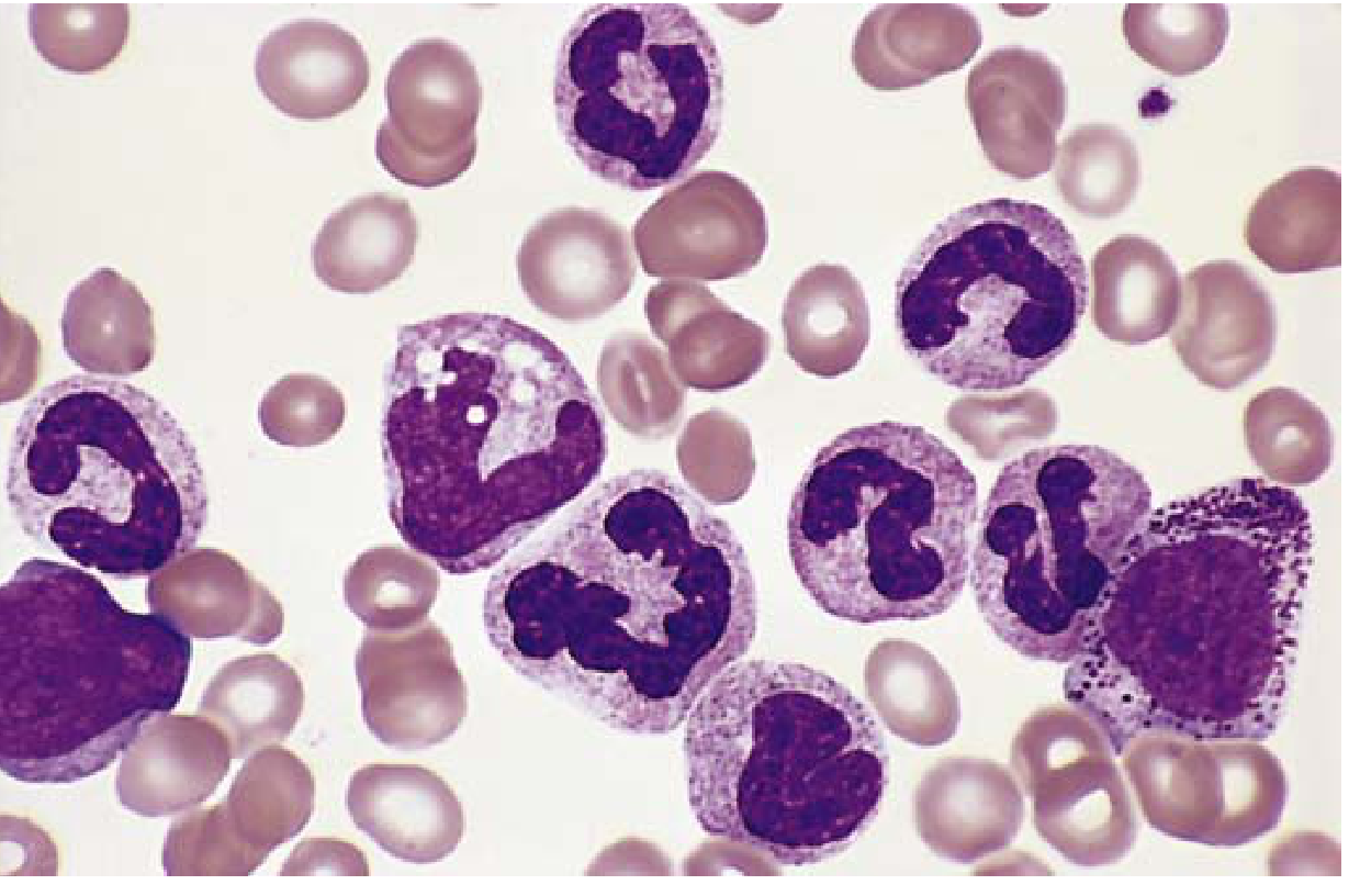
Peripheral Blood Smear
- Leukocytosis — WBC often >100,000 cells/μL
- Spectrum of maturing granulocytes at all stages (myeloblasts → metamyelocytes → neutrophils)
- Basophilia and eosinophilia (characteristic)
- Thrombocytosis (elevated platelets)
- Low LAP score (Leukocyte Alkaline Phosphatase) — distinguishes from leukemoid reaction
Bone Marrow
- Hypercellular (markedly)
- Increased maturing granulocytic and megakaryocytic precursors
- M:E ratio markedly increased
- Extramedullary haematopoiesis in spleen (red pulp resembles bone marrow) → massive splenomegaly
Clinical Features
- Onset insidious — fatigue, weakness, weight loss
- Dragging sensation in abdomen (splenomegaly)
- Distinguish from leukemoid reaction: CML has low LAP score; leukemoid reaction has high LAP
- Blast crisis: transformation to AML/ALL (acquisition of additional mutations)
Treatment
- Imatinib (Gleevec) — BCR-ABL tyrosine kinase inhibitor; revolutionary treatment
-
80% achieve durable remission
4. CHRONIC LYMPHOCYTIC LEUKEMIA (CLL)
Epidemiology
- Most common leukemia of adults in Western world
- Mainly >50 years; males > females
- Less common in Asian populations
Pathogenesis
- Tumour of mature B cells (CD20+, CD5+)
- Increased survival > proliferation — key mechanism
- High BCL-2 (anti-apoptotic) → due to deletion of 13q (loss of miRNAs that negatively regulate BCL-2)
- BTK (Bruton tyrosine kinase) mediates BCR signalling → growth/survival
- CLL/SLL — same disease; CLL = peripheral blood lymphocyte count >5000/μL
Peripheral Blood Smear
- Lymphocytosis — absolute (small, mature-looking lymphocytes)
- Smudge cells (Basket cells) — fragile CLL cells disrupted during smear preparation (pathognomonic)
- Variable larger activated lymphocytes
Bone Marrow / Lymph Node
- Lymph nodes diffusely effaced by sheets of small lymphocytes
- Proliferation centres (foci of larger mitotically active cells) — pathognomonic for CLL/SLL
- Bone marrow, spleen, liver also involved
Immunophenotype
- CD20+, CD5+, surface Ig+ (B-cell marker)
- Only CLL/SLL and mantle cell lymphoma co-express CD5 among B-cell lymphomas
Cytogenetics
- Trisomy 12; deletions of 11q, 13q, 17q
- Chromosomal translocations rare (unlike other B-cell neoplasms)
Clinical Features
- Often asymptomatic initially
- Fatigue, weight loss, anorexia
- Generalised lymphadenopathy + hepatosplenomegaly (50–60%)
- Hypogammaglobulinaemia → bacterial infections
- Autoimmune haemolytic anaemia (warm type), autoimmune thrombocytopenia (~15%)
- WBC can range from mildly elevated to >200,000/μL
- Richter transformation — conversion to aggressive DLBCL (median survival <1 year)
Treatment
- BTK inhibitors (ibrutinib), BCL-2 inhibitors (venetoclax)
- Cure only with haematopoietic stem cell transplant
5. ACUTE LYMPHOBLASTIC LEUKEMIA (ALL)
Epidemiology
- Most common cancer of children (peak age 3 years)
- ~2500 new cases/year; slightly more in boys
- Hispanic/Latino children have highest incidence
- 85% B-ALL, 15% T-ALL
Pathogenesis
- Neoplasm of immature B (pre-B) or T (pre-T) lymphoblasts
- T-ALL: NOTCH1 mutations (50–70%) — essential for T-cell development
- B-ALL: mutations in PAX5, TCF3, ETV6, RUNX1, BCR-ABL1, KMT2A, PBX1
- Maturation arrest + increased self-renewal → stem cell-like phenotype
- ~90% have chromosomal changes: hyperdiploidy (>50 chr, good prognosis), hypodiploidy (poor)
- Key translocation in B-ALL: t(12;21) ETV6-RUNX1 (25% — most common, good prognosis)
- Ph+ B-ALL [t(9;22)] — worse prognosis
FAB Classification of ALL
| FAB | Morphology | Predominant Type |
|---|---|---|
| L1 | Small uniform blasts, high N:C ratio, inconspicuous nucleoli, scant cytoplasm | Common childhood ALL |
| L2 | Large heterogeneous blasts, nuclear clefting/irregularity, prominent nucleoli, more cytoplasm | Adult ALL |
| L3 | Large blasts with deeply basophilic vacuolated cytoplasm, prominent nucleoli | Burkitt-type (mature B-cell) |
(Note: FAB classification of ALL is now largely superseded by immunophenotyping + genetics but still tested in exams)
Peripheral Blood / Bone Marrow Findings
- Bone marrow replaced by >20% lymphoblasts
- Peripheral blood: lymphoblasts present
- Pancytopenia (anaemia, thrombocytopenia, neutropenia) from marrow replacement
- Lymphoblasts: high N:C ratio, immature chromatin, nuclear irregularity (L2)
Cytochemical Stains
| Stain | ALL Result |
|---|---|
| PAS (Periodic Acid-Schiff) | Positive — coarse block-like granules (glycogen) |
| Myeloperoxidase (MPO) | Negative |
| Sudan Black B (SBB) | Negative |
| Non-specific esterase (NSE) | Negative |
| TdT (Terminal deoxynucleotidyl transferase) | Positive (nuclear) — key marker |
Immunophenotype
- B-ALL: CD19+, CD10 (CALLA)+, CD34+, TdT+, HLA-DR+, PAX5+
- T-ALL: CD7+, CD3+, TdT+, CD34±
Clinical Features
- Symptoms of bone marrow failure: fatigue, fever, bleeding (petechiae, ecchymoses)
- Bone pain (marrow expansion)
- Lymphadenopathy, hepatosplenomegaly
- CNS involvement (headache, vomiting, cranial nerve palsies) — more common than AML
- T-ALL: anterior mediastinal mass (thymus), may cause SVC syndrome
- Testicular involvement — sanctuary site
Prognosis
- B-ALL children: 80–90% cure with chemotherapy (best prognosis of all leukemias)
- Ph+ ALL: poor prognosis (treated with TKI + chemo)
6. ACUTE MYELOID LEUKEMIA (AML)
Epidemiology
- Most common acute leukemia in adults (median age >60 years)
- ~13,000–20,000 new cases/year in USA
Pathogenesis
Driver mutations fall into 4 categories:
- Transcription factor mutations → block myeloid differentiation
- t(8;21) → RUNX1::RUNX1T1 — disturbs RUNX1/CBFB transcription factor
- inv(16) → CBFB::MYH11
- t(15;17) → PML::RARα (acute promyelocytic leukemia, APL, M3)
- Tyrosine kinase/RAS signalling mutations → promote proliferation (FLT3, RAS)
- Epigenome regulators (TET2, DNMT3A, IDH1/2)
- Tumour suppressor mutations (TP53 — therapy-related AML)
FAB Classification of AML
| FAB | Name | Key Features |
|---|---|---|
| M0 | AML minimally differentiated | No maturation, no Auer rods; MPO negative by light microscopy |
| M1 | AML without maturation | >90% blasts; MPO/SBB positive; rare Auer rods |
| M2 | AML with maturation | Blasts + maturing granulocytes; t(8;21); Auer rods common |
| M3 | Acute promyelocytic leukemia (APL) | Hypergranular promyelocytes; Auer rod bundles (faggot cells); t(15;17); DIC risk |
| M4 | Acute myelomonocytic leukemia | Myeloid + monocytic blasts; NSE+, MPO+ |
| M5 | Acute monocytic leukemia | Monoblasts/promonocytes; NSE strongly+; gum infiltration (leukemia cutis) |
| M6 | Acute erythroleukaemia | Erythroblasts predominate; PAS+ erythroblasts; TP53 mutations |
| M7 | Acute megakaryoblastic leukemia | Megakaryoblasts; CD41+, CD61+; marrow fibrosis |
Peripheral Blood / Bone Marrow Findings
- ≥20% myeloid blasts in bone marrow (WHO criterion) — some cases with <20% if defining genetics present
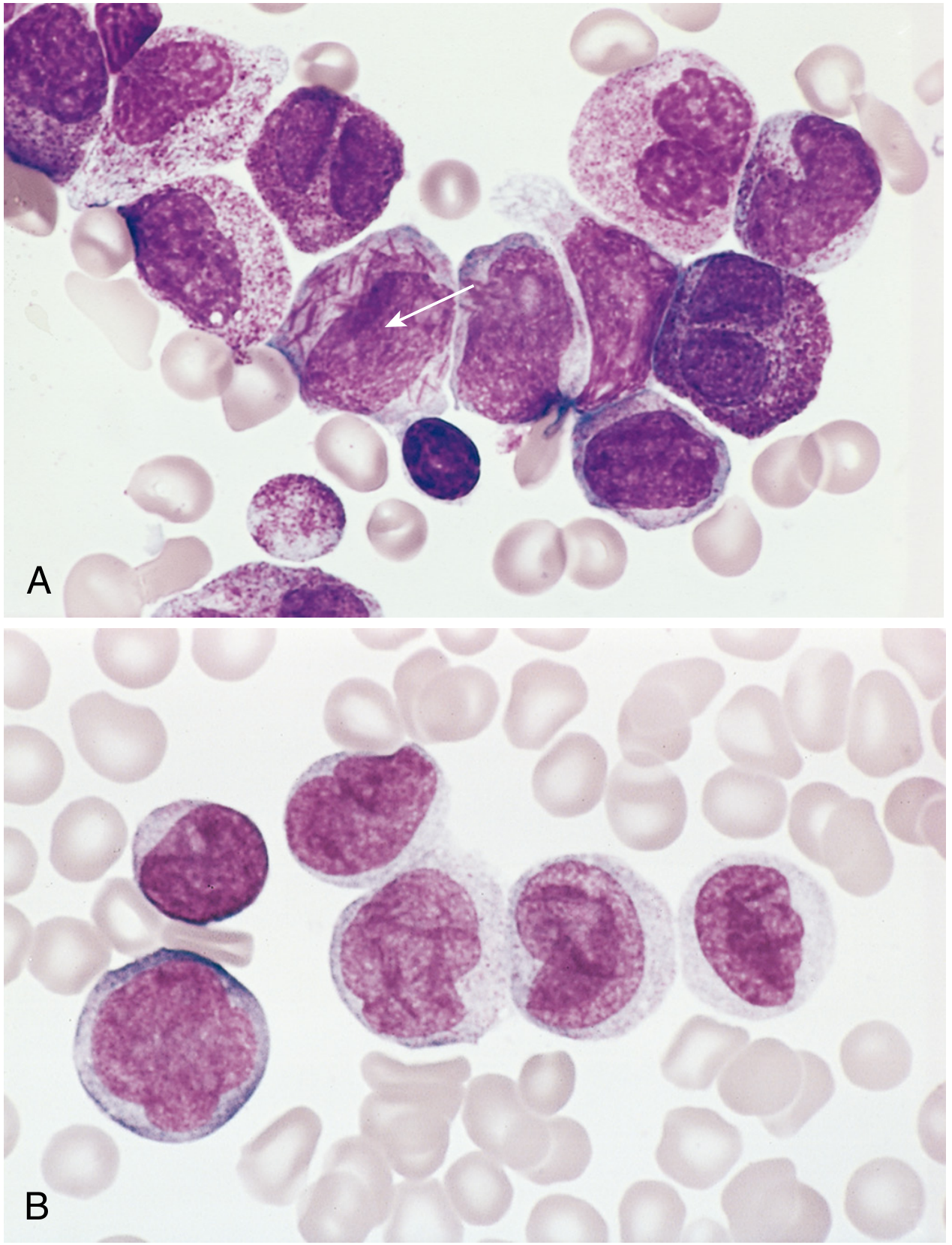
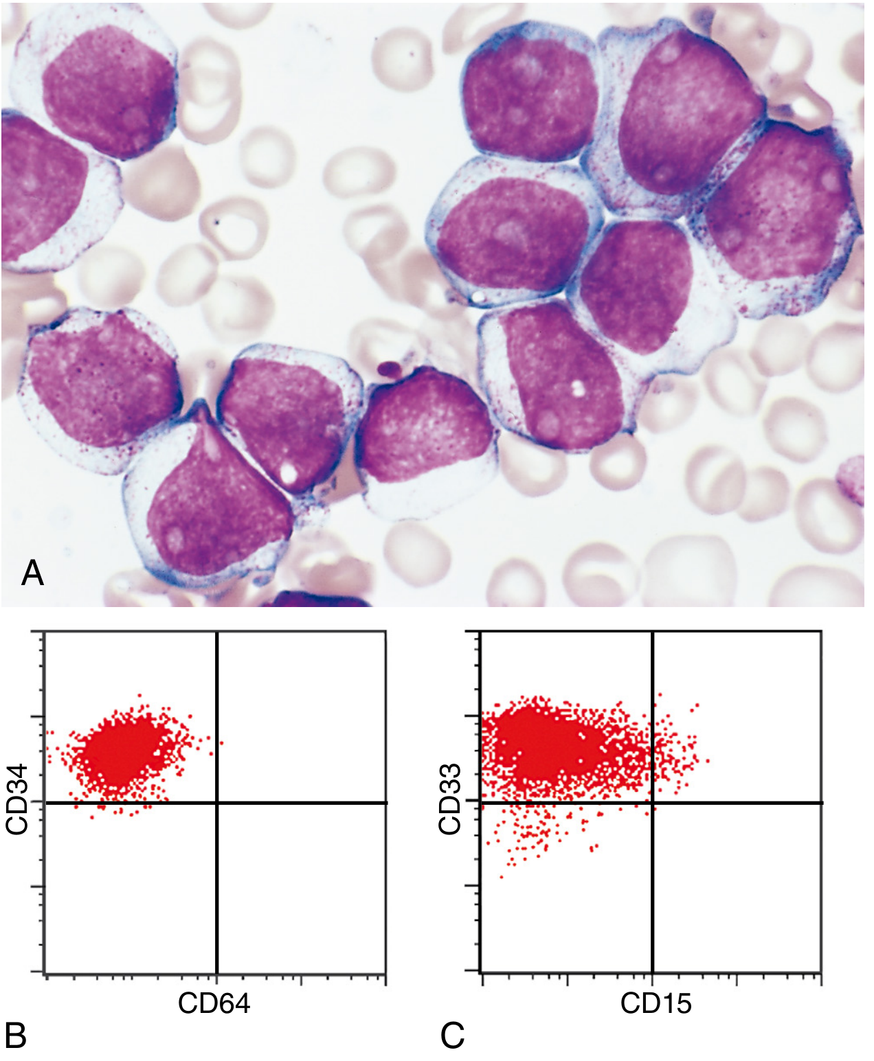
- Myeloblasts: delicate nuclear chromatin, 2–4 nucleoli, voluminous cytoplasm with azurophilic granules
- Auer rods — needle-like azurophilic granules in cytoplasm; pathognomonic for AML; most numerous in M3 (APL)
- Monoblasts (M4/M5): folded/lobulated nuclei, no Auer rods, NSE+
- Occasional aleukemic leukemia — blasts absent from blood; bone marrow essential
Cytochemical Stains in AML
| Stain | AML | Significance |
|---|---|---|
| Myeloperoxidase (MPO) | Positive (myeloblasts) | Best single stain for myeloid lineage |
| Sudan Black B (SBB) | Positive (myeloblasts) | Mirrors MPO; stains lipid granules |
| Non-specific esterase (NSE) | Strongly positive in M4/M5 (monocytic) | Monocytic differentiation |
| PAS | Weak diffuse (myeloid); Strong in M6 erythroblasts | |
| Chloroacetate esterase (CAE) | Positive in neutrophilic lineage | |
| TdT | Negative | (Positive = lymphoid) |
Clinical Features
- Rapid onset over weeks–months
- Fatigue, fever, spontaneous mucosal/cutaneous bleeding (thrombocytopenia)
- Petechiae, ecchymoses, gingival/urinary tract haemorrhage
- Infections — skin, lungs, urinary tract (opportunists: fungi, Pseudomonas)
- M5: gum hypertrophy (leukemia cutis), skin infiltration
- M3 (APL): severe DIC (disseminated intravascular coagulation) — release of procoagulants
- Myeloid sarcoma (chloroma) — localised soft tissue mass
- CNS spread less common than ALL
Cytogenetics in AML
| Translocation | Gene | Prognosis |
|---|---|---|
| t(8;21) | RUNX1::RUNX1T1 | Favourable |
| inv(16) | CBFB::MYH11 | Favourable |
| t(15;17) | PML::RARα (APL) | Favourable with ATRA + arsenic |
| t(9;11) | KMT2A-MLLT3 | Intermediate |
| Complex/del(5,7) | — | Unfavourable |
Treatment
- Standard: 7+3 chemotherapy (cytarabine + anthracycline)
- APL (M3): ATRA (all-trans retinoic acid) + arsenic trioxide → induces differentiation of promyelocytes → excellent outcomes
- ~60% achieve complete remission; only 15–30% disease-free at 5 years overall
7. FAB CLASSIFICATION SUMMARY — ACUTE LEUKEMIAS
AML — FAB (M0–M7)
| FAB | Name | Auer Rods | MPO | NSE | Key Feature |
|---|---|---|---|---|---|
| M0 | Min. differentiated | − | −(EM+) | − | MPO- by light microscopy |
| M1 | Without maturation | ± | + | − | |
| M2 | With maturation | ++ | + | − | t(8;21) |
| M3 | APL | +++ (faggot) | ++ | − | t(15;17), DIC |
| M4 | Myelomonocytic | + | + | + | Both MPO and NSE+ |
| M5 | Monocytic | − | − | +++ | Gum hypertrophy |
| M6 | Erythroleukemia | − | − | − | PAS+ erythroblasts |
| M7 | Megakaryoblastic | − | − | − | CD41+, fibrosis |
ALL — FAB (L1–L3)
| FAB | Size | N:C ratio | Nucleoli | Cytoplasm | Type |
|---|---|---|---|---|---|
| L1 | Small, uniform | Very high | Indistinct | Scant | Common childhood |
| L2 | Large, heterogeneous | Lower | Prominent | More | Adult ALL |
| L3 | Large | Low | Prominent | Deeply basophilic, vacuolated | Burkitt type |
ALL cytochemistry: PAS+ (block pattern), MPO−, SBB−, TdT+
8. COMPARISON TABLE: CML vs CLL vs ALL vs AML
| Feature | CML | CLL | ALL | AML |
|---|---|---|---|---|
| Type | Chronic myeloid | Chronic lymphoid | Acute lymphoid | Acute myeloid |
| Age | 25–60 yrs (peak 4th–5th decade) | >50 yrs (adults) | Children (peak 3 yrs); adults | >60 yrs (adults) |
| Cell of origin | Myeloid stem cell | Mature B cell (CD5+) | Immature B (85%) or T lymphoblast | Myeloid progenitor |
| Key mutation / cytogenetics | t(9;22) BCR-ABL (Ph chromosome) | del(13q), trisomy 12, del(11q) | t(12;21) ETV6-RUNX1 (B-ALL); NOTCH1 (T-ALL); t(9;22) (Ph+ ALL) | t(15;17) APL; t(8;21); inv(16) |
| WBC count | Markedly ↑ (>100,000) | Markedly ↑ (>5000, often >200,000) | ↑ (variable) | Variable (↑, normal, or ↓) |
| Peripheral smear | All stages of granulocytes, basophilia, eosinophilia | Small mature lymphocytes + smudge cells | Lymphoblasts (high N:C ratio) | Myeloblasts ± Auer rods |
| Blast % in marrow | <10% (chronic phase) | <10% | ≥20% | ≥20% |
| Bone marrow | Hypercellular; ↑ granulocytes + megakaryocytes | Infiltrated by small lymphocytes; proliferation centres | Replaced by lymphoblasts; hypercellular | Replaced by myeloid blasts; Auer rods |
| Splenomegaly | Massive (hallmark) | Moderate | Moderate | Variable (less prominent) |
| Lymphadenopathy | Mild/absent | Generalised | Generalised | Mild |
| LAP score | Low | Normal/↑ | — | — |
| Cytochemistry MPO | + (mature cells) | − | − | + |
| Cytochemistry SBB | + | − | − | + |
| Cytochemistry NSE | − | − | − | + (M4/M5) |
| PAS stain | − | − | + (block pattern) | + (M6 erythroblasts) |
| TdT | − | − | + | − |
| Immunophenotype | CD13+, CD33+ (mature markers) | CD20+, CD5+, CD23+ | B-ALL: CD19+, CD10+, TdT+; T-ALL: CD7+, TdT+ | CD13+, CD33+, CD117+, CD34± |
| Hallmark | Philadelphia chromosome | Smudge cells; proliferation centres | PAS+; TdT+; most common childhood cancer | Auer rods; MPO+ |
| Complications | Blast crisis (→AML/ALL) | Richter transformation; autoimmune haemolysis | CNS involvement; testicular disease | DIC (M3/APL); leukemia cutis (M5) |
| Prognosis | Good with imatinib | Variable (indolent); cure with SCT | Children 80–90% cure; Ph+ poor | 60% CR; 15–30% cure |
| Key treatment | Imatinib (BCR-ABL inhibitor) | BTK inhibitors (ibrutinib); venetoclax | Multiagent chemo; TKI if Ph+ | 7+3 chemo; ATRA + arsenic (M3) |
9. KEY MNEMONICS
Auer Rods = AML (never in ALL)
Smudge cells = CLL
Philadelphia chromosome = CML (also Ph+ ALL)
TdT = ALL (and AML M0 rarely)
PAS block pattern = ALL
MPO / SBB = Myeloid (AML positive, ALL negative)
NSE = Monocytic (M4/M5)
ATRA cures APL (M3) — differentiation therapy
Sources: Robbins & Kumar Basic Pathology, 10e; Robbins, Cotran & Kumar Pathologic Basis of Disease, 10e; Quick Compendium of Clinical Pathology 5e; Washington Manual of Medical Therapeutics
---Your credits are exhausted. Your daily credits reset tomorrow, or you can purchase a top-up for more.
AIDS - etiology, routes of transmission, natural history (phases), diagnosis
Your credits are exhausted. Your daily credits reset tomorrow, or you can purchase a top-up for more.
This is a shared conversation. Sign in to Orris to start your own chat.