
Examination of spine
Reading File
Searching sources...
Finding Sources
Reading File
Finding Sources
Reading File
Reading File
Now let me read the thoracic and lumbar spine sections and the special tests section:
Reading File
Searching Images
straight leg raise test lumbar spine Lasegue
Reading File
Reading File
Loading Image
I now have comprehensive content to compile a thorough answer. Let me also fetch a couple more key images.
Loading Image
Loading Image
I now have comprehensive content. Here is the complete structured overview of the examination of the spine:
Examination of the Spine
Source: Bailey and Love's Short Practice of Surgery, 28th Edition
The spinal column consists of 33 vertebrae with 23 intervertebral discs, supported by numerous ligaments and paraspinal muscles. Examination follows the standard orthopaedic sequence: Look - Feel - Move - Neurological - Special Tests, applied separately to the cervical, thoracic, and lumbar regions.
General Inspection (Standing Patient)
Coronal plane (front and back): A plumb line dropped from the spinous process of C7 should fall through the gluteal crease. The ear, shoulder and greater trochanter should lie in the same vertical plane.
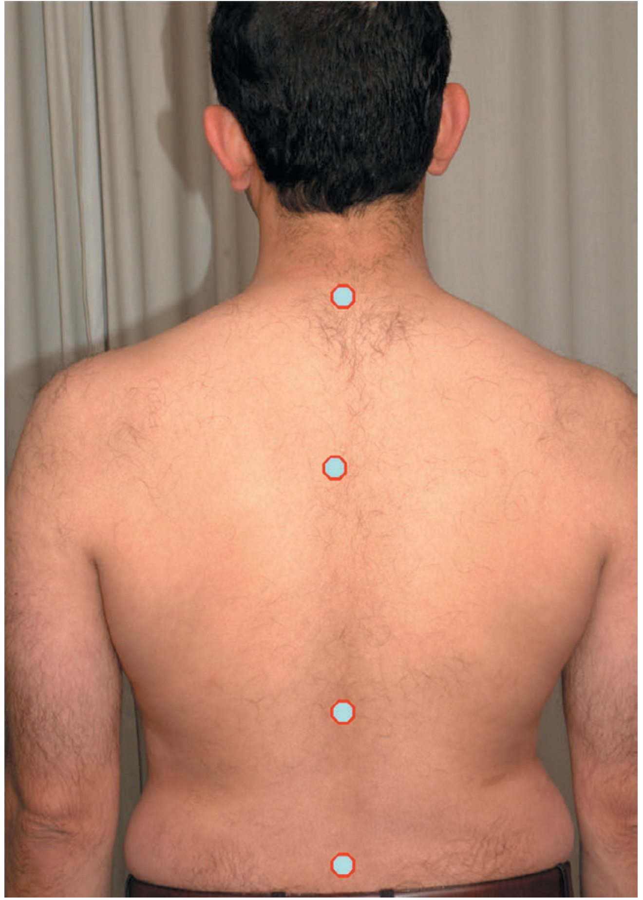
Sagittal plane (side view): Assess the four physiological curves - cervical lordosis (20-40°), thoracic kyphosis (20-45°), lumbar lordosis (40-60°), and sacral kyphosis.
Cervical Spine
Look
- Expose shoulders, back muscles and scapulae
- Look for muscle wasting and asymmetry of neck creases
- Check shoulders are level and there is a normal cervical lordosis (20-40°)
Feel
Stand behind the patient, support the chin.
- Soft tissues: Feel for spasm of the paraspinal muscles
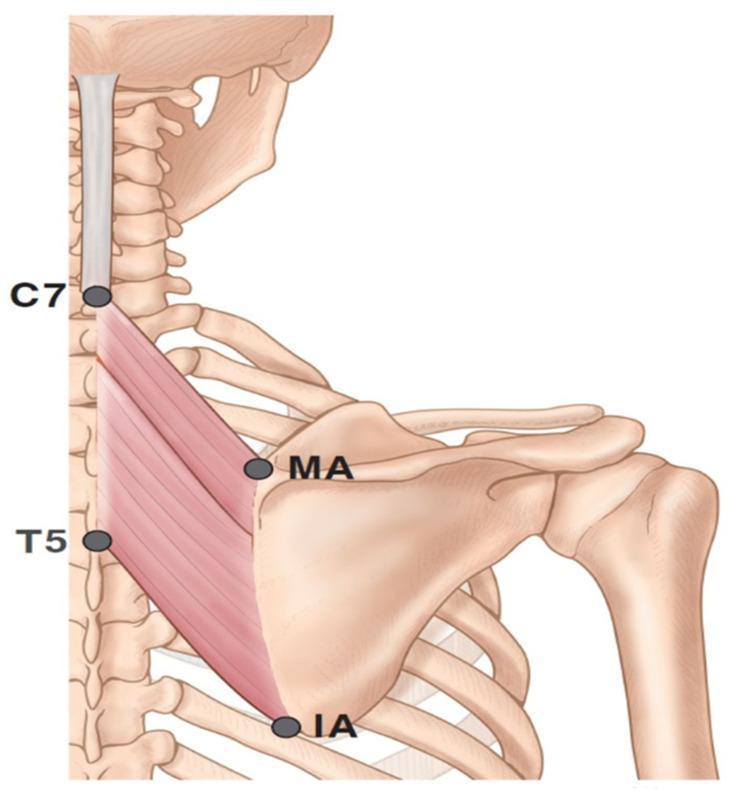
- Bone: Palpate the spinous processes for tenderness and alignment. C7 (vertebra prominens) and T1 are easily palpable at the base of the neck
Move
Motion occurs in three planes:
| Movement | Normal Range | Method |
|---|---|---|
| Flexion | 45° | Chin to chest; measure chin-sternum distance |
| Extension | 55° | Patient looks up at ceiling |
| Rotation (R/L) | 70° | Patient looks over each shoulder without moving chest |
| Lateral bending (R/L) | 40° | Patient lays ear on ipsilateral shoulder |
Neurological
Focus on C5 to T1 nerve roots, which supply the upper extremities. Assess sensation, tone, power, reflexes, proprioception and coordination.
Special Test - Spurling's Test
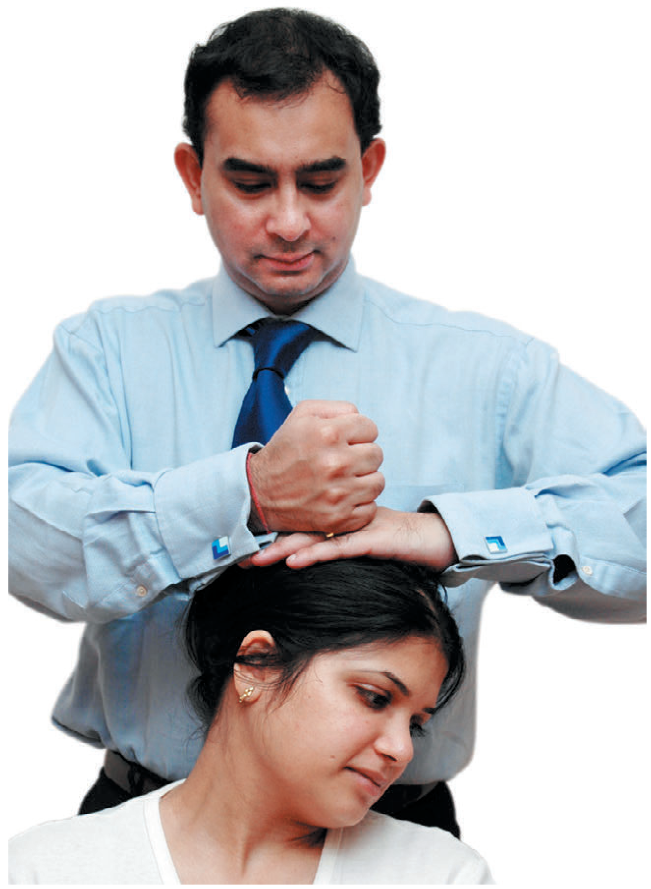
Used to detect cervical nerve root entrapment. The neck is extended and the head rotated toward the symptomatic side, then downward pressure is applied to the top of the head. Reproduction of radicular arm pain is a positive test.
Thoracic Spine
Pathology commonly presents with pain and deformity. The thoracic spine is normally convex with a gentle kyphosis (20-45°).
Look
Ensure front and back from neck to gluteal cleft are visible.
- Skin: Check for cafe-au-lait spots, hairy patches (may suggest occult neurology or bony pathology)
- Front: Check for asymmetry of shoulder and ribcage suggesting scoliosis
- Back: Check for difference in iliac crest height (pelvic tilt); look for coronal plane deformity (scoliosis); a rib hump suggests structural scoliosis
- Side: Assess for increased kyphosis (sagittal plane deformity)
Feel
Palpate with one hand supporting the patient's pelvis.
Move
Forward bending test (Adam's test): Ask the patient to bend forwards to touch their toes:
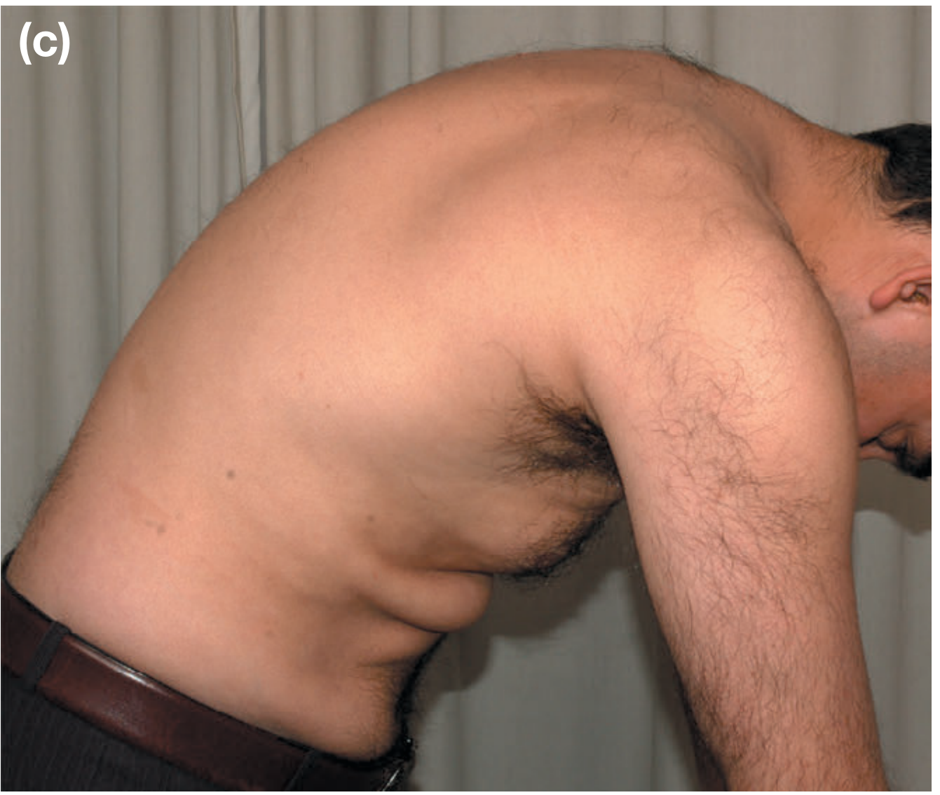
- Structural scoliosis: Rib hump increases as patient bends forwards (bulges posteriorly on thoracic convex side) - diagnostic of idiopathic thoracic scoliosis with rotatory deformity
- Functional scoliosis: Spine straightens on forward bending, no rib hump - secondary to abnormal leg lengths or lumbar muscle spasm
Lateral bending: Assesses flexibility of scoliosis; radiographs can be taken in this position.
Lumbar Spine
Always examine the pelvis, hips, lower limbs, gait, and peripheral vascular system alongside the lumbar region. Always consider referred pain - nerve irritation in the lumbar spine can mimic lower limb problems.
Look
- Back: Check skin at base of spine for hairy tufts and dimples (underlying spina bifida); unilateral prominence of spinal muscles suggests muscle spasm
- Side: The lumbar lordosis (normal 40-60°) should have a smooth concavity; loss of lordosis suggests muscle spasm
Feel
Feel for any 'step-off' in the spinous processes - this may indicate forward slippage of one vertebra on another (spondylolisthesis).
Move
| Movement | Normal Range | Method |
|---|---|---|
| Forward flexion | 40-60° | Schober's test: thumb on T12/L1 junction, index on lumbosacral junction - measure separation as patient bends; also note finger-floor distance |
| Lateral bending | - | Slide hand down outer thigh; compare each side |
| Rotation | 3-18° | Hold pelvis still; ask patient to twist and look over shoulder |
Neurological
Focus on L1 to S1 nerve roots. Assess sensation, tone, power, reflexes (particularly knee jerk L3/4, ankle jerk S1), proprioception and coordination.
Special Tests
Lasègue's Straight Leg Raise (SLR) Test - increases tension along the sciatic nerve (L5 and S1 roots):
- Patient supine - elevate the leg with the knee bent to check pain-free hip movement
- Straighten the knee and note the angle at which hamstrings allow hip to flex
- Lower the hip until tension is removed from hamstrings, then firmly dorsiflex the ankle
- Positive test: pain running down the leg (sciatica) on ankle dorsiflexion
Contralateral stretch test: Elevate the asymptomatic leg; if pain is reproduced in the other (symptomatic) leg, the test is positive (indicates significant nerve root compression, e.g. large disc prolapse).
Summary: Spine Examination at a Glance
| Step | Cervical | Thoracic | Lumbar |
|---|---|---|---|
| Look | Lordosis, wasting, alignment | Scoliosis, rib hump, kyphosis, skin marks | Lordosis, hairy tufts, dimples, muscle prominence |
| Feel | Paraspinal spasm, spinous processes | Palpate with pelvis supported | Step-off deformity |
| Move | Flex/Ext (45°/55°), Lat bend (40°), Rotation (70°) | Forward bending test, lateral bending | Flex (40-60°), Lat bend, Rotation (3-18°) |
| Neuro | C5-T1 roots (upper limb) | Thoracic roots (trunk) | L1-S1 roots (lower limb) |
| Special tests | Spurling's test | Forward bending (Adam's) test | SLR (Lasègue's), Contralateral stretch |
- Bailey and Love's Short Practice of Surgery, 28th Ed., Chapter 35, pp. 497-500
This is a shared conversation. Sign in to Orris to start your own chat.