
PowerPoint for postgraduate internal medicine students on SLE
systemic lupus erythematosus malar butterfly rash face
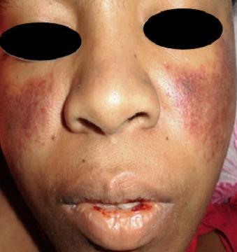
A clinical photograph of a patient's face demonstrating dermatological and mucosal manifestations of Systemic Lupus Erythematosus (SLE). A classic malar rash (butterfly rash) is present, characterized by symmetric, erythematous-to-violaceous patchy lesions over the malar eminences and the bridge of the nose, notably sparing the nasolabial folds. Additionally, the perioral region exhibits significant erythema and mucosal involvement. The lips show evidence of hemorrhagic lesions, with visible blood crusting and a small fissure on the lower lip, suggestive of vasculitis or active systemic inflammation. These visual findings are key diagnostic indicators for SLE, particularly when associated with hematological abnormalities such as thrombocytopenia. The photograph provides a clear example of cutaneous lupus manifestations for clinical diagnosis and medical education.
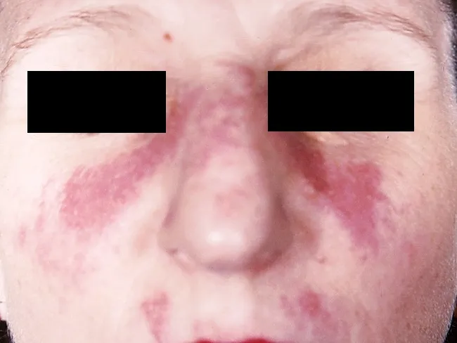
This clinical dermatology photograph displays the classic malar or butterfly rash of systemic lupus erythematosus (SLE) on the central face. Modality is clinical photography using standard white-light illumination; frontal/anterior view; color-balanced, high-resolution capture to depict superficial erythema and patchy hyperemia across the malar eminences and nasal bridge. The rash forms a bilateral, 'butterfly' distribution that typically reaches the cheeks and bridge of the nose while sparing the nasolabial folds. The observed features include confluent to patchy, erythematous macules and plaques with uniform erythema, mild perivascular edema, and subtle textural change without overt crusting or scaling in this image. The clinical morphology is characteristic for acute cutaneous lupus erythematosus; photosensitivity may exacerbate lesions. This cutaneous finding is one of the diagnostic criteria for SLE when aligned with serologic abnormalities (ANA, anti-dsDNA) and systemic features; its presence increases diagnostic probability in a compatible patient. Differential considerations include rosacea, seborrheic dermatitis, contact dermatitis, and dermatomyositis rash; however, the malar distribution and nasal bridge involvement help distinguish lupus. Clinically, this image supports SLE workup and educational reference for recognizing lupus-associated facial rash in medical students, residents, and researchers; useful for pattern-recognition training and multimodal data repository indexing. This image emphasizes clinical-context interpretation and education.
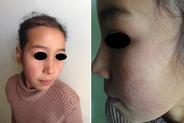
This clinical photograph displays a pediatric patient with dermatological manifestations consistent with Systemic Lupus Erythematosus (SLE). The image consists of two panels: a frontal view and a lateral close-up of the face. A distinctive malar rash, often referred to as a 'butterfly rash,' is visible across the nasal bridge and bilateral cheeks, notably sparing the nasolabial folds. The affected skin exhibits erythematous discoloration and an irregular texture. Higher magnification reveals atrophic scarring within the malar region, characterized by thin, slightly depressed skin with altered pigmentation. The forehead and peri-orbital regions appear relatively unaffected. These visual features are classic diagnostic markers for lupus-related skin involvement, illustrating the chronic inflammatory and sequela phases of the disease. The content is relevant for medical students and clinicians studying rheumatology, dermatology, and autoimmune connective tissue disorders.
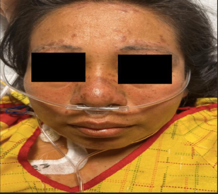
A clinical photograph of a female patient's face demonstrating classic dermatological manifestations of systemic lupus erythematosus (SLE). A confluent, erythematous malar rash is visible in a characteristic 'butterfly distribution,' spanning the bridge of the nose and both cheeks. Notably, the nasolabial folds are spared from the erythema. In addition to the malar rash, there are multiple discrete, raised, hyperpigmented, and round discoid lesions located along the hairline and eyebrows, suggesting discoid lupus involvement. Mild peri-orbital edema is present. The patient is shown with a nasal cannula in place, indicating respiratory support, consistent with potential systemic complications such as pleural effusion. This image serves as a teaching tool for identifying pathognomonic skin signs of autoimmune connective tissue diseases, specifically highlighting the distinction between the malar rash and lesions that involve the forehead and scalp.
lupus nephritis renal biopsy wire loop lesion histology
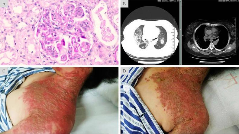
A composite medical image featuring four panels (A-D) illustrating clinical, radiological, and pathological findings of a complex case involving Lupus Nephritis and severe Herpes Zoster. Panel A is a light microscopy image (H&E stain) of a renal biopsy showing glomerular endothelial and mesangial hypercellularity with characteristic 'wire-loop' lesions, consistent with Class IV-G Lupus Nephritis. Panel B presents axial chest CT scans (lung and mediastinal windows) displaying bilateral, scattered patchy and nodular opacities with ill-defined borders, suggestive of diffuse alveolar hemorrhage. Panels C and D are clinical photographs of the neck and chest region showing the progression of skin lesions. Panel C depicts the acute phase of Herpes Zoster, characterized by confluent, grouped vesicles on an intense erythematous base extending across multiple dermatomes. Panel D shows the same region post-antiviral treatment, demonstrating resolving lesions with dried-up exudate and crusting on a fading erythematous base. This figure serves as an educational resource for multi-organ manifestations in immunocompromised patients, specifically highlighting systemic lupus erythematosus complications and disseminated viral infection.
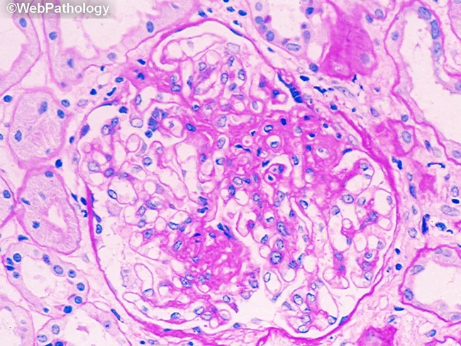
Imaging modality: light microscopy of renal tissue. A single glomerulus is shown in cross section from a renal cortical biopsy. The specimen is stained with Hematoxylin and Eosin (H&E) and viewed at high magnification. The glomerular tuft demonstrates increased cellularity within the capillary loops, consistent with endocapillary proliferation. Mesangial matrix appears expanded, and there are scattered infiltrating cells in the capillary lumens. Capillary lumina are variably narrowed, suggesting early glomerular inflammation. The basement membranes are not clearly visible as thickened in this field, and no overt segmental sclerosis or crescent formation is evident in this slice. The surrounding renal parenchyma shows mild tubulointerstitial involvement, including tubules with pale cytoplasm and interstitial edema, but the primary focus is the glomerulus. The staining highlights nuclei in blue and the extracellular matrix and cytoplasm in pink, typical of routine H&E preparation. This histopathology raises suspicion for a proliferative glomerulonephritis pattern, which can be seen in diseases such as postinfectious GN, IgA nephropathy, or lupus nephritis depending on ancillary tests. Clinically, findings may include hematuria, variable proteinuria, and potential decline in renal function; biopsy helps classify severity and guide therapy. Documentation of periglomerular inflammation and capillary loop involvement supports timely treatment decisions and assessment.
SLE antinuclear antibody ANA immunofluorescence pattern
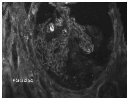
This diagnostic image shows a direct immunofluorescence (DIF) microscopy of a percutaneous kidney biopsy specimen at 20X magnification. The image demonstrates a hallmark pathognomonic finding: strong, continuous, linear immunofluorescence staining along the glomerular basement membrane (GBM). The staining outlines the capillary loops of the glomerulus with high intensity and uniformity, indicating the deposition of immunoglobulin G (IgG) antibodies directly against the basement membrane. The surrounding mesangial regions and Bowman's space show significantly less fluorescence, creating a sharp contrast that emphasizes the linear pattern. This visual presentation is characteristic of anti-glomerular basement membrane (anti-GBM) antibody disease, often associated with Goodpasture syndrome or rapidly progressive (crescentic) glomerulonephritis. The clinical significance of this finding is the confirmation of autoimmune-mediated renal damage where autoantibodies target Type IV collagen within the GBM.
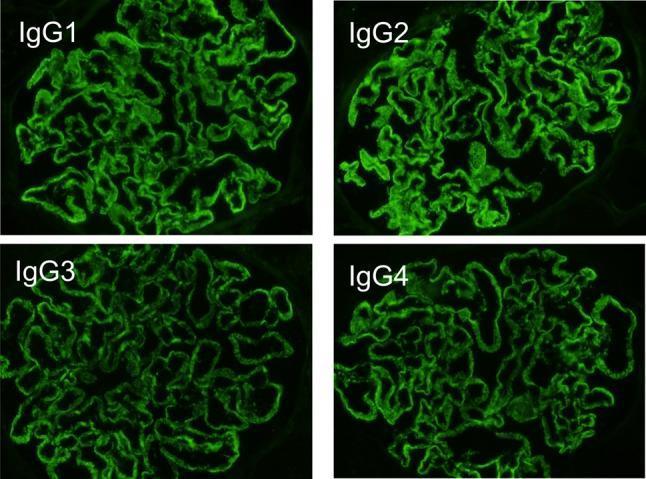
This diagnostic image consists of a four-panel comparison of direct immunofluorescence (DIF) microscopy findings from a renal biopsy. Each panel displays a glomerulus stained for a specific IgG subclass: IgG1, IgG2, IgG3, and IgG4. All four subclasses demonstrate a positive, bright green fluorescent signal. The staining pattern is diffuse and granular, localized predominantly along the glomerular capillary loops, outlining the basement membrane. The distribution is global and relatively uniform across all four images, which is a characteristic finding in lupus-like membranous nephropathy (MN). This multi-subclass deposition (full-house pattern for IgG) helps clinically differentiate secondary causes of membranous nephropathy, such as Systemic Lupus Erythematosus (SLE), from primary membranous nephropathy, which typically shows a predominance of IgG4. The intensity of the fluorescence indicates significant immune complex deposition within the subepithelial space of the glomerular filtration barrier.
discoid lupus erythematosus scalp scarring alopecia skin lesion
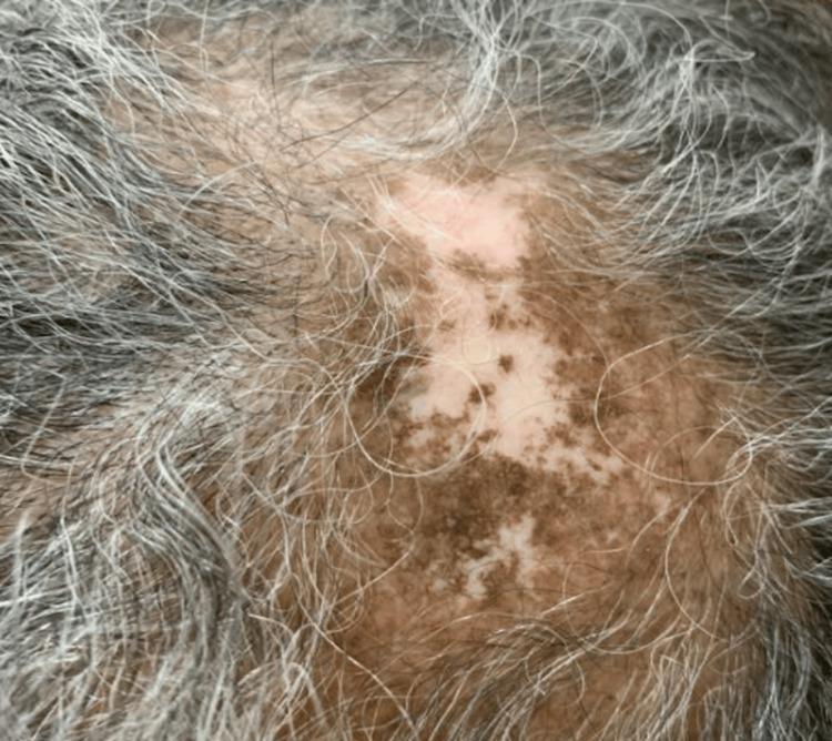
This clinical photograph shows a close-up view of the scalp demonstrating a healed lesion of discoid lupus erythematosus (DLE). The primary finding is a large, irregularly shaped patch of cicatricial (scarring) alopecia characterized by significant hair thinning and permanent hair loss. The affected skin exhibits prominent pigmentary changes, including central hypopigmentation (leukoderma) interspersed with mottled, dark brown hyperpigmented macules, creating a 'salt-and-pepper' appearance. The surrounding scalp skin appears normally pigmented but shows age-related graying (canities) of the remaining hair. The texture within the lesion appears smooth and atrophic, consistent with post-inflammatory scarring from chronic cutaneous lupus. There are no signs of active inflammation such as erythema, scaling, or follicular plugging, indicating the lesion is currently in a quiescent or healed state. This visual is representative of the long-term sequelae of DLE in patients with Systemic Lupus Erythematosus (SLE), highlighting the importance of early intervention to prevent permanent follicular destruction and disfiguring dyschromia.
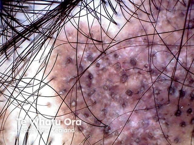
This in vivo scalp dermoscopy/trichoscopy image shows a localized discoid lupus erythematosus lesion presenting with scarring alopecia. The study field is the scalp skin, captured in a close‑up en‑face view at moderate magnification. The scalp surface demonstrates patchy erythema and diffuse hypopigmentation with conspicuous follicular plugging (keratotic follicular openings) and reduced hair follicle ostia consistent with cicatricial alopecia. The numerous dark dot-like structures correspond to follicular openings and keratin plugs, while atrophic scarring regions reveal diminished follicular density and linear scarring tracts. Perifollicular scaling and milky red zones reflect underlying inflammation and vascular changes; crusts or hyperkeratotic scales may be present. These trichoscopic findings, together with clinical history of discoid lupus erythematosus, support a diagnosis of DLE‑related cicatricial alopecia. The imaging is particularly useful for distinguishing scarring alopecias from non-scarring types and for monitoring disease activity or response to antimalarial therapy. Clinically, this pattern raises considerations of photodistributed disease, potential progression to extensive scalp involvement, and need for biopsy correlation and serologic assessment. In educational contexts, this image serves as a reference for dermatology, trichology, and rheumatology collaborations, illustrating hallmark DLE features and differential diagnoses such as lichen planopilaris and inflammatory scarring alopecias.
SLE lupus organ system involvement multisystem diagram
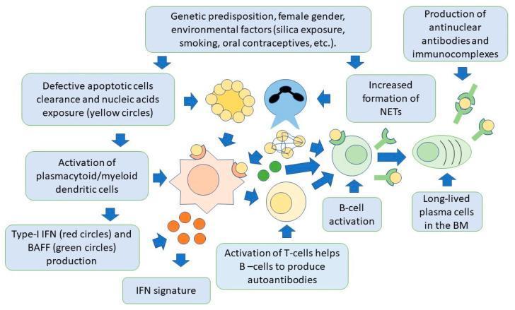
A pathophysiology diagram illustrating the integrated innate and adaptive immune pathways in the pathogenesis of Systemic Lupus Erythematosus (SLE). The process begins with genetic predisposition and environmental triggers (e.g., UV exposure, smoking), leading to defective clearance of apoptotic cells and increased formation of Neutrophil Extracellular Traps (NETs). These processes expose self-nucleic acids (represented by yellow circles) to the immune system. Plasmacytoid and myeloid dendritic cells are activated via toll-like receptors, resulting in the secretion of Type-I Interferon (IFN, red circles) and B-cell activating factor (BAFF, green circles). This cytokine milieu promotes an 'IFN signature' and stimulates self-reactive T-cells and B-cells. Activated B-cells differentiate into long-lived plasma cells within the bone marrow, which continuously produce antinuclear antibodies (ANA). The diagram concludes with the formation of immune complexes (antibodies bound to nuclear antigens) that precipitate in tissues, leading to systemic inflammation and organ damage. The flowchart uses color-coded icons to distinguish molecular components and arrows to denote the sequential activation pathways and cellular interactions.
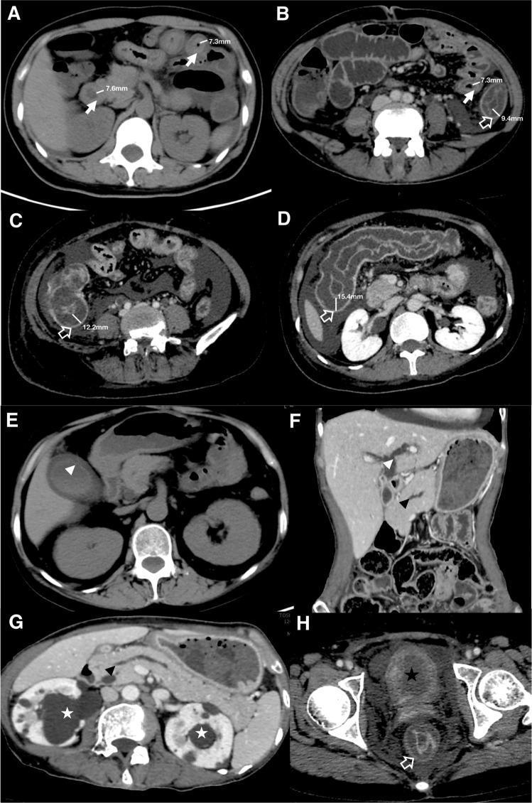
This composite of eight CT images (A–H) illustrates a clinical scoring system for gastrointestinal (GI) and extra-GI involvement in Systemic Lupus Erythematosus (SLE). Images A–D and H demonstrate abnormal bowel wall thickening across various segments: the duodenum and jejunum (A, 7.3–7.6 mm), ileum and descending colon (B, 7.3–9.4 mm), ileocecum (C, 12.2 mm), transverse colon (D, 15.4 mm), and rectum (H). White arrows indicate the small intestine, while hollow arrows denote the large intestine. Extra-GI manifestations are also highlighted: Image E shows gallbladder wall thickening (white arrowhead), and F shows biliary duct dilatation (white arrowhead) alongside pancreatic duct dilatation (black arrowhead in F and G). Urinary system involvement is evidenced by ureterohydronephrosis (white asterisks in D and G) and bladder wall thickening (black asterisk in H). These findings collectively facilitate a CT-based assessment of intestinal pseudo-obstruction (VPO) severity and the presence of multi-organ inflammation characteristic of vasculitic complications in lupus patients.
lupus libman sacks endocarditis echocardiography
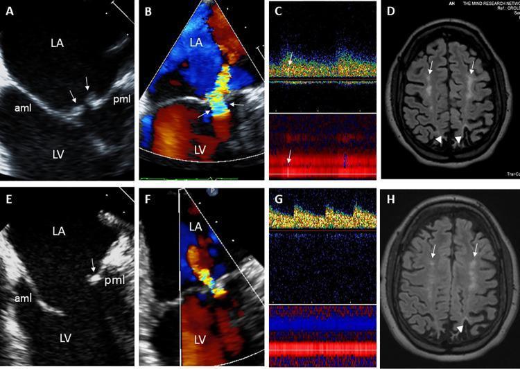
This composite figure illustrates the multi-organ manifestations and treatment response of Libman-Sacks endocarditis. Panels A and B are transesophageal echocardiography (TEE) images showing sessile, oval-shaped vegetations (arrows) on the atrial side of the anterior (aml) and posterior (pml) mitral leaflets, accompanied by a large flow convergence zone indicating moderate to severe eccentric mitral regurgitation. Panel C displays a transcranial Doppler (TCD) showing high-intensity transient signals suggestive of microemboli in the left middle cerebral artery. Panel D is an axial T2-FLAIR brain MRI demonstrating white matter hyperintensities (arrows) and cortical infarcts (arrowheads). Panels E through H represent follow-up imaging after 3 months of immunosuppressive and anticoagulant therapy. These show partial resolution of the mitral vegetations (E), a significant reduction in the mitral regurgitation jet (F), clearance of cerebral microemboli on TCD (G), and a reduction in the total burden and size of white matter lesions and infarct visibility on MRI (H). This clinical comparison highlights the systemic embolic potential of non-bacterial thrombotic endocarditis (NBTE) in the context of systemic lupus erythematosus.
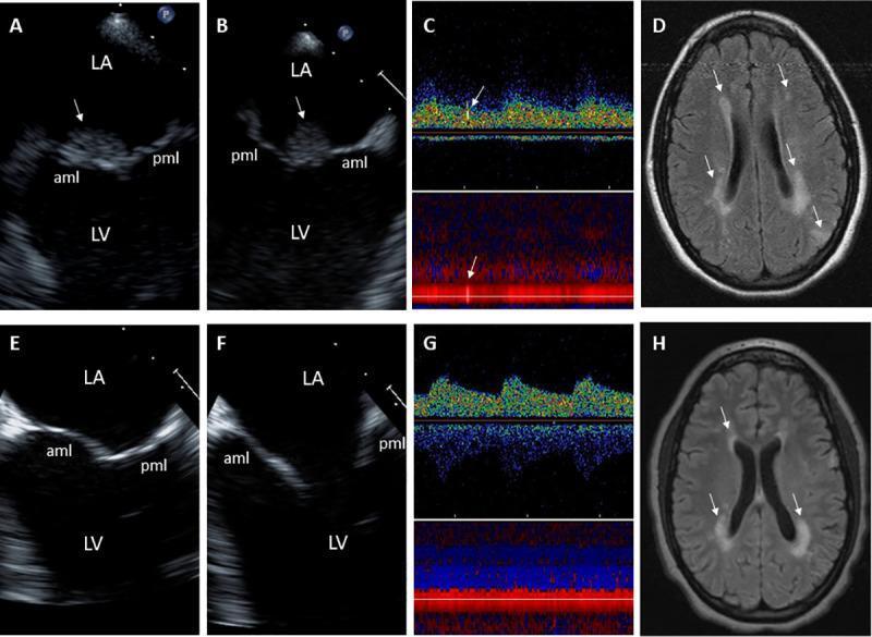
This composite figure illustrates the clinical progression and treatment response of Libman-Sacks endocarditis in an 18-year-old female. Panels A and B are 2-D transesophageal echocardiography (TEE) orthogonal views showing a large, sessile, oval-shaped vegetation (arrows) on the atrial side of the anterior mitral leaflet (aml). Panel C displays a Transcranial Doppler (TCD) of the right middle cerebral artery, identifying microemboli (arrows). Panel D is a brain Magnetic Resonance Imaging (MRI) axial Diffusion-Weighted Image (DWI) demonstrating multiple bilateral periventricular hyperintense lesions indicative of acute cerebral infarcts. Panels E through H show follow-up imaging after 9 weeks of immunosuppressive and anticoagulant therapy. Panels E and F (TEE) reveal complete resolution of the mitral valve vegetations and restoration of smooth leaflet morphology. Panel G (TCD) shows the absence of cerebromicroembolism. Panel H (MRI DWI) demonstrates a significant reduction in the lesion load and intensity of the periventricular white matter infarcts. The set serves as a comprehensive example of the multi-organ involvement of systemic lupus erythematosus-related endocarditis and the efficacy of targeted medical management in reducing embolic sources and secondary neurological complications.
systemic lupus erythematosus management guidelines
lupus erythematosus oral ulcers painless mouth
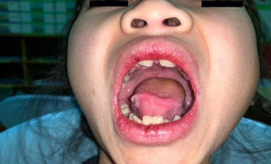
This clinical photograph shows a close-up intraoral view of a pediatric patient's oral cavity and perioral region. The image demonstrates mucocutaneous manifestations typical of systemic inflammatory or autoimmune conditions. Key visible findings include multiple shallow, erythematous ulcers with ill-defined borders located on the dorsal and lateral surfaces of the tongue and the hard palate. The lips (cheilitis) appear significantly erythematous and edematous, with evidence of crusting and irregularity along the vermillion border of the upper and lower lips. The oral mucosa shows generalized hyperemia. In the clinical context of malar rash and systemic symptoms, these findings are highly suggestive of systemic lupus erythematosus (SLE) mucosal involvement. The image serves as an educational resource for identifying painless oral ulcerations and inflammatory lip changes as diagnostic criteria in pediatric rheumatology.
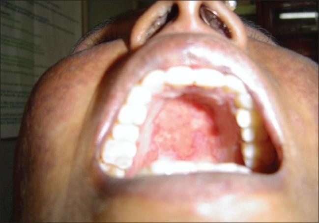
This clinical intraoral photograph displays the hard and soft palate of a patient, showing significant mucosal pathology. The image reveals multiple, scattered erythematous ulcers and erosions localized to the palatal mucosa. These lesions are characterized by a red, inflamed base with irregular borders; some appear to be coalescing into larger patches. The surrounding palatal tissue shows signs of diffuse inflammation and disruption of the normal smooth texture. Such findings are a key clinical manifestation of Systemic Lupus Erythematosus (SLE), where painless oral ulcers often serve as a non-specific but important diagnostic criterion. The photograph provides a clear educational example of autoimmune-related mucosal damage, useful for medical students and clinicians in identifying oral signs of systemic connective tissue diseases. The dental arches and teeth are visible and appear largely unaffected by the primary pathology.
mkdir -p /tmp/workspace/SLE_presentation && ls /tmp/workspace/SLE_presentation/
Where is the presentation
~/SLE_presentation/build_sle_pptx.py
import json, subprocess, base64, textwrap
from io import BytesIO
from pptx import Presentation
from pptx.util import Inches, Pt, Emu
from pptx.dml.color import RGBColor
from pptx.enum.text import PP_ALIGN, MSO_ANCHOR
from pptx.oxml.ns import qn
from lxml import etree

# ── Colour palette ──────────────────────────────────────────────────────────
NAVY      = RGBColor(0x0A, 0x2A, 0x5C)   # dark navy – slide titles / header BG
TEAL      = RGBColor(0x00, 0x7A, 0x87)   # accent / sub-headings
GOLD      = RGBColor(0xE8, 0xA1, 0x00)   # highlight / icons
WHITE     = RGBColor(0xFF, 0xFF, 0xFF)
LIGHT_BG  = RGBColor(0xF4, 0xF7, 0xFC)   # slide background
DARK_TXT  = RGBColor(0x1A, 0x1A, 0x2E)
MID_GREY  = RGBColor(0x6B, 0x7A, 0x99)
RED_ALERT = RGBColor(0xC0, 0x39, 0x2B)
GREEN_OK  = RGBColor(0x1A, 0x7A, 0x4A)

W = Inches(13.333)
H = Inches(7.5)

# ── Helper: fetch images ─────────────────────────────────────────────────────
IMAGE_URLS = {
    "malar_rash":      "https://cdn.orris.care/cdss_images/Pathology_1760052272739_5d26310f-f900-4ecf-ad1f-83f7a618bc35.jpg",
    "malar_rash2":     "https://cdn.orris.care/cdss_images/pmc_clinical_VQA_0b4e1b51b007615806cc3a1d6dcf8520d34e446c5f6904a8048b957ad3e3a70a.jpg",
    "discoid":         "https://cdn.orris.care/cdss_images/pmc_clinical_VQA_0d7bb1e0f8c1514ed23a0bd45bc17a975a074e9166d1420f2487b8e0e499b031.jpg",
    "oral_ulcers":     "https://cdn.orris.care/cdss_images/pmc_clinical_VQA_a6369d39475c39f4efa49cd342f1afb901779dd7120cfc094c9b7197b4c90215.jpg",
    "pathogenesis":    "https://cdn.orris.care/cdss_images/pmc_clinical_VQA_0dd08c3bc1131bde40fc5ecdf7ab6f319182b7ff677ebd42b2bcc62ec4d155e7.jpg",
    "nephritis_histo": "https://cdn.orris.care/cdss_images/pmc_clinical_VQA_92cc809ac0c810c6819fd2ae2abca485602cfbc4d6be79d02e2d817463ecaeea.jpg",
    "libman_sacks":    "https://cdn.orris.care/cdss_images/pmc_clinical_VQA_6689fcaabc4ca805f6b63a1da004e1726e00e676ee66aa10a796198084233c4e.jpg",
    "full_house_if":   "https://cdn.orris.care/cdss_images/pmc_clinical_VQA_77f37c536b3be1dcf921cc5d5026e14e8ef7a8c83ede5ad031327cd613e0c07b.jpg",
    "discoid_trich":   "https://cdn.orris.care/cdss_images/DermNetNZ_1760030887040_790df1ea-720d-4c8c-bd40-199addafd792.jpg",
    "malar_rash3":     "https://cdn.orris.care/cdss_images/pmc_clinical_VQA_1840306c3bee083a0905954bc9db424cbbc7ff22c6954b71f0e6ca8069aab78a.jpg",
}

print("Fetching images …")
urls_list = list(IMAGE_URLS.values())
raw = subprocess.check_output(
    ["python", "/tmp/skills/shared/scripts/fetch_images.py"] + urls_list,
    timeout=60
)
fetched = json.loads(raw)
img_data = {}
for key, url in IMAGE_URLS.items():
    for item in fetched:
        if item["url"] == url and item.get("base64"):
            img_data[key] = item["base64"]
            break

print(f"Fetched {len(img_data)} / {len(IMAGE_URLS)} images")

# ── Utility functions ────────────────────────────────────────────────────────
def add_img(slide, key, left, top, width, height=None):
    if key not in img_data:
        return
    mime, b64 = img_data[key].split(";base64,", 1)
    ext = "jpeg" if "jpeg" in mime or "jpg" in mime else "png"
    raw_bytes = base64.b64decode(b64)
    bio = BytesIO(raw_bytes)
    if height:
        slide.shapes.add_picture(bio, left, top, width, height)
    else:
        slide.shapes.add_picture(bio, left, top, width)

def rect(slide, left, top, width, height, fill_rgb, alpha=None):
    shape = slide.shapes.add_shape(1, left, top, width, height)
    shape.line.fill.background()
    shape.fill.solid()
    shape.fill.fore_color.rgb = fill_rgb
    return shape

def textbox(slide, text, left, top, width, height,
            size=18, bold=False, italic=False, color=DARK_TXT,
            align=PP_ALIGN.LEFT, wrap=True, anchor=MSO_ANCHOR.TOP,
            font="Calibri"):
    tb = slide.shapes.add_textbox(left, top, width, height)
    tf = tb.text_frame
    tf.word_wrap = wrap
    tf.vertical_anchor = anchor
    tf.margin_left = Inches(0.05)
    tf.margin_right = Inches(0.05)
    tf.margin_top = Pt(2)
    tf.margin_bottom = Pt(2)
    p = tf.paragraphs[0]
    p.alignment = align
    run = p.add_run()
    run.text = text
    run.font.name = font
    run.font.size = Pt(size)
    run.font.bold = bold
    run.font.italic = italic
    run.font.color.rgb = color
    return tb

def multiline_textbox(slide, lines, left, top, width, height,
                      size=16, color=DARK_TXT, leading_bold=False,
                      font="Calibri", line_spacing=1.15):
    tb = slide.shapes.add_textbox(left, top, width, height)
    tf = tb.text_frame
    tf.word_wrap = True
    tf.auto_size = None
    tf.margin_left = Inches(0.08)
    tf.margin_right = Inches(0.05)
    tf.margin_top = Pt(2)
    tf.margin_bottom = Pt(2)
    first = True
    for line in lines:
        if first:
            p = tf.paragraphs[0]
            first = False
        else:
            p = tf.add_paragraph()
        p.space_after = Pt(3)
        if isinstance(line, tuple):
            # (bold_part, normal_part)
            r1 = p.add_run()
            r1.text = line[0]
            r1.font.bold = True
            r1.font.size = Pt(size)
            r1.font.color.rgb = color
            r1.font.name = font
            if len(line) > 1:
                r2 = p.add_run()
                r2.text = line[1]
                r2.font.bold = False
                r2.font.size = Pt(size)
                r2.font.color.rgb = color
                r2.font.name = font
        else:
            run = p.add_run()
            run.text = line
            run.font.size = Pt(size)
            run.font.color.rgb = color
            run.font.name = font
            if leading_bold and first:
                run.font.bold = True
    return tb

def slide_header(slide, title, subtitle=None):
    """Top navy bar + title."""
    rect(slide, 0, 0, W, Inches(1.1), NAVY)
    textbox(slide, title, Inches(0.35), Inches(0.08), Inches(12.5), Inches(0.8),
            size=28, bold=True, color=WHITE, align=PP_ALIGN.LEFT, font="Calibri")
    if subtitle:
        textbox(slide, subtitle, Inches(0.35), Inches(0.78), Inches(12.5), Inches(0.35),
                size=14, italic=True, color=RGBColor(0xCC, 0xDD, 0xFF), font="Calibri")
    # teal accent line
    rect(slide, 0, Inches(1.1), W, Pt(4), TEAL)

def slide_bg(slide):
    rect(slide, 0, 0, W, H, LIGHT_BG)

def bullet_slide(slide, title, bullets, subtitle=None, two_col=False,
                 img_key=None, img_caption=None, notes=None):
    slide_bg(slide)
    slide_header(slide, title, subtitle)
    content_top = Inches(1.25)
    content_h   = Inches(5.9)
    if img_key and img_key in img_data:
        left_w = Inches(7.8)
        img_l  = Inches(8.0)
        img_w  = Inches(5.0)
        add_img(slide, img_key, img_l, content_top, img_w)
        if img_caption:
            textbox(slide, img_caption, img_l, Inches(6.7), img_w, Inches(0.45),
                    size=11, italic=True, color=MID_GREY, align=PP_ALIGN.CENTER)
    else:
        left_w = Inches(12.7)

    lines = []
    for b in bullets:
        if isinstance(b, str):
            lines.append("  \u2022  " + b)
        else:
            lines.append(b)  # pass-through tuple
    multiline_textbox(slide, lines, Inches(0.35), content_top, left_w, content_h,
                      size=17, color=DARK_TXT)
    if notes:
        notes_slide = slide.notes_slide
        notes_slide.notes_text_frame.text = notes

def section_divider(prs, section_title, section_num, color=TEAL):
    blank = prs.slide_layouts[6]
    slide = prs.slides.add_slide(blank)
    rect(slide, 0, 0, W, H, NAVY)
    # big accent block
    rect(slide, 0, Inches(2.8), W, Inches(1.9), color)
    textbox(slide, f"Section {section_num}", Inches(1), Inches(1.5), Inches(11), Inches(1),
            size=22, color=RGBColor(0xCC, 0xDD, 0xFF), bold=False, align=PP_ALIGN.CENTER)
    textbox(slide, section_title, Inches(0.5), Inches(2.9), Inches(12.3), Inches(1.7),
            size=40, bold=True, color=WHITE, align=PP_ALIGN.CENTER, anchor=MSO_ANCHOR.MIDDLE)
    return slide

# ── Build presentation ───────────────────────────────────────────────────────
prs = Presentation()
prs.slide_width  = W
prs.slide_height = H
blank = prs.slide_layouts[6]

# ════════════════════════════════════════════════════════════════════════════
# SLIDE 1 – TITLE
# ════════════════════════════════════════════════════════════════════════════
s = prs.slides.add_slide(blank)
rect(s, 0, 0, W, H, NAVY)
rect(s, 0, Inches(4.2), W, Inches(3.3), RGBColor(0x06, 0x1A, 0x40))
rect(s, 0, Inches(4.15), W, Pt(6), GOLD)

# large SLE text
textbox(s, "Systemic Lupus", Inches(0.6), Inches(0.5), Inches(9), Inches(1.35),
        size=58, bold=True, color=WHITE, font="Calibri")
textbox(s, "Erythematosus", Inches(0.6), Inches(1.75), Inches(9), Inches(1.3),
        size=58, bold=True, color=GOLD, font="Calibri")
textbox(s, "A Comprehensive Review for Postgraduate Internal Medicine",
        Inches(0.6), Inches(3.2), Inches(9), Inches(0.8),
        size=20, italic=True, color=RGBColor(0xAA, 0xCC, 0xFF), font="Calibri")

textbox(s, "Department of Internal Medicine  |  Postgraduate Education Series",
        Inches(0.6), Inches(4.5), Inches(12), Inches(0.5),
        size=15, color=RGBColor(0x88, 0xAA, 0xCC), font="Calibri")
textbox(s, "2026  |  Based on ACR 2025, EULAR/ACR 2019, KDIGO 2024 Guidelines",
        Inches(0.6), Inches(5.0), Inches(12), Inches(0.5),
        size=13, italic=True, color=RGBColor(0x88, 0xAA, 0xCC), font="Calibri")

# malar rash image on right
if "malar_rash" in img_data:
    add_img(s, "malar_rash", Inches(9.4), Inches(0.3), Inches(3.7), Inches(3.7))
    rect(s, Inches(9.35), Inches(0.25), Inches(3.8), Inches(3.8), GOLD)
    add_img(s, "malar_rash", Inches(9.4), Inches(0.3), Inches(3.7), Inches(3.7))

# ════════════════════════════════════════════════════════════════════════════
# SLIDE 2 – LEARNING OBJECTIVES
# ════════════════════════════════════════════════════════════════════════════
s = prs.slides.add_slide(blank)
slide_bg(s)
slide_header(s, "Learning Objectives")
objectives = [
    "Understand the epidemiology and aetiopathogenesis of SLE",
    "Apply the EULAR/ACR 2019 classification criteria in clinical practice",
    "Recognise the diverse multisystem clinical manifestations of SLE",
    "Interpret the immunological investigations (ANA, anti-dsDNA, complement)",
    "Classify lupus nephritis using ISN/RPS 2018 criteria",
    "Design a stepwise management plan incorporating hydroxychloroquine, immunosuppressives, and biologics",
    "Identify and manage major complications: nephritis, NPSLE, cardiovascular, antiphospholipid syndrome",
    "Monitor disease activity using SLEDAI and BILAG scoring tools",
    "Manage SLE in special populations: pregnancy, paediatric, elderly",
]
rect(s, Inches(0.3), Inches(1.25), Inches(12.7), Inches(5.9), WHITE)
for i, obj in enumerate(objectives):
    y = Inches(1.35) + i * Inches(0.62)
    rect(s, Inches(0.35), y + Pt(6), Inches(0.4), Inches(0.38), TEAL)
    textbox(s, f"{i+1}", Inches(0.35), y + Pt(4), Inches(0.4), Inches(0.4),
            size=13, bold=True, color=WHITE, align=PP_ALIGN.CENTER)
    textbox(s, obj, Inches(0.85), y, Inches(12.1), Inches(0.58),
            size=16, color=DARK_TXT)

# ════════════════════════════════════════════════════════════════════════════
# SECTION 1
# ════════════════════════════════════════════════════════════════════════════
section_divider(prs, "Epidemiology & Aetiopathogenesis", 1, TEAL)

# SLIDE 3 – EPIDEMIOLOGY
bullet_slide(prs.slides.add_slide(blank),
    "Epidemiology of SLE",
    [
        "Global prevalence: 20–150 per 100,000 population; incidence 1–25 per 100,000/year",
        "Female predominance: F:M ratio ~ 9:1 (reproductive age); narrows in childhood & elderly",
        "Peak onset: 15–45 years; can occur at any age",
        "Ethnic disparities: higher severity & prevalence in Afro-Caribbean, Hispanic, Asian populations",
        "African Americans: 3× higher incidence, earlier onset, more severe renal/CNS disease vs. Caucasians",
        "Mortality: standardised mortality ratio 2–3× general population; leading causes: infection, cardiovascular, renal failure",
        "Morbidity: chronic, relapsing-remitting course; significant impact on quality of life",
    ],
    subtitle="Prevalence, Demographics & Disease Burden"
)

# SLIDE 4 – AETIOLOGY & RISK FACTORS
bullet_slide(prs.slides.add_slide(blank),
    "Aetiology & Risk Factors",
    [
        ("Genetic (30%): ", "HLA-DR2, HLA-DR3; C1q, C2, C4 complement deficiencies; IRF5, STAT4, BLK, PTPN22 polymorphisms"),
        ("Hormonal: ", "Oestrogen promotes B-cell activation; prolactin elevates disease activity; testosterone protective"),
        ("Environmental triggers: ", "UV light (induces apoptosis → nuclear antigen release), infections (EBV cross-reactivity), silica dust"),
        ("Drugs: ", "Procainamide, hydralazine, isoniazid, minocycline → drug-induced SLE (anti-histone antibodies)"),
        ("Epigenetic: ", "DNA hypomethylation of immune genes (CD11a, CD70); microRNA dysregulation"),
        ("Defective apoptosis clearance: ", "Impaired clearance of apoptotic debris → exposure of nuclear antigens → autoantibody production"),
        "Concordance in identical twins: 25–50% (indicates strong but not exclusive genetic role",
    ],
    subtitle="Multifactorial: Genetic + Hormonal + Environmental"
)

# SLIDE 5 – PATHOGENESIS
s = prs.slides.add_slide(blank)
slide_bg(s)
slide_header(s, "Pathogenesis of SLE", subtitle="Loss of Tolerance to Self-Nuclear Antigens")
add_img(s, "pathogenesis", Inches(0.3), Inches(1.25), Inches(7.6), Inches(5.8))
textbox(s, "Key Pathogenic Steps:", Inches(8.1), Inches(1.3), Inches(5.0), Inches(0.5),
        size=17, bold=True, color=TEAL)
steps = [
    "1. Defective clearance of apoptotic cells exposes nuclear antigens (dsDNA, histones, Sm)",
    "2. NETs from neutrophils release chromatin → triggers pDC via TLR7/9",
    "3. pDCs produce Type I Interferons (IFN-α) → 'IFN signature'",
    "4. BAFF production promotes self-reactive B-cell survival",
    "5. T-cell help (Tfh cells) drives autoantibody class switching",
    "6. Autoantibodies (anti-dsDNA, anti-Sm) form immune complexes",
    "7. IC deposition in kidneys, skin, vessels → complement activation → inflammation",
    "8. Chronic inflammation → organ damage (nephritis, vasculitis, CNS disease)",
]
multiline_textbox(s, steps, Inches(8.0), Inches(1.85), Inches(5.1), Inches(5.1),
                  size=14, color=DARK_TXT)

# ════════════════════════════════════════════════════════════════════════════
# SECTION 2
# ════════════════════════════════════════════════════════════════════════════
section_divider(prs, "Classification Criteria", 2, GOLD)

# SLIDE 6 – ACR 1997 CRITERIA
s = prs.slides.add_slide(blank)
slide_bg(s)
slide_header(s, "ACR 1997 Classification Criteria", subtitle="≥4 of 11 criteria (serially or simultaneously)")
criteria = [
    ("1. Malar rash", "Fixed erythema over malar eminences, sparing nasolabial folds"),
    ("2. Discoid rash", "Erythematous raised patches with keratotic scaling; follicular plugging"),
    ("3. Photosensitivity", "Skin rash from unusual reaction to sunlight"),
    ("4. Oral ulcers", "Oral or nasopharyngeal ulcers (painless)"),
    ("5. Arthritis", "Non-erosive arthritis ≥2 peripheral joints"),
    ("6. Serositis", "Pleuritis or pericarditis"),
    ("7. Renal", "Proteinuria >0.5 g/day or cellular casts"),
    ("8. Neurologic", "Seizures or psychosis (without other cause)"),
    ("9. Haematologic", "Haemolytic anaemia, leukopenia <4000, lymphopenia <1500, thrombocytopenia <100×10⁹/L"),
    ("10. Immunologic", "Anti-dsDNA, anti-Sm, or antiphospholipid antibodies"),
    ("11. ANA", "Abnormal ANA titre at any point"),
]
for i, (c, d) in enumerate(criteria):
    col = 0 if i < 6 else 1
    row = i if i < 6 else i - 6
    lft = Inches(0.3) + col * Inches(6.55)
    top = Inches(1.3) + row * Inches(0.95)
    rect(s, lft, top, Inches(6.3), Inches(0.88), WHITE)
    rect(s, lft, top, Inches(0.08), Inches(0.88), TEAL)
    textbox(s, c, lft + Inches(0.15), top + Pt(2), Inches(6.0), Inches(0.38),
            size=14, bold=True, color=NAVY)
    textbox(s, d, lft + Inches(0.15), top + Inches(0.35), Inches(6.0), Inches(0.5),
            size=12, color=MID_GREY)

# SLIDE 7 – SLICC 2012
bullet_slide(prs.slides.add_slide(blank),
    "SLICC 2012 Classification Criteria",
    [
        "Requires ≥4 criteria (at least 1 clinical + 1 immunological), OR biopsy-proven lupus nephritis + ANA or anti-dsDNA",
        "CLINICAL (11): Acute cutaneous lupus, chronic cutaneous lupus, oral ulcers, non-scarring alopecia, synovitis ≥2 joints, serositis, renal, neurological, haemolytic anaemia, leukopenia <4000, thrombocytopenia <100×10⁹/L",
        "IMMUNOLOGICAL (6): ANA, anti-dsDNA (×2 above reference), anti-Sm, antiphospholipid antibody, low complement (C3, C4, CH50), direct Coombs test positive",
        "Greater sensitivity (97%) vs ACR 1997 (83%); specificity ~84%",
        "Better captures patients with fewer but definite features",
        "KEY ADVANTAGE: Allows diagnosis with biopsy-proven nephritis alone (+ ANA/anti-dsDNA)",
    ],
    subtitle="Higher sensitivity for earlier/incomplete disease"
)

# SLIDE 8 – EULAR/ACR 2019
s = prs.slides.add_slide(blank)
slide_bg(s)
slide_header(s, "EULAR/ACR 2019 Classification Criteria", subtitle="Entry criterion: ANA ≥1:80 on HEp-2 cells (mandatory) | Score ≥10 = SLE")
rect(s, Inches(0.25), Inches(1.22), W - Inches(0.5), Inches(5.95), WHITE)
domains = [
    ("Constitutional", "Fever (2)"),
    ("Haematological", "Leucopenia (3), Thrombocytopenia (4), Autoimmune haemolysis (4)"),
    ("Neuropsychiatric", "Delirium (2), Psychosis (3), Seizure (5)"),
    ("Mucocutaneous", "Non-scarring alopecia (2), Oral ulcers (2), SCLE/DLE (4), Acute cutaneous lupus (6)"),
    ("Serosal", "Pleural/pericardial effusion (5), Acute pericarditis (6)"),
    ("Musculoskeletal", "Joint involvement (6)"),
    ("Renal", "Proteinuria >0.5 g/24h (4), Biopsy Class II/V (8), Biopsy Class III/IV (10)"),
    ("Antiphospholipid Ab", "Anti-cardiolipin or anti-β2GPI or lupus anticoagulant (2)"),
    ("Complement proteins", "Low C3 or C4 (3), Low C3 and C4 (4)"),
    ("SLE-specific antibodies", "Anti-dsDNA or anti-Sm (6)"),
]
colors_d = [TEAL, RGBColor(0x1A,0x6A,0x4A), RGBColor(0x7A,0x1A,0x6A),
            RGBColor(0xC0,0x6A,0x00), RGBColor(0x1A,0x4A,0x8A),
            RGBColor(0x8A,0x1A,0x1A), RGBColor(0x6A,0x00,0xC0),
            RGBColor(0x00,0x6A,0x6A), NAVY, RED_ALERT]
for i, (domain, details) in enumerate(domains):
    col = 0 if i < 5 else 1
    row = i if i < 5 else i - 5
    lft = Inches(0.35) + col * Inches(6.55)
    top = Inches(1.32) + row * Inches(1.12)
    rect(s, lft, top, Inches(0.18), Inches(0.9), colors_d[i % len(colors_d)])
    textbox(s, domain, lft + Inches(0.25), top, Inches(6.0), Inches(0.4),
            size=13, bold=True, color=NAVY)
    textbox(s, details, lft + Inches(0.25), top + Inches(0.38), Inches(6.0), Inches(0.55),
            size=12, color=DARK_TXT)
textbox(s, "Score ≥10 = classified as SLE (without scoring the entry criterion)",
        Inches(0.35), Inches(7.0), Inches(12.5), Inches(0.4),
        size=13, bold=True, color=RED_ALERT)

# ════════════════════════════════════════════════════════════════════════════
# SECTION 3
# ════════════════════════════════════════════════════════════════════════════
section_divider(prs, "Clinical Manifestations", 3, RGBColor(0x7A, 0x1A, 0x6A))

# SLIDE 9 – OVERVIEW OF MANIFESTATIONS
bullet_slide(prs.slides.add_slide(blank),
    "Overview of Clinical Manifestations",
    [
        "SLE is the prototype multisystem autoimmune disease; virtually any organ can be involved",
        ("Constitutional (95%): ", "Fatigue, fever, weight loss, lymphadenopathy — often the presenting complaint"),
        ("Musculoskeletal (95%): ", "Arthralgia/arthritis (Jaccoud arthropathy — non-erosive, reducible deformity)"),
        ("Mucocutaneous (80%): ", "Malar rash, discoid lupus, SCLE, photosensitivity, oral ulcers, alopecia"),
        ("Renal (50%): ", "Lupus nephritis — most important determinant of prognosis"),
        ("Neuropsychiatric (50%): ", "Headache, seizures, psychosis, cognitive dysfunction, stroke, myelitis"),
        ("Cardiovascular (30–50%): ", "Pericarditis, myocarditis, Libman-Sacks endocarditis, accelerated atherosclerosis"),
        ("Pulmonary (30–50%): ", "Pleuritis, pneumonitis, shrinking lung syndrome, pulmonary hypertension"),
        ("Haematological (50–80%): ", "Anaemia (AIHA or ACD), leukopenia, lymphopenia, thrombocytopenia"),
        ("GI (30–50%): ", "Nausea, serositis, hepatitis, lupus enteritis, pancreatitis"),
    ],
    subtitle="Affects virtually every organ system"
)

# SLIDE 10 – SKIN
s = prs.slides.add_slide(blank)
slide_bg(s)
slide_header(s, "Cutaneous Manifestations", subtitle="4 of 11 ACR criteria are mucocutaneous")
# left column text
left_bullets = [
    "ACUTE CLE (malar/butterfly rash):",
    "  • Symmetric erythema over malar eminences + nasal bridge",
    "  • Spares nasolabial folds (distinguishes from rosacea)",
    "  • Photosensitive; resolves without scarring",
    "  • Interface dermatitis on biopsy",
    "",
    "SUBACUTE CLE (SCLE):",
    "  • Papulosquamous or annular/polycyclic lesions",
    "  • Trunk & upper limbs; strongly anti-Ro/SSA positive",
    "  • Heals without scarring but may leave dyspigmentation",
    "",
    "DISCOID LUPUS (DLE):",
    "  • Erythematous plaques with keratotic scaling",
    "  • Follicular plugging → PERMANENT scarring alopecia",
    "  • 5–10% of DLE patients develop systemic SLE",
    "",
    "BULLOUS SLE:",
    "  • Sub-epidermal blisters; anti-type VII collagen antibodies",
    "  • Dramatic response to dapsone",
]
multiline_textbox(s, left_bullets, Inches(0.35), Inches(1.25), Inches(6.2), Inches(5.9),
                  size=14, color=DARK_TXT)

# right images
if "malar_rash2" in img_data:
    add_img(s, "malar_rash2", Inches(6.75), Inches(1.3), Inches(3.1), Inches(2.8))
    textbox(s, "Malar rash (butterfly distribution)", Inches(6.75), Inches(4.1), Inches(3.1), Inches(0.4),
            size=11, italic=True, color=MID_GREY, align=PP_ALIGN.CENTER)
if "discoid" in img_data:
    add_img(s, "discoid", Inches(10.0), Inches(1.3), Inches(3.1), Inches(2.8))
    textbox(s, "Discoid lupus – scarring alopecia", Inches(10.0), Inches(4.1), Inches(3.1), Inches(0.4),
            size=11, italic=True, color=MID_GREY, align=PP_ALIGN.CENTER)
if "oral_ulcers" in img_data:
    add_img(s, "oral_ulcers", Inches(6.75), Inches(4.6), Inches(3.1), Inches(2.6))
    textbox(s, "Painless oral ulcers – palatal", Inches(6.75), Inches(7.15), Inches(3.1), Inches(0.3),
            size=11, italic=True, color=MID_GREY, align=PP_ALIGN.CENTER)
if "discoid_trich" in img_data:
    add_img(s, "discoid_trich", Inches(10.0), Inches(4.6), Inches(3.1), Inches(2.6))
    textbox(s, "Trichoscopy: DLE-related cicatricial alopecia", Inches(10.0), Inches(7.15), Inches(3.1), Inches(0.3),
            size=11, italic=True, color=MID_GREY, align=PP_ALIGN.CENTER)

# SLIDE 11 – MUSCULOSKELETAL
bullet_slide(prs.slides.add_slide(blank),
    "Musculoskeletal Manifestations",
    [
        "Most common manifestation (>90% of patients at some point during the disease)",
        ("Arthralgia/arthritis: ", "Symmetric, migratory; small joints of hands, wrists, knees; morning stiffness"),
        ("Jaccoud arthropathy: ", "Non-erosive, reducible deformities (swan-neck, ulnar deviation); X-ray shows no erosions"),
        ("Differentiating from RA: ", "No periarticular erosions on X-ray; reducibility of deformities; positive anti-CCP uncommon"),
        ("Avascular necrosis (AVN): ", "Femoral head most common; risk with high-dose steroids + antiphospholipid antibodies"),
        ("Myositis: ", "Occurs in 5–10%; elevated CK; may overlap with inflammatory myopathy"),
        ("Fibromyalgia: ", "Common comorbidity (~25%); contributes significantly to pain and fatigue"),
        ("Osteoporosis: ", "Due to disease activity, vitamin D deficiency, and chronic corticosteroid use"),
    ],
    subtitle="Arthritis is the most frequent presenting symptom"
)

# SLIDE 12 – RENAL
s = prs.slides.add_slide(blank)
slide_bg(s)
slide_header(s, "Lupus Nephritis – Overview & Classification",
             subtitle="ISN/RPS 2018 Classification | Occurs in ~50% of SLE patients")

rect(s, Inches(0.25), Inches(1.22), W - Inches(0.5), Inches(5.95), WHITE)
lnclasses = [
    ("Class I",   "Minimal mesangial LN",       "Normal by LM; mesangial IC by IF/EM",                 "No specific therapy needed",          GREEN_OK),
    ("Class II",  "Mesangial proliferative LN",  "Mesangial hypercellularity; mesangial IC",             "HCQ ± low-dose steroids",             GREEN_OK),
    ("Class III", "Focal LN",                    "<50% glomeruli; subendothelial IC; active/inactive",   "Steroids + MMF or CYC",               GOLD),
    ("Class IV",  "Diffuse LN",                  "≥50% glomeruli; wire-loop lesions; IV-S or IV-G",      "High-dose steroids + MMF or IV CYC",  RED_ALERT),
    ("Class V",   "Membranous LN",               "Subepithelial IC; can overlap with III/IV",            "HCQ + ACEi + MMF if active",          GOLD),
    ("Class VI",  "Advanced sclerosis LN",       "≥90% glomeruli sclerosed; no residual activity",       "Consider dialysis/transplantation",   MID_GREY),
]
textbox(s, "Class", Inches(0.35), Inches(1.28), Inches(0.9), Inches(0.35), size=13, bold=True, color=NAVY)
textbox(s, "Name", Inches(1.3), Inches(1.28), Inches(2.2), Inches(0.35), size=13, bold=True, color=NAVY)
textbox(s, "Pathology", Inches(3.6), Inches(1.28), Inches(3.5), Inches(0.35), size=13, bold=True, color=NAVY)
textbox(s, "Management Principle", Inches(7.2), Inches(1.28), Inches(5.8), Inches(0.35), size=13, bold=True, color=NAVY)
rect(s, Inches(0.3), Inches(1.63), Inches(12.7), Pt(2), TEAL)
for i, (cls, name, path, mgmt, col) in enumerate(lnclasses):
    top = Inches(1.68) + i * Inches(0.82)
    bg_c = RGBColor(0xF9, 0xF9, 0xF9) if i % 2 == 0 else WHITE
    rect(s, Inches(0.3), top, Inches(12.7), Inches(0.8), bg_c)
    rect(s, Inches(0.3), top, Inches(0.06), Inches(0.8), col)
    textbox(s, cls, Inches(0.42), top + Pt(5), Inches(0.85), Inches(0.7), size=13, bold=True, color=col)
    textbox(s, name, Inches(1.3), top + Pt(5), Inches(2.2), Inches(0.7), size=13, color=DARK_TXT)
    textbox(s, path, Inches(3.6), top + Pt(5), Inches(3.5), Inches(0.7), size=12, color=MID_GREY)
    textbox(s, mgmt, Inches(7.2), top + Pt(5), Inches(5.8), Inches(0.7), size=12, color=DARK_TXT)

# SLIDE 13 – RENAL HISTOLOGY IMAGE SLIDE
s = prs.slides.add_slide(blank)
slide_bg(s)
slide_header(s, "Lupus Nephritis – Histopathology", subtitle="Renal biopsy is essential for classification and guiding therapy")
if "nephritis_histo" in img_data:
    add_img(s, "nephritis_histo", Inches(0.35), Inches(1.25), Inches(6.5), Inches(5.8))
    textbox(s, "Class IV-G LN: Endocapillary hypercellularity with 'wire-loop' lesions (H&E)",
            Inches(0.35), Inches(7.0), Inches(6.5), Inches(0.45),
            size=12, italic=True, color=MID_GREY, align=PP_ALIGN.CENTER)
if "full_house_if" in img_data:
    add_img(s, "full_house_if", Inches(7.0), Inches(1.25), Inches(6.1), Inches(5.8))
    textbox(s, "'Full-house' immunofluorescence: IgG1-4 subclass deposits — hallmark of SLE nephritis",
            Inches(7.0), Inches(7.0), Inches(6.1), Inches(0.45),
            size=12, italic=True, color=MID_GREY, align=PP_ALIGN.CENTER)

textbox(s, "KEY TEACHING POINTS: Wire-loop = diffuse subendothelial IC deposits (Class IV) | Full-house IF (IgG+IgA+IgM+C3+C1q) virtually diagnostic of lupus nephritis",
        Inches(0.35), Inches(7.08), W - Inches(0.7), Inches(0.38),
        size=12, bold=True, color=RED_ALERT)

# SLIDE 14 – NEUROPSYCHIATRIC SLE
bullet_slide(prs.slides.add_slide(blank),
    "Neuropsychiatric SLE (NPSLE)",
    [
        "Occurs in 50–75% of SLE patients (ACR lists 19 neuropsychiatric syndromes)",
        "CENTRAL NERVOUS SYSTEM manifestations:",
        "  • Headache (>50%) — tension, migraine, intracranial hypertension",
        "  • Cognitive dysfunction — most common (memory, attention impairment)",
        "  • Seizures (9–27%): generalised > focal; associated with aPL, anti-Sm, low C3",
        "  • Cerebrovascular disease: ischaemic stroke (90%), haemorrhagic (10%); twofold risk",
        "  • Psychosis, mood disorders, acute confusional state",
        "  • Myelitis (longitudinal), PRES, CNS vasculitis",
        "PERIPHERAL NERVOUS SYSTEM:",
        "  • Cranial neuropathy (optic neuropathy 1%, trigeminal 2.2%)",
        "  • Peripheral polyneuropathy (axonal, demyelinating)",
        "INVESTIGATIONS: MRI brain (T2/FLAIR hyperintensities), CSF analysis, aPL antibodies, EEG",
        "TREATMENT: immunosuppression + anticoagulation (if aPL+) + anticonvulsants",
    ],
    subtitle="ACR defines 19 NPSLE syndromes; exclusion of infection/metabolic causes mandatory"
)

# SLIDE 15 – CARDIOVASCULAR
s = prs.slides.add_slide(blank)
slide_bg(s)
slide_header(s, "Cardiovascular Manifestations", subtitle="Leading cause of late mortality: accelerated atherosclerosis")
left_bullets = [
    "PERICARDITIS (most common cardiac manifestation ~30%):",
    "  • Pleuritic chest pain; pericardial rub; ECG — saddle-shaped ST elevation",
    "  • Treat with NSAIDs/hydroxychloroquine; steroids for refractory cases",
    "",
    "LIBMAN-SACKS ENDOCARDITIS:",
    "  • Non-bacterial verrucous endocarditis; mitral valve most common",
    "  • Sessile, small vegetations on ATRIAL side of leaflets",
    "  • Risk of systemic embolism, stroke; requires anticoagulation if aPL+",
    "",
    "MYOCARDITIS:",
    "  • Rare (<10%); tachycardia, HF; Troponin↑, Echo shows LV dysfunction",
    "",
    "ACCELERATED ATHEROSCLEROSIS:",
    "  • 50× higher risk of MI in young women (20–44 yrs) vs. general population",
    "  • Multifactorial: chronic inflammation, immune complex deposition, dyslipidaemia,",
    "    steroid-induced metabolic effects, renal disease",
    "  • Aggressive CV risk factor modification essential",
]
multiline_textbox(s, left_bullets, Inches(0.35), Inches(1.25), Inches(6.9), Inches(5.9),
                  size=14, color=DARK_TXT)
if "libman_sacks" in img_data:
    add_img(s, "libman_sacks", Inches(7.4), Inches(1.3), Inches(5.7), Inches(5.5))
    textbox(s, "TEE: Libman-Sacks vegetations on mitral leaflets with resolution after immunosuppression",
            Inches(7.4), Inches(6.85), Inches(5.7), Inches(0.55),
            size=11, italic=True, color=MID_GREY, align=PP_ALIGN.CENTER)

# SLIDE 16 – PULMONARY
bullet_slide(prs.slides.add_slide(blank),
    "Pulmonary Manifestations",
    [
        ("Pleuritis/pleural effusion (45%): ", "Most common; exudative; pleuritic chest pain; treat with NSAIDs/steroids"),
        ("Pneumonitis (1–4%): ", "Acute (fever, cough, hypoxia, bilateral infiltrates) — treat with high-dose steroids"),
        ("Diffuse alveolar haemorrhage (DAH): ", "Rare but life-threatening; haemoptysis, anaemia, ground-glass opacities; high-dose steroids + CYC"),
        ("Shrinking lung syndrome: ", "Diaphragmatic dysfunction (not fibrosis); dyspnoea with preserved parenchyma; treated with beta-agonists/steroids"),
        ("Pulmonary arterial hypertension (PAH): ", "2–14%; insidious onset; echo + RHC to confirm; treat with vasodilators (sildenafil, ambrisentan)"),
        ("Pulmonary embolism: ", "Increased risk with antiphospholipid syndrome; lifelong anticoagulation required"),
        ("ILD: ", "Less common than in SSc/MCTD; UIP or NSIP pattern on HRCT"),
    ],
    subtitle="Pleuritis most common; DAH most life-threatening"
)

# SLIDE 17 – HAEMATOLOGICAL
bullet_slide(prs.slides.add_slide(blank),
    "Haematological Manifestations",
    [
        ("Anaemia (50–80%): ", "Most common haematological feature"),
        "  • Anaemia of chronic disease (most common type — normocytic, normochromic, low reticulocyte)",
        "  • Autoimmune haemolytic anaemia (AIHA): positive direct Coombs test; elevated reticulocytes, LDH",
        "  • Iron deficiency (GI loss, heavy periods), drug-induced aplasia",
        "",
        ("Leukopenia (<4000/µL): ", "Occurs in ~50%; lymphopenia more specific than neutropenia for SLE"),
        "  • Anti-lymphocyte antibodies, type I IFN-mediated bone marrow suppression",
        "",
        ("Thrombocytopenia (<100×10⁹/L): ", "Immune destruction (anti-platelet antibodies), TTP, aPL syndrome"),
        "  • Severe thrombocytopenia (<20×10⁹/L): IVIg, high-dose steroids, rituximab",
        "",
        ("Antiphospholipid syndrome (APS): ", "20–30% of SLE patients; arterial/venous thrombosis, recurrent pregnancy loss"),
        "  • LA, anti-cardiolipin, anti-β2GPI antibodies on 2 occasions ≥12 weeks apart",
    ],
    subtitle="Cytopenias are common; APS is a major complication"
)

# ════════════════════════════════════════════════════════════════════════════
# SECTION 4
# ════════════════════════════════════════════════════════════════════════════
section_divider(prs, "Investigations & Diagnosis", 4, RGBColor(0x1A, 0x6A, 0x4A))

# SLIDE 18 – AUTOANTIBODIES
s = prs.slides.add_slide(blank)
slide_bg(s)
slide_header(s, "Immunological Investigations – Autoantibodies", subtitle="Central to SLE diagnosis, monitoring, and prognosis")
rect(s, Inches(0.25), Inches(1.22), W - Inches(0.5), Inches(5.95), WHITE)
ab_data = [
    ("ANA (Anti-nuclear Ab)",     "Sensitivity 95–99%", "Specificity <70% — best screening test; entry criterion in EULAR/ACR 2019", NAVY),
    ("Anti-dsDNA",                "Sensitivity ~70%", "Specificity >95%; correlates with disease activity, especially nephritis; titre follows flares", RED_ALERT),
    ("Anti-Sm (Smith)",           "Sensitivity 25–30%", "Specificity >99% — HIGHLY SPECIFIC for SLE; does not correlate with activity", RED_ALERT),
    ("Anti-Ro/SSA",               "Sensitivity ~30%", "Neonatal lupus, SCLE, photosensitivity, secondary Sjogren's; crosses placenta → neonatal heart block", GOLD),
    ("Anti-La/SSB",               "Sensitivity ~15%", "Associated with anti-Ro; secondary Sjogren's; neonatal lupus (lower risk than anti-Ro alone)", GOLD),
    ("Anti-Histone",              "Sensitivity ~70%",   "Drug-induced SLE (procainamide, hydralazine, isoniazid) — virtually diagnostic of drug-induced SLE", TEAL),
    ("Anti-Phospholipid (aPL)",   "Sensitivity ~30%",   "Anticardiolipin, anti-β2GPI, lupus anticoagulant; APS thrombosis + pregnancy loss", GOLD),
    ("Anti-C1q",                  "Sensitivity ~40%",   "Associated with lupus nephritis and hypocomplementaemia", MID_GREY),
    ("Complement (C3, C4, CH50)", "Low = active disease","Consumption by immune complexes; C4 null allele in genetic predisposition", TEAL),
]
cols_w = [Inches(2.9), Inches(2.2), Inches(7.3)]
headers = ["Antibody", "Sensitivity/Note", "Clinical Significance"]
for ci, (hw, hdr) in enumerate(zip(cols_w, headers)):
    lft = Inches(0.35) + sum(cols_w[:ci])
    textbox(s, hdr, lft + Pt(4), Inches(1.28), hw, Inches(0.35), size=13, bold=True, color=NAVY)
rect(s, Inches(0.3), Inches(1.64), Inches(12.7), Pt(2), TEAL)
for ri, (ab, sens, sig, col) in enumerate(ab_data):
    top = Inches(1.67) + ri * Inches(0.61)
    bg_c = RGBColor(0xF4,0xF7,0xFC) if ri % 2 == 0 else WHITE
    rect(s, Inches(0.3), top, Inches(12.7), Inches(0.59), bg_c)
    rect(s, Inches(0.3), top, Inches(0.05), Inches(0.59), col)
    textbox(s, ab, Inches(0.4), top + Pt(3), Inches(2.8), Inches(0.55), size=13, bold=True, color=NAVY)
    textbox(s, sens, Inches(3.3), top + Pt(3), Inches(2.1), Inches(0.55), size=12, color=col)
    textbox(s, sig, Inches(5.55), top + Pt(3), Inches(7.4), Inches(0.55), size=12, color=DARK_TXT)

# SLIDE 19 – DISEASE ACTIVITY INDICES
bullet_slide(prs.slides.add_slide(blank),
    "Disease Activity Measurement",
    [
        "SLEDAI (SLE Disease Activity Index): validated, widely used; scores 24 items (range 0–105)",
        "  • Mild: 1–5 | Moderate: 6–11 | Severe: 12–19 | Very severe: ≥20",
        "  • SLEDAI-2K: includes persistent items; updated version for clinical practice",
        "BILAG (British Isles Lupus Assessment Group): organ-based A–E grading; A=severe active, E=never involved",
        "  • More sensitive to change in individual organs; useful in clinical trials",
        "ECLAM (European Consensus Lupus Activity Measure): used in European cohort studies",
        "SLICC/ACR Damage Index (SDI): measures IRREVERSIBLE damage; scores 0–47 across 12 organs",
        "  • Each point increase in SDI independently associated with mortality",
        "  • Important distinction: damage ≠ activity (damage does not improve with treatment)",
        "Patient-reported outcomes: LupusQoL, SLAQ — capture fatigue, pain, quality of life",
        "Treat-to-target: aim for SLEDAI ≤4 with no BILAG A or B organ involvement",
    ],
    subtitle="SLEDAI, BILAG, and SLICC Damage Index in routine practice"
)

# SLIDE 20 – LABORATORY INVESTIGATIONS
bullet_slide(prs.slides.add_slide(blank),
    "Laboratory Investigations in SLE",
    [
        "INITIAL WORKUP:",
        "  • CBC with differential (cytopenias), reticulocytes, direct Coombs",
        "  • ESR (often elevated), CRP (typically low in active SLE — high CRP suggests infection/serositis)",
        "  • Renal: urinalysis + microscopy (RBC casts), PCR or 24h urine protein, eGFR, creatinine",
        "  • Liver function, albumin, lipid profile",
        "  • ANA (HEp-2 cells), anti-dsDNA, anti-Sm, anti-Ro/La, anti-histone, aPL panel",
        "  • Complement: C3, C4, CH50",
        "",
        "MONITORING:",
        "  • Anti-dsDNA + C3/C4: 'lupus trio' — rising anti-dsDNA + falling complement precedes renal flare",
        "  • CBC every 3 months (cytopenia monitoring, drug toxicity)",
        "  • Urinalysis at every visit (proteinuria surveillance)",
        "  • Hydroxychloroquine eye screen: annual from year 5 (Humphrey visual field + OCT)",
        "  • Bone density (DEXA): baseline if on steroids; repeat every 1–2 years",
    ],
    subtitle="'Lupus trio': anti-dsDNA + C3 + C4 for disease monitoring"
)

# ════════════════════════════════════════════════════════════════════════════
# SECTION 5
# ════════════════════════════════════════════════════════════════════════════
section_divider(prs, "Management of SLE", 5, NAVY)

# SLIDE 21 – GENERAL PRINCIPLES
bullet_slide(prs.slides.add_slide(blank),
    "Management Principles",
    [
        "GOALS: Achieve and maintain remission (SLEDAI ≤4), prevent organ damage, minimise drug toxicity",
        "TREAT-TO-TARGET (T2T): EULAR/ACR 2025 supports targeting remission or low disease activity state (LLDAS)",
        ("Hydroxychloroquine (HCQ) — Backbone for ALL SLE patients: ", "200–400 mg/day; reduces flares, renal damage, thrombosis, CV events; decreases mortality by ~50%"),
        "Sun protection: year-round SPF ≥50 sunscreen; protective clothing; avoid peak UV hours",
        "Smoking cessation: accelerates atherosclerosis, reduces HCQ efficacy, worsens disease activity",
        "Contraception counselling: avoid oestrogen-containing OCP in active disease or aPL+; progestogen or IUD preferred",
        "Vaccinations: pneumococcal, influenza, HBV, COVID-19 — avoid live vaccines while on immunosuppression",
        "Osteoporosis prophylaxis: calcium + vitamin D for all steroid users; bisphosphonates if on long-term steroids",
        "Cardiovascular risk reduction: statin, antihypertensive (ACEi/ARB preferred in nephritis), glycaemic control",
    ],
    subtitle="ACR 2025 Guidelines: HCQ for all, treat-to-target approach"
)

# SLIDE 22 – PHARMACOLOGICAL MANAGEMENT
s = prs.slides.add_slide(blank)
slide_bg(s)
slide_header(s, "Pharmacological Management — Step Approach", subtitle="ACR 2025 | EULAR/ACR Guidelines")
rect(s, Inches(0.25), Inches(1.22), W - Inches(0.5), Inches(5.95), WHITE)
steps_rx = [
    ("All patients",   "Hydroxychloroquine",  "200–400 mg/day (max 5 mg/kg ideal body weight/day)\nMust screen for retinopathy (annual from year 5)", TEAL),
    ("Mild–Moderate",  "NSAIDs / Low-dose GC","NSAIDs for arthralgia/serositis (short-term)\nPrednisolone ≤7.5 mg/day for chronic control; aim to taper/stop", GREEN_OK),
    ("Moderate",       "Conventional ISTs",   "Methotrexate (MTX): skin, joints 7.5–25 mg/week\nAzathioprine (AZA): 2–3 mg/kg/day — maintenance, pregnancy compatible\nMycophenolate mofetil (MMF): 2–3 g/day — renal and non-renal manifestations", GOLD),
    ("Severe/Renal",   "Cyclophosphamide (CYC)", "IV pulses: Euro-Lupus regimen (500 mg Q2w × 6) or NIH regimen (0.5–1 g/m² monthly × 6)\nFor Class III/IV nephritis, NPSLE, severe haematological", RED_ALERT),
    ("Add-on Biologic","Belimumab",           "Anti-BAFF; IV 10 mg/kg monthly or SC 200 mg weekly\nFor active extrarenal SLE + renal maintenance; BLISS trial evidence\nNOT recommended for severe NPSLE", NAVY),
    ("Refractory",     "Rituximab",           "Anti-CD20; 1g × 2 doses (or 375 mg/m²×4)\nRefractory haematological disease, NPSLE, nephritis off-label; used where biologics fail", RGBColor(0x7A,0x1A,0x6A)),
    ("IFN-pathway",    "Anifrolumab",         "Anti-IFNAR1; IV 300 mg monthly\nFor active musculocutaneous/articular disease despite HCQ ± IST; 2024 approved add-on\nNOT recommended for severe NPSLE", RGBColor(0x00,0x6A,0x6A)),
]
col_widths = [Inches(1.7), Inches(2.0), Inches(9.1)]
headers2 = ["Indication", "Agent", "Dose / Notes"]
for ci, (cw, hdr) in enumerate(zip(col_widths, headers2)):
    lft = Inches(0.35) + sum(col_widths[:ci])
    textbox(s, hdr, lft + Pt(4), Inches(1.28), cw, Inches(0.35), size=13, bold=True, color=NAVY)
rect(s, Inches(0.3), Inches(1.64), Inches(12.7), Pt(2), TEAL)
for ri, (ind, agent, notes, col) in enumerate(steps_rx):
    top = Inches(1.67) + ri * Inches(0.83)
    bg_c = RGBColor(0xF4,0xF7,0xFC) if ri % 2 == 0 else WHITE
    rect(s, Inches(0.3), top, Inches(12.7), Inches(0.81), bg_c)
    rect(s, Inches(0.3), top, Inches(0.05), Inches(0.81), col)
    textbox(s, ind, Inches(0.4), top + Pt(3), Inches(1.6), Inches(0.75), size=12, bold=True, color=col)
    textbox(s, agent, Inches(2.1), top + Pt(3), Inches(1.9), Inches(0.75), size=13, bold=True, color=NAVY)
    textbox(s, notes, Inches(4.15), top + Pt(3), Inches(9.0), Inches(0.75), size=12, color=DARK_TXT)

# SLIDE 23 – LUPUS NEPHRITIS TREATMENT
bullet_slide(prs.slides.add_slide(blank),
    "Lupus Nephritis – Treatment (KDIGO 2024 / ACR 2024)",
    [
        "INDUCTION (Classes III/IV):",
        "  • HCQ for all patients throughout",
        "  • EURO-LUPUS regimen preferred: IV CYC 500 mg Q2w × 6 pulses (less toxicity, equivalent efficacy to NIH)",
        "  • OR MMF 2–3 g/day + steroids (IV methylprednisolone 0.5–1 g × 3 days then oral prednisolone 0.5 mg/kg/day)",
        "  • VOCASRIT (Voclosporin + MMF + steroids): approved by FDA/EMA; triple combination for Classes III/IV",
        "",
        "MAINTENANCE:",
        "  • MMF 1–2 g/day preferred over AZA for maintaining renal remission (MAINTAIN, ALMS trials)",
        "  • Continue for minimum 3 years (KDIGO 2024); reduce steroids to <7.5 mg/day prednisolone",
        "  • Belimumab add-on: shown to reduce renal outcomes (BLISS-LN trial)",
        "",
        "CLASS V (Membranous):",
        "  • HCQ + ACEi/ARB; add MMF if proteinuria persists >3.5 g/day",
        "",
        "TARGET: PCR <50 mg/mmol, stable eGFR; complete renal remission by 12 months",
    ],
    subtitle="KDIGO 2024 and ACR 2024 guidelines — PMID 38182286, 40331662"
)

# SLIDE 24 – STEROID SPARING & MONITORING
bullet_slide(prs.slides.add_slide(blank),
    "Steroid Use & Minimisation Strategy",
    [
        "Corticosteroids are indispensable but contribute heavily to damage accrual (SDI)",
        "Goal: minimise to <7.5 mg/day prednisolone equivalent and taper to zero if possible",
        "Steroid-associated damage: cataracts (>30%), osteoporosis, AVN, DM, hypertension, infection",
        "IV methylprednisolone pulses (0.5–1 g × 3 days): effective bridge during severe flares to allow lower maintenance doses",
        "ALWAYS combine with steroid-sparing IST (MMF, AZA, MTX) to facilitate tapering",
        "Bone protection: ALL patients on >3 months steroids should receive:",
        "  • Calcium 1000–1500 mg/day + Vitamin D 800–1000 IU/day",
        "  • Bisphosphonate (alendronate, risedronate, zoledronic acid) if T-score ≤ -1.5",
        "Adrenal insufficiency: risk if >10 mg/day for >3 weeks; counsel patients on sick-day rules",
        "Monitoring on steroids: fasting glucose, blood pressure, lipids, eye review, DEXA",
    ],
    subtitle="Steroid toxicity is a leading contributor to long-term morbidity"
)

# ════════════════════════════════════════════════════════════════════════════
# SECTION 6
# ════════════════════════════════════════════════════════════════════════════
section_divider(prs, "Complications & Special Scenarios", 6, RED_ALERT)

# SLIDE 25 – APS
bullet_slide(prs.slides.add_slide(blank),
    "Antiphospholipid Syndrome (APS) in SLE",
    [
        "Occurs in 20–30% of SLE patients; may also occur in isolation (primary APS)",
        "DIAGNOSIS (Revised Sapporo/Sydney 2006 criteria):",
        "  • Clinical: arterial or venous thrombosis OR morbidity pregnancy (3 early miscarriages, ≥1 unexplained foetal deaths at ≥10 wk, ≥1 premature births ≤34 wk with preeclampsia/placental insufficiency)",
        "  • Laboratory: LA, anti-cardiolipin IgG/IgM, anti-β2GPI IgG/IgM — confirmed on 2 occasions ≥12 weeks apart",
        "CLINICAL FEATURES: DVT, PE, stroke, TIA, livedo reticularis, thrombocytopenia, migraine",
        "CATASTROPHIC APS (CAPS): multi-organ thrombosis over days; life-threatening; treat with anticoagulation + steroids + IVIG/plasmapheresis",
        "MANAGEMENT:",
        "  • First VTE: LMWH then warfarin (INR 2–3)",
        "  • Arterial thrombosis or recurrent VTE: warfarin (INR 2–3 to 3–4) ± aspirin",
        "  • DOACs: RAPS trial showed rivaroxaban inferior to warfarin for high-risk triple-positive aPL — AVOID",
        "  • Pregnancy + aPL: aspirin 75 mg + LMWH throughout pregnancy",
    ],
    subtitle="A major cause of thrombosis, stroke, and pregnancy loss in SLE"
)

# SLIDE 26 – PREGNANCY & SLE
bullet_slide(prs.slides.add_slide(blank),
    "SLE in Pregnancy",
    [
        "Fertility generally preserved; CONCEPTION should be planned during quiescent disease (SLEDAI ≤4 for ≥6 months)",
        "RISKS to mother: disease flares (30–50%), pre-eclampsia (10–30%), VTE, anaemia, renal deterioration",
        "RISKS to foetus: preterm birth, IUGR, miscarriage, neonatal lupus (anti-Ro/La → CHB in 2%)",
        "SAFE DRUGS IN PREGNANCY:",
        "  • Hydroxychloroquine (CONTINUE — reduces flares, protects against neonatal CHB)",
        "  • Prednisolone <20 mg/day (minimal placental transfer)",
        "  • Azathioprine ≤2 mg/kg/day (preferred steroid-sparer)",
        "  • LMWH (for APS or thrombosis)",
        "CONTRAINDICATED in pregnancy:",
        "  • MMF, CYC, MTX, belimumab, anifrolumab — STOP before conception (MMF: 3 months, CYC/MTX: 3–6 months)",
        "Anti-Ro+ mothers: foetal echocardiography at 18–26 weeks (weekly or fortnightly) for CHB",
        "POST-PARTUM flares common: monitor closely; breastfeeding compatible with HCQ, prednisolone, AZA",
    ],
    subtitle="Planned pregnancy in remission; HCQ throughout; avoid teratogenic immunosuppressants"
)

# SLIDE 27 – FLARE MANAGEMENT
bullet_slide(prs.slides.add_slide(blank),
    "Managing SLE Flares",
    [
        "IDENTIFY THE FLARE: new or worsening symptoms + objective evidence (serological, clinical, organ function)",
        "Distinguish from: infection, medication side effects, overlap/unrelated pathology",
        "SEROLOGICAL FLARE PREDICTORS: rising anti-dsDNA + falling C3/C4 — may precede clinical flare by weeks",
        "MILD FLARE (skin, joints, fatigue): optimise HCQ, short course NSAIDs, topical steroids, brief oral prednisolone pulse",
        "MODERATE FLARE (serositis, haematological): prednisolone 0.5 mg/kg/day + optimise IST; step up if needed",
        "SEVERE FLARE (nephritis, NPSLE, haematological crisis, alveolitis, myocarditis):",
        "  • IV methylprednisolone 0.5–1 g/day × 3 days",
        "  • Initiate/intensify induction IST (CYC or MMF)",
        "  • Consider rituximab for refractory cases",
        "  • IVIG for severe thrombocytopenia or haemolysis",
        "BIOLOGIC ESCALATION per ACR 2025: belimumab or anifrolumab for non-renal, non-NPSLE active disease",
    ],
    subtitle="Match treatment intensity to flare severity; exclude infection before escalating immunosuppression"
)

# SLIDE 28 – MONITORING & FOLLOW-UP
s = prs.slides.add_slide(blank)
slide_bg(s)
slide_header(s, "Monitoring & Follow-Up Schedule", subtitle="Regular structured review to detect disease activity, damage & drug toxicity")
rect(s, Inches(0.25), Inches(1.22), W - Inches(0.5), Inches(5.95), WHITE)
monitoring = [
    ("Every Visit (min. 3-monthly)",  "SLEDAI score, BP, weight\nUrinalysis (PCR if proteinuric)\nMedications reconciliation\nVaccination / infection review",   TEAL),
    ("Every 3–6 months",             "CBC, renal/liver function, ESR, CRP\nanti-dsDNA, C3, C4 (lupus trio)\nFasting glucose & lipids (if on steroids)\nVitamin D level",  NAVY),
    ("Annually",                     "HCQ ophthalmological review (from year 5)\nDEXA scan (if on steroids/at risk)\nBlood pressure + CVD risk assessment\nCervical smear (HPV risk ↑ with IST)\nPneumococcal booster (5-yearly)",  GREEN_OK),
    ("As Clinically Indicated",       "Renal biopsy (new/worsening nephritis)\nEchocardiography (PAH, Libman-Sacks)\nBrain MRI (NPSLE)\nObstetric liaison (anti-Ro+, pregnancy planning)\nElectrolytes, thyroid function",  GOLD),
]
for i, (timing, items, col) in enumerate(monitoring):
    lft = Inches(0.35) + (i % 2) * Inches(6.55)
    top = Inches(1.3) + (i // 2) * Inches(2.85)
    rect(s, lft, top, Inches(6.3), Inches(2.7), RGBColor(0xF4,0xF7,0xFC))
    rect(s, lft, top, Inches(0.12), Inches(2.7), col)
    textbox(s, timing, lft + Inches(0.18), top + Pt(5), Inches(5.9), Inches(0.5),
            size=14, bold=True, color=col)
    textbox(s, items, lft + Inches(0.18), top + Inches(0.52), Inches(6.0), Inches(2.1),
            size=13, color=DARK_TXT)

# SLIDE 29 – PROGNOSIS & DAMAGE
bullet_slide(prs.slides.add_slide(blank),
    "Prognosis & Damage Accrual",
    [
        "10-year survival >90% in developed countries; lower in resource-limited settings and severe renal/CNS disease",
        "BIMODAL MORTALITY PATTERN:",
        "  • Early (within 5 years): active SLE — renal failure, infection (often opportunistic), CNS disease",
        "  • Late (>5 years): cardiovascular disease (MI, stroke) — accelerated atherosclerosis",
        "POOR PROGNOSTIC FACTORS:",
        "  • Lupus nephritis (especially Class IV), NPSLE, thrombocytopenia <20×10⁹/L",
        "  • Afro-Caribbean or Hispanic ethnicity, male sex, childhood-onset",
        "  • Delay to diagnosis >1 year, high baseline SLEDAI, early SDI accrual",
        "  • Anti-dsDNA positivity, persistent hypocomplementaemia",
        "DAMAGE INDEX (SDI): each unit increase independently associated with future damage and mortality",
        "KEY PREDICTORS OF RENAL FAILURE: Class IV nephritis, persistent proteinuria >1 g/day, hypertension, elevated creatinine at diagnosis, delayed treatment",
    ],
    subtitle="Treat-to-target reduces long-term damage accrual"
)

# SLIDE 30 – DIFFERENTIAL DIAGNOSIS
bullet_slide(prs.slides.add_slide(blank),
    "Differential Diagnosis",
    [
        ("Rheumatoid arthritis: ", "Erosive arthritis, RF+/anti-CCP+; SLE arthritis non-erosive (Jaccoud); overlap possible"),
        ("Drug-induced lupus: ", "Anti-histone Ab+, resolves on drug withdrawal; anti-dsDNA usually negative; rarely causes nephritis"),
        ("Mixed connective tissue disease (MCTD): ", "Overlap SLE/SSc/PM; anti-U1RNP strongly positive; Raynaud's prominent"),
        ("Undifferentiated CTD (UCTD): ", "ANA+ with features not meeting full SLE criteria; 20–30% evolve to SLE over 5 years"),
        ("Sjögren's syndrome: ", "Sicca symptoms, anti-Ro/La+; secondary Sjögren's occurs in 10–30% of SLE"),
        ("Vasculitis (ANCA-associated): ", "Renal + pulmonary + ENT; ANCA (PR3/MPO) vs ANA; may coexist"),
        ("Viral mimics: ", "EBV (ANA+ transiently), Parvovirus B19 (arthralgia, rash, cytopenias), COVID-19 (can trigger SLE or mimic)"),
        ("Thrombotic microangiopathy: ", "TTP (ADAMTS13↓), HUS — distinguish from SLE thrombocytopenia; plasma exchange if TTP"),
        ("Fibromyalgia: ", "Common comorbidity; can mimic SLE flares; normal inflammatory markers and complement"),
    ],
    subtitle="ANA positivity alone is insufficient — always correlate clinically"
)

# SLIDE 31 – SPECIAL POPULATIONS
bullet_slide(prs.slides.add_slide(blank),
    "SLE in Special Populations",
    [
        ("CHILDHOOD SLE (<18 years): ", "More severe; higher rates of nephritis, NPSLE, haematological disease; growth retardation with steroids; EULAR/PReS 2023 guidelines"),
        ("ELDERLY-ONSET SLE (≥50 years): ", "Milder disease; higher rates of interstitial lung disease, serositis; lower rates of malar rash, nephritis; drug-induced lupus more common"),
        ("MALE SLE: ", "Less common (10%); more severe; higher SDI accrual; greater renal and CNS involvement"),
        ("RENAL IMPAIRMENT: ", "Adjust MMF and CYC doses; avoid NSAIDs; ACEi/ARB for proteinuria; monitor tacrolimus levels if used"),
        ("SLE + INFECTION: ", "Opportunistic infections (PCP, CMV, fungal) with immunosuppression; Cotrimoxazole prophylaxis when CD4-equivalent <200 or CYC + steroids used"),
        ("SURGICAL PATIENTS: ", "Maintain HCQ perioperatively; stress-dose steroids if on chronic steroids; VTE prophylaxis; aPL screening preoperatively"),
        ("VACCINATION: ", "All non-live vaccines recommended; live vaccines CONTRAINDICATED on significant immunosuppression; ideally vaccinate before starting IST"),
    ],
    subtitle="Age, sex, and comorbidities significantly modify disease expression and management"
)

# SLIDE 32 – EMERGING THERAPIES
bullet_slide(prs.slides.add_slide(blank),
    "Emerging & Future Therapies",
    [
        ("Anifrolumab (approved): ", "Anti-IFNAR1 (type I IFN receptor blocker); IV 300 mg monthly; TULIP-2 trial: superior to placebo for skin/joint disease; now in ACR 2025 guidelines as add-on for refractory non-renal SLE"),
        ("Voclosporin (approved): ", "Calcineurin inhibitor; AURORA 1 trial: MMF+voclosporin+low-dose GC vs MMF+GC; superior complete renal response in Class III/IV LN; EMA/FDA approved"),
        ("Obinutuzumab: ", "Anti-CD20 (glycoengineered); NOBILITY trial: superior renal response vs placebo in LN; Phase III ongoing"),
        ("CAR-T cell therapy: ", "CD19-targeted CAR-T (pilot studies 2023–2024); remarkable drug-free remission in small cohorts of refractory SLE; needs further safety/efficacy data"),
        ("Deucravacitinib: ", "TYK2 inhibitor; oral; Phase II PAISLEY trial: dose-dependent improvement vs placebo in non-renal SLE; Phase III ongoing"),
        ("Iberdomide / CC-220: ", "Cereblon modulator; degrades IKZF2/4; Phase II positive results in skin/joint disease"),
        ("BTK inhibitors: ", "Fenebrutinib, evobrutinib — targeting B-cell activation; early-phase data promising"),
    ],
    subtitle="Rapidly expanding armamentarium — IFN pathway, B-cell, and T-cell targets"
)

# SLIDE 33 – KEY TAKE-HOME MESSAGES
s = prs.slides.add_slide(blank)
rect(s, 0, 0, W, H, NAVY)
rect(s, 0, Inches(0.95), W, Pt(5), GOLD)
textbox(s, "Key Take-Home Messages", Inches(0.5), Inches(0.1), Inches(12.3), Inches(0.85),
        size=34, bold=True, color=WHITE, align=PP_ALIGN.CENTER)
messages = [
    ("1. ", "SLE is a multisystem autoimmune disease — think of it in any young woman with multi-organ involvement and positive ANA"),
    ("2. ", "Anti-dsDNA is most specific for SLE; rising titre + falling complement predicts renal flares (the 'lupus trio')"),
    ("3. ", "Hydroxychloroquine is the cornerstone — prescribe for ALL patients regardless of disease severity"),
    ("4. ", "Renal biopsy is mandatory for proper ISN/RPS classification and targeted treatment selection"),
    ("5. ", "Immunosuppressive choice: MMF for proliferative nephritis maintenance; CYC for induction; AZA/MTX for non-renal"),
    ("6. ", "Belimumab and anifrolumab are approved add-on biologics for inadequately controlled non-severe/non-NPSLE disease"),
    ("7. ", "Monitor for the 3 'Ds': Disease Activity (SLEDAI), Damage (SDI), Drug Toxicity (CBC, LFTs, eyes)"),
    ("8. ", "APS complicates 20–30%; avoid DOACs in triple-positive aPL; warfarin remains standard"),
    ("9. ", "Pregnancy requires planning in remission, continuing HCQ, and stopping teratogenic agents 3–6 months before conception"),
    ("10.", "Late mortality is cardiovascular — aggressive CV risk factor management is as important as immunosuppression"),
]
for i, (num, msg) in enumerate(messages):
    col_offset = 0 if i < 5 else Inches(6.55)
    row = i if i < 5 else i - 5
    lft = Inches(0.35) + col_offset
    top = Inches(1.08) + row * Inches(1.21)
    rect(s, lft, top, Inches(6.3), Inches(1.15), RGBColor(0x10, 0x35, 0x70))
    rect(s, lft, top, Inches(0.08), Inches(1.15), GOLD)
    textbox(s, num, lft + Inches(0.15), top + Pt(5), Inches(0.45), Inches(1.0),
            size=20, bold=True, color=GOLD, align=PP_ALIGN.CENTER)
    textbox(s, msg, lft + Inches(0.65), top + Pt(5), Inches(5.5), Inches(1.05),
            size=14, color=WHITE)

# SLIDE 34 – REFERENCES
s = prs.slides.add_slide(blank)
slide_bg(s)
slide_header(s, "Key References & Guidelines")
refs = [
    "1. Sammaritano LR et al. 2025 ACR Guideline for Treatment of SLE. Arthritis Care Res 2026; Jul. PMID: 41182321",
    "2. Sammaritano LR et al. 2024 ACR Guideline for Lupus Nephritis. Arthritis Rheumatol 2025; Sep. PMID: 40331662",
    "3. KDIGO 2024 Clinical Practice Guideline for Lupus Nephritis. Kidney Int 2024. PMID: 38182286",
    "4. Fanouriakis A et al. 2023 EULAR Recommendations for SLE Management. Ann Rheum Dis 2024;83:15.",
    "5. Aringer M et al. 2019 EULAR/ACR Classification Criteria for SLE. Ann Rheum Dis 2019;78:1151.",
    "6. ISN/RPS 2018 Revision of Lupus Nephritis Classification. J Am Soc Nephrol 2018;29:596.",
    "7. Merrill JT et al. Anifrolumab (TULIP-2 trial). N Engl J Med 2020;383:211.",
    "8. Rovin BH et al. Voclosporin in Lupus Nephritis (AURORA). Lancet 2021;397:2070.",
    "9. Furie R et al. Belimumab in Lupus Nephritis (BLISS-LN). N Engl J Med 2020;383:1117.",
    "10. Hochberg MC. Updating the ACR Revised Criteria for SLE. Arthritis Rheum 1997;40:1725.",
    "11. Petri M et al. SLICC 2012 Classification Criteria for SLE. Arthritis Rheum 2012;64:2677.",
    "12. Textbook sources: Rheumatology 2022 (Elsevier), Harrison's Principles of Internal Medicine 22E (2025), Andrews' Diseases of the Skin, Braunwald's Heart Disease, Bradley and Daroff's Neurology.",
]
multiline_textbox(s, refs, Inches(0.35), Inches(1.25), Inches(12.6), Inches(5.9),
                  size=14, color=DARK_TXT)

# ── Save ─────────────────────────────────────────────────────────────────────
out_path = "/tmp/workspace/SLE_presentation/SLE_Comprehensive_IM_Postgraduate.pptx"
prs.save(out_path)
print(f"\nSaved: {out_path}")
print(f"Slides: {len(prs.slides)}")
cd /tmp/workspace/SLE_presentation && python build_sle_pptx.py 2>&1
find /tmp/skills -name "fetch_images.py" 2>/dev/null; ls /tmp/skills/ 2>/dev/null
/tmp/skills
/tmp/skills/shared
/tmp/skills/shared/scripts
~/SLE_presentation/build_sle_pptx.py
Successfully replaced 1 instance(s) of the string in '/tmp/workspace/SLE_presentation/build_sle_pptx.py'