
What are the symptoms clinical features of typhoid
typhoid fever clinical features rose spots rash
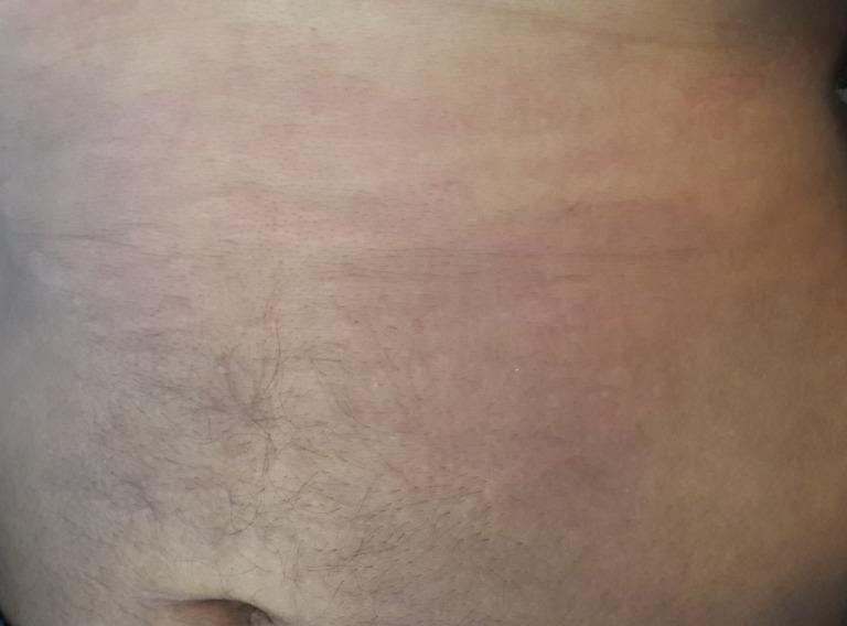
This clinical photograph shows the abdominal region of a 31-year-old male exhibiting a classic cutaneous manifestation of Adult-onset Still's disease (AOSD). The image demonstrates a faint, salmon-pink (rose-salmon) maculopapular rash. The lesions consist of small, non-confluent macules and slightly raised papules with indistinct borders, distributed across the trunk and peri-umbilical area. The rash is characteristically evanescent, often coinciding with febrile peaks. Key clinical features include the subtle coloration and the diffuse, non-scaling texture of the eruption. This visual is an essential diagnostic reference for rheumatology and dermatology, illustrating one of the major Yamaguchi criteria for AOSD. The clinical context involves a patient with systemic inflammatory symptoms, including high-grade fever, polyarthritis, and significantly elevated ferritin levels, where this specific dermatological finding helps differentiate AOSD from other systemic inflammatory or infectious diseases.
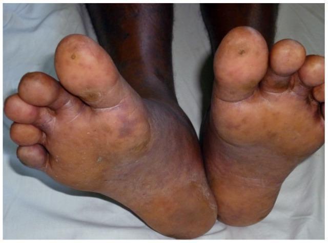
This clinical photograph displays the plantar surfaces and toes of both feet, demonstrating a vasculitic rash characteristic of Spotted Fever Group (SFG) rickettsiosis. The lesions are distributed across the soles and toes, appearing as discrete, dusky red to dark brown macules and papules against a dark skin background. Some lesions exhibit necrotic changes, particularly visible as darkened, crust-like centers on the distal phalanges of the great toes. Morphologically, the spots are predominantly round to oval, ranging approximately 2–10 mm in size, with areas of healthy, normal-looking skin interspersed between the affected sites. Secondary skin features include generalized dryness, focal desquamation (peeling) on the toes, and mild pedal edema. Clinically, these findings represent cutaneous small-vessel vasculitis and microvascular thrombosis typical of rickettsial infection, providing an important diagnostic clue for systemic infectious diseases presenting with dermatological manifestations in the extremities.
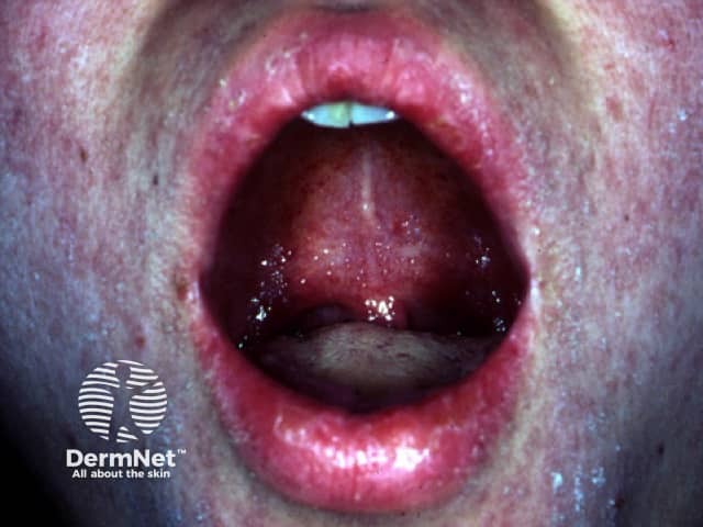
Clinical photograph of the oral cavity showing Koplik spots, a classic mucosal manifestation of measles. Modality: Clinical photography; technique: intraoral close‑up of the buccal mucosa. Location: buccal mucosa near the molar teeth, on an erythematous background. Visual features: numerous small, discrete, bluish‑white to white centers surrounded by red mucosa; lesions measure approximately 1–3 mm in diameter and may appear vesicular or punctate. The mucosa surrounding the spots is inflamed and moist, with mild hyperemia and glossed surfaces in this region. Significance: Koplik spots are highly suggestive of acute measles infection, often appearing during the prodromal phase before the maculopapular rash; they are considered pathognomonic when present. Pathology: viral cytopathic changes in the oropharyngeal epithelium with focal desquamation; clinically, the finding helps confirm a diagnosis of measles in the appropriate clinical context. Diagnostic significance: supports presumptive diagnosis of measles; prompts public health actions, isolation, and antiviral/public health precautions. Differential considerations include aphthous ulcers, herpangina, other viral exanthems, or mucosal lesions in systemic illness, though Koplik spots possess a distinctive bluish center on erythematous mucosa. Clinical correlation: correlate with fever, cough, coryza, conjunctivitis, and rash onset. Utility: educational for clinicians, nurses, medical/dental students; aids recognition and differential diagnosis of febrile rash illness.
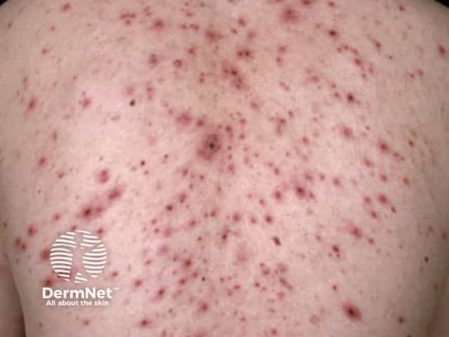
Imaging modality: Clinical photography of an in vivo human back demonstrates a varicella-zoster infection with a classic chickenpox eruption. The posterior trunk shows numerous vesicular and papular lesions on an erythematous base, distributed in crops at different developmental stages from macules and papules to tense vesicles, some becoming crusted. The vesicles often have a dew-drop on a rose petal appearance and are interspersed with non-vesicular papules, producing a mixed morphology pattern. The rash is trunk-predominant with relative sparing of the face in early illness, and lesions are in various evolutionary phases within close proximity. Visual features include clustered, fluid-filled lesions, surrounding erythema, mild edema, and crust formation as vesicles rupture and heal. Clinically, this presentation is highly suggestive of primary varicella-zoster virus infection, with contagiousness until crusts form and fever or malaise commonly accompanying rash. Diagnostic significance rests on lesion morphology, distribution, temporal sequence, and epidemiologic context, distinguishing varicella from herpes zoster, disseminated HSV infection, pityriasis rosea, or dermatitis. Management implications include isolation, supportive care, and consideration of antiviral therapy in children or adults at risk. This image serves educational value for dermatology, infectious diseases, medical students, and clinical documentation. Documentation supports clinical teaching and patient education on contagious spread.
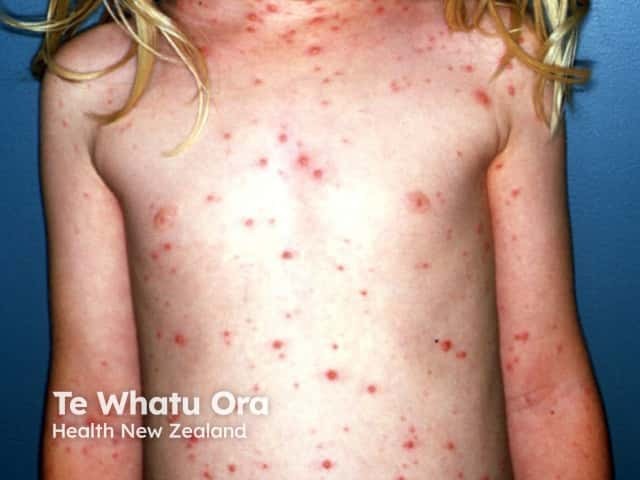
Imaging modality: Clinical photography of a pediatric patient with varicella (chickenpox) rash. Anterior frontal full-body view documenting widespread vesicular lesions on an erythematous base, most prominent on the trunk with scattered involvement of the limbs and face. The lesions appear in crops and at different stages, including macules, pruritic papules, clear-vesicles (dew drop on a rose petal), pustules, and crusted crusts. The distribution is generalized and centripetal, with higher density on the trunk and chest relative to the peripheral extremities. Skin texture shows mild erythema surrounding vesicles; some lesions show crusting as healing begins. No mucosal involvement visible in this image. Lighting is even and natural, color rendition preserved to aid lesion characterization. This dermatologic eruption is characteristic of varicella-zoster virus infection: contagious, self-limited in healthy children, typically presenting with fever, malaise, and pruritic vesicular rash that evolves over several days. Clinically, the image demonstrates classic features used for diagnosis: vesicles at multiple stages on erythematous base, cropped distribution, and trunk predominance. The image is useful for educational purposes, differential diagnosis training (including disseminated herpes simplex, pityriasis rosea-like viral exanthem), and patient counseling about contagion control and rash progression. It may guide teledermatology triage and documentation of clinical course in dermatology and pediatrics.
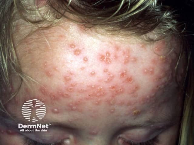
Clinical photography of a pediatric facial skin eruption performed in frontal close-up view. The image documents multiple small vesicular and papular lesions on the forehead with surrounding erythema and several crusted plaques, representing variable disease stages. Findings are characteristic of varicella-zoster virus infection (chickenpox): crops of vesicles and papules at different evolution points, dew-drop on rose petal appearance, superficial epidermal involvement, and mild surface crusting. The distribution is predominantly on the face with potential trunk involvement not shown here; lesions are discretely grouped with some coalescence possible as they crust. This modality captures surface morphology, color change, and lesion configuration to support clinical diagnosis without histology. Pathognomonic clinical features include pruritus, fever, malaise, and contagion potential; differential considerations include disseminated herpes simplex, pityriasis rosea, contact dermatitis, and other vesicular exanthems. Diagnostic significance lies in recognizing crops and stage variability, enabling timely isolation, supportive care, and parental guidance. This image is valuable for dermatology education, pediatrics training, telemedicine triage, and reference in clinical case discussions. Use cases include differential dx practice, image-based teaching, and documentation of characteristic vesicular rash patterns seen in primary varicella. Images of similar presentation aid rapid recognition in clinics, schools, and remote learning platforms worldwide for education.
"typhoid fever" clinical features symptoms
Typhoid Fever - Symptoms and Clinical Features
Incubation Period
- Mean: 10-14 days (range 5-21 days, depending on inoculum size and host immunity)
- Harrison's Principles of Internal Medicine 22E, p.1360
The Classic Three-Week Course
Week 1 - Bacteremic Phase
| Symptom | Frequency |
|---|---|
| Prolonged fever (38.8-40.5°C / 101.8-104.9°F) | >75% at presentation |
| Headache | ~80% |
| Anorexia | ~55% |
| Chills | 35-45% |
| Cough (dry) | ~30% |
| Sweating | 20-25% |
| Myalgias | ~20% |
| Nausea | 18-24% |
| Vomiting | ~18% |
| Malaise | ~10% |
| Arthralgia | 2-4% |
- Harrison's Principles, p.1360
Week 2 - Clinical Manifestation Phase
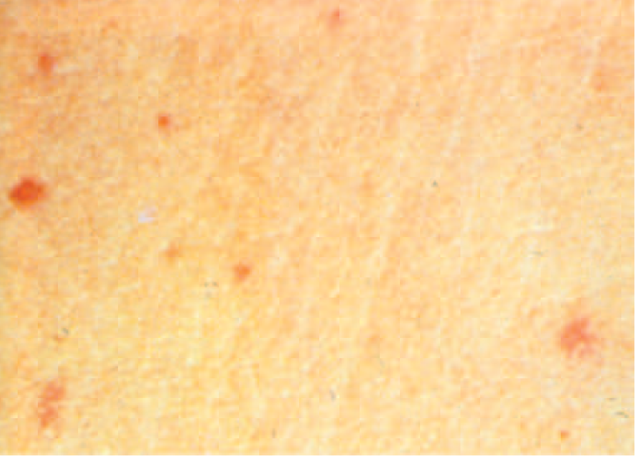
- Rose spots - the pathognomonic rash of typhoid (see image below): faint, salmon-colored, blanching maculopapular lesions, 2-4 mm, primarily on the trunk and chest. Appear in ~30% of patients at end of the first week. Resolve without a trace in 2-5 days. Multiple crops (2-3) can occur. Difficult to detect in dark-skinned patients.
- Relative bradycardia - pulse rate inappropriately slow for the degree of fever (Faget's sign); present in <50% of patients
- Hepatosplenomegaly - splenomegaly in 5-6%; abdominal tenderness in 4-5%
- Coated (furred) tongue - in 51-56%
- Epistaxis
- Diarrhea in 22-28% OR constipation in 13-16% (constipation in 30% overall; diarrhea is more prominent in epidemic settings)
- Abdominal distension
Week 3 and Beyond - Complications Phase
- Abdominal pain worsens
- Intestinal perforation (~1%) - at the ileocecal Peyer's patches; life-threatening
- GI bleeding (~6%) - from ulceration of Peyer's patches
- High fever may lead to toxic appearance, delirium, and even coma ("typhoid state")
- Worsening hepatosplenomegaly
Neurological Manifestations
-
Meningitis
-
Guillain-Barré syndrome
-
Peripheral neuritis
-
Neuropsychiatric symptoms ("muttering delirium," psychosis)
-
Ataxia, seizures
-
Deafness (sequela)
-
Tintinalli's Emergency Medicine, p.1377
Musculoskeletal Features
- Arthralgia and myalgia in ~30% of patients
- Septic sacroiliitis and osteomyelitis (uncommon but recognized)
- Thoracolumbar junction most affected when vertebral involvement occurs
- Campbell's Operative Orthopaedics 15th Ed 2026
Complications Summary
| System | Complication |
|---|---|
| GI | Intestinal perforation, GI hemorrhage, cholecystitis |
| Hematologic | Anemia, DIC, bone marrow suppression |
| Cardiovascular | Myocarditis, mycotic aneurysm |
| Pulmonary | Pneumonia |
| Renal | Renal failure |
| Neurologic | Meningitis, encephalopathy, psychosis, ataxia, deafness |
| Musculoskeletal | Osteomyelitis, septic arthritis |
- Harrison's Principles 22E, p.1360
Key Distinguishing Features at a Glance
| Feature | Detail |
|---|---|
| Fever pattern | Step-ladder, continuous, up to 40.5°C |
| Relative bradycardia | Pulse-temperature dissociation (Faget's sign) |
| Rose spots | Salmon-colored blanching macules on trunk (~30%) |
| GI | More constipation than diarrhea (non-epidemic); anorexia prominent |
| Tongue | Coated/furred |
| Spleen | Splenomegaly by week 2 |
| Sensorium | Delirium/toxic state in severe cases |
Sources: Harrison's Principles of Internal Medicine 22E (2025), p.1359-1360 | Tintinalli's Emergency Medicine | Sleisenger and Fordtran's GI & Liver Disease | Campbell's Operative Orthopaedics 15th Ed 2026 | Park's Textbook of Preventive & Social Medicine
How is typhoid diagnosed and what tests are used?
"typhoid fever" diagnosis laboratory
Diagnosis of Typhoid Fever - Laboratory Tests
1. Microbiological (Culture) Methods - The Gold Standard
Blood Culture
- Primary diagnostic test and most widely used
- Sensitivity: 40-80% (variability due to prior antibiotics, blood volume drawn, timing)
- Only ~15 organisms/mL of blood are typically present - a small inoculum
- Yield is highest in Week 1 of illness and declines thereafter
- Sensitivity falls further if the patient has already taken antibiotics
- Centrifugation and buffy coat culture reduces time to isolation but does not improve sensitivity
- Harrison's Principles 22E, p.1361; Sleisenger & Fordtran's GI Disease, p.2199
Bone Marrow Culture - Most Sensitive Single Test
- Sensitivity: ~80-90%, even after up to 5 days of prior antibiotic therapy
- Considered the gold standard by many authorities
- Yield is NOT reduced by antibiotic use (unlike blood culture)
- Invasive and impractical for routine use
- Harrison's 22E, p.1361; Sleisenger & Fordtran's, p.2199
Stool Culture
- Negative in 60-70% of cases during Week 1
- Becomes positive during Week 2 and Week 3 of untreated illness
- Useful for detecting chronic carriers (repeated cultures required)
- Harrison's 22E, p.1361
Urine Culture
- Positive in approximately 25% of patients by the third week
- Less sensitive than blood or stool
- Sleisenger & Fordtran's, p.2199
Duodenal String Test (Intestinal Secretions)
- A noninvasive test - string is swallowed, retrieves duodenal bile
- Sensitivity: ~70% (positive even when bone marrow culture is negative)
- If blood + bone marrow + intestinal secretions are all cultured together: combined yield >90%
- Harrison's 22E, p.1361
Rose Spot Biopsy
- S. Typhi can be cultured directly from punch biopsies of rose spots
- Rarely done in practice but confirms bacteremia-driven skin seeding
Yield of Cultures by Week of Illness
| Test | Week 1 | Week 2 | Week 3 | Notes |
|---|---|---|---|---|
| Blood culture | 60-80% | Declining | Low | Highest early; affected by antibiotics |
| Bone marrow | 80-90% | 80-90% | 80-90% | Unaffected by prior antibiotics |
| Stool culture | Low (30-40%) | Rising | Positive | Best in weeks 2-3 |
| Urine culture | Negative | Low | ~25% | Only useful in week 3+ |
| Duodenal string | 70% | 70% | -- | Not reduced by antibiotics |
2. Serology
Widal Test (Felix-Widal Test)
- Measures agglutinating antibodies against somatic O antigen and flagellar H antigen of S. Typhi
- O antibodies appear on day 6-8 after onset
- H antibodies appear on day 10-12 after onset
- Diagnostic criterion: A 4-fold rise in titer between acute and convalescent sera (taken 10-14 days apart) is considered strong evidence
- Single titer thresholds (e.g., O ≥1:80 or 1:160) vary by country and endemic background
- Widely used in developing countries due to low cost
- Sensitivity: only moderate - can be negative in up to 30% of culture-proven cases
- May be suppressed by early antibiotic therapy
- False positives from cross-reactions with: other Salmonella serotypes, other Enterobacteriaceae, malaria, typhus, bacteremia from other organisms, cirrhosis
- A single titer is unreliable - paired sera are needed
- Park's Preventive & Social Medicine, p.278; Sleisenger & Fordtran's, p.2199
3. Rapid Diagnostic Tests (RDTs)
| Test | Principle | Performance |
|---|---|---|
| Typhidot | Detects IgM and IgG against a 50 kDa S. Typhi outer membrane protein | Takes ~3 hours; sensitivity ~70-80%, specificity ~80-90% |
| Typhidot-M | Detects IgM only (more specific for acute infection) | Improved specificity over standard Typhidot |
| TUBEX (IDL Tubex) | Detects IgM anti-O9 antibodies; results in minutes | Rapid; sensitivity ~70-80%, specificity ~80-90% |
| Dipstick test | IgM antibodies against S. Typhi LPS; colloidal dye staining | Simple; suitable for field use |
4. Non-Specific Laboratory Findings
| Test | Typical Finding | Notes |
|---|---|---|
| CBC | Leukopenia + neutropenia in 15-25% | Leukocytosis in children, or with perforation/secondary infection |
| Liver enzymes (ALT/AST) | Mildly elevated (2-3x normal) | Hepatitis-like picture |
| ESR | Elevated | Non-specific |
| CRP | Elevated | Non-specific |
| Anemia | Mild | From bone marrow suppression or GI loss |
| Thrombocytopenia | Can occur | Especially in severe disease |
5. Molecular Diagnosis (PCR)
- PCR for S. Typhi/Paratyphi in blood
- Sensitivity: ~40-100% depending on gene targets
- Previously limited to research labs; expanding post-COVID-19 pandemic due to improved molecular diagnostic infrastructure
- Not yet widely available in resource-limited endemic settings
- Harrison's 22E, p.1361
6. Chronic Carrier Detection
- Carriers shed S. Typhi intermittently - repeated stool and urine cultures required
- Vi antigen antibody testing - used as a serologic screening test for carriers; limited value in low-prevalence settings
- Sleisenger & Fordtran's, p.2199
Diagnostic Approach Summary
Clinical suspicion (fever + travel history)
           |
    Blood culture (x3, >10 mL each)
           |
   ┌── Positive → Identify S. Typhi/Paratyphi, do sensitivity testing
   └── Negative → Consider:
           ├── Bone marrow culture (best if antibiotics given)
           ├── Stool/urine culture (weeks 2-3)
           ├── Duodenal string test
           └── Rapid tests (Typhidot/TUBEX) if culture unavailable
Key rule: The only definitive diagnosis is culture isolation. Serology and RDTs support but cannot confirm diagnosis on their own.
Sources: Harrison's Principles of Internal Medicine 22E (2025), p.1360-1361 | Sleisenger & Fordtran's Gastrointestinal and Liver Disease, p.2199 | Park's Textbook of Preventive & Social Medicine, p.278 | Tintinalli's Emergency Medicine
How is typhoid diagnosed and what tests are used?
"typhoid fever" diagnosis rapid test accuracy
Diagnosis of Typhoid Fever - Tests & Methods
1. Culture Methods - Definitive Diagnosis
Blood Culture
- Primary diagnostic test - most widely performed first
- Sensitivity: 40-80% (study settings give higher figures; real-world yield closer to 40% when prior antibiotics have been given)
- Why it's limited: only <15 organisms/mL of blood are typically present
- Almost all S. Typhi in blood are attached to the mononuclear cell/platelet fraction - buffy coat culture reduces time to isolation but does NOT improve sensitivity
- Yield is highest in Week 1 and declines as the disease progresses
- 3 sets of blood cultures (>10 mL each) maximize yield
- Harrison's Principles 22E, p.1361; Sleisenger & Fordtran's GI Disease, p.2199
Bone Marrow Culture - Most Sensitive Overall
- Sensitivity: ~80-90%, even after up to 5 days of prior antibiotic therapy
- Considered the gold standard - yield is not reduced by antibiotics (unlike blood culture)
- Invasive and impractical for routine/field use; reserved for culture-negative cases where suspicion remains high
- Harrison's 22E, p.1361; Sleisenger & Fordtran's, p.2199
Stool Culture
- Negative in 60-70% of cases in Week 1
- Becomes positive in Weeks 2-3 of untreated illness as the organism is shed into the gut
- Essential for detecting chronic carriers (repeated cultures needed as shedding is intermittent)
- Harrison's 22E, p.1361
Urine Culture
- Positive in approximately 25% of patients by the third week
- Less sensitive; useful as an adjunct in later disease
- Sleisenger & Fordtran's, p.2199
Duodenal String Test (Intestinal Secretions Culture)
- A swallowed string retrieves duodenal bile containing the organism
- Sensitivity: ~70% - can be positive even when bone marrow is negative
- If blood + bone marrow + intestinal secretions are all cultured together: combined yield >90%
- Harrison's 22E, p.1361
Rose Spot Culture
- S. Typhi can be cultured directly from punch biopsies of rose spots
- Rarely performed in practice but confirms bacteremia
Culture Yield by Week of Illness
| Specimen | Week 1 | Week 2 | Week 3 | Key Note |
|---|---|---|---|---|
| Blood culture | 60-80% | Declining | Low | Most affected by prior antibiotics |
| Bone marrow | ~80-90% | ~80-90% | ~80-90% | Unaffected by 5 days of antibiotics |
| Stool | 30-40% | Rising | Positive | Best in weeks 2-3 |
| Urine | Negative | Low | ~25% | Only useful week 3 onwards |
| Duodenal string | ~70% | ~70% | -- | Not reduced by antibiotics |
2. Serology
Widal Test (Felix-Widal Test)
- Measures agglutinating antibodies against:
- O antigen (somatic) - appears day 6-8 after onset
- H antigen (flagellar) - appears day 10-12 after onset
- Paired sera method (gold standard interpretation): a 4-fold rise in titer between acute and convalescent samples (taken 10-14 days apart) is considered strong evidence
- Single titer thresholds vary by country (e.g., O ≥1:80 or 1:160 in endemic areas)
- Cheap, widely available, used at point-of-care in endemic countries
| Problem | Detail |
|---|---|
| False negatives | Negative in up to 30% of culture-proven cases; early sampling before antibody rise; prior antibiotics blunting response |
| False positives | Cross-reaction with other Salmonella serotypes, other Enterobacteriaceae; also in malaria, typhus, bacteremia, cirrhosis |
| Moderate accuracy | Only moderate sensitivity AND specificity |
| Single titer unreliable | Must use paired sera for reliable interpretation |
- Park's Preventive & Social Medicine, p.278; Sleisenger & Fordtran's, p.2199
3. Rapid Diagnostic Tests (RDTs)
| Test | Principle | Time | Performance |
|---|---|---|---|
| TUBEX (IDL Tubex) | Detects IgM anti-O9 antibodies by color inhibition assay | Minutes | Sensitivity ~70-80%, specificity ~80-90% |
| Typhidot | Detects IgM + IgG against a 50 kDa outer membrane protein of S. Typhi | ~3 hours | Sensitivity ~70-80%, specificity ~80-90% |
| Typhidot-M | Detects IgM only (newer, more specific for acute infection) | ~3 hours | Improved specificity over standard Typhidot |
| Dipstick test | IgM binding to S. Typhi LPS antigen; colloidal dye staining | Rapid | Simple, field-suitable |
Key limitation: A 2017 systematic review found rapid diagnostic test sensitivities of ~70-80% and specificities of ~80-90% - not accurate enough to replace blood cultures as the primary approach.
- Harrison's Principles 22E, p.1361; Park's Preventive Medicine, p.278
4. Non-Specific Laboratory Findings
| Test | Typical Finding | Notes |
|---|---|---|
| CBC - WBC | Leukopenia + neutropenia (15-25% of cases) | Leukocytosis in children, or if intestinal perforation/secondary infection |
| Liver enzymes (ALT/AST) | Mildly elevated (2-3× normal) | Hepatitis-like picture; very common |
| Muscle enzymes | Mildly elevated | Non-specific |
| Anemia | Mild | Bone marrow suppression or GI blood loss |
| Thrombocytopenia | Can occur | Especially in severe/complicated disease |
| ESR / CRP | Elevated | Non-specific markers of inflammation |
- Harrison's 22E, p.1361
5. Molecular Diagnosis (PCR)
- PCR targeting S. Typhi/Paratyphi genes in blood
- Sensitivity: ~40-100% depending on gene targets - wide range reflects variation in methodology
- Previously limited to research/reference labs
- Expanded availability post-COVID-19 pandemic due to investment in molecular diagnostics infrastructure
- Not yet standard of care in most resource-limited endemic settings
- Harrison's 22E, p.1361; Sleisenger & Fordtran's, p.2199
6. Carrier Detection
- Chronic carriers shed S. Typhi intermittently - repeated stool and urine cultures required
- Vi antigen antibody testing - serologic screening for carriers; limited diagnostic value in low-prevalence settings, not widely adopted as a public health tool
- Sleisenger & Fordtran's, p.2199
Differential Diagnosis to Exclude
- Malaria (most important to exclude first)
- Viral hepatitis
- Dengue fever
- Rickettsial infections
- Leptospirosis
- Amebic liver abscess
- Acute HIV infection
Practical Diagnostic Approach
Febrile patient + endemic travel history
            |
    ┌── Send Blood Cultures (x3, ≥10 mL each)
    ├── Baseline bloods: CBC, LFTs, CRP, ESR
    ├── Malaria smear / RDT (rule out first)
    │
    ├── If blood culture negative but suspicion persists:
    │       ├── Bone marrow culture (especially if antibiotics given)
    │       ├── Stool + urine culture (weeks 2-3)
    │       └── Duodenal string test
    │
    └── In resource-limited settings:
            └── Rapid tests (Typhidot / TUBEX) as adjunct
                (not a replacement for culture)
Bottom line: Blood culture is the practical first-line test. Bone marrow culture is the most sensitive single test. The Widal test has major limitations and should be interpreted cautiously. RDTs are useful in endemic low-resource settings but cannot replace culture.