Give me differentials for right adnexal lesion on usg

Reading File
Finding Sources
Finding Sources
Finding Sources
Reading File
Finding Sources
Finding Sources
Reading File
Reading File
Reading File
Reading File
Reading File
Searching Images

adnexal mass ultrasound differential diagnosis ovarian cyst

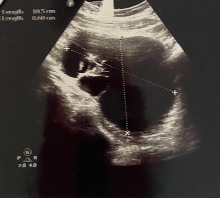
This diagnostic image is a transvaginal ultrasound scan of a right adnexal mass, identified in clinical context as an ovarian dermoid cyst (struma ovarii). The image demonstrates a large, complex cystic structure with multiple internal thin septations. The cyst exhibits a predominantly hypoechoic internal fluid appearance, interspersed with focal areas of heterogeneous echogenicity and echogenic components, suggestive of solid elements or specialized tissue within the cyst. Dotted electronic calipers and plus-sign markers indicate two-dimensional measurements of the lesion: a vertical length of 10.5 cm and a diagonal length of 8.60 cm. This ultrasound is used in gynecology and obstetrics to evaluate adnexal masses for signs of complexity, such as septations and solid components, which inform the differential diagnosis between benign functional cysts and germ cell tumors or other neoplasms. The educational focus is on the ultrasonographic characterization of complex ovarian pathology.

This diagnostic image is a transvaginal ultrasound scan of a right adnexal mass, identified in clinical context as an ovarian dermoid cyst (struma ovarii). The image demonstrates a large, complex cystic structure with multiple internal thin septations. The cyst exhibits a predominantly hypoechoic internal fluid appearance, interspersed with focal areas of heterogeneous echogenicity and echogenic components, suggestive of solid elements or specialized tissue within the cyst. Dotted electronic calipers and plus-sign markers indicate two-dimensional measurements of the lesion: a vertical length of 10.5 cm and a diagonal length of 8.60 cm. This ultrasound is used in gynecology and obstetrics to evaluate adnexal masses for signs of complexity, such as septations and solid components, which inform the differential diagnosis between benign functional cysts and germ cell tumors or other neoplasms. The educational focus is on the ultrasonographic characterization of complex ovarian pathology.

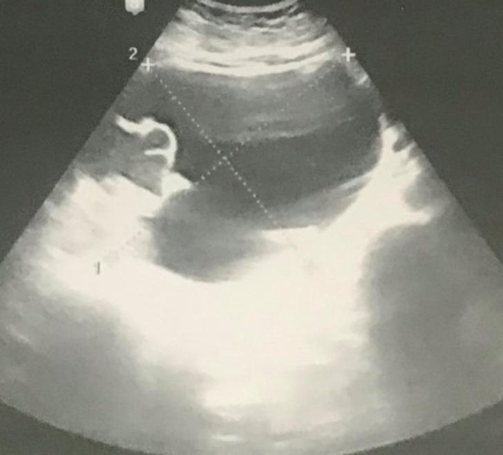
This diagnostic ultrasound image in the pelvic region displays a large, complex adnexal mass measuring approximately 15x12 cm. The mass exhibits a multicystic appearance characterized by multiple internal septations and cystic compartments of varying sizes. The internal compartments are primarily anechoic, consistent with fluid-filled spaces, while the septations and certain peripheral areas show increased echogenicity, suggesting thickened walls or solid components. Measurement calipers are visible on the screen, delineating the extent of the mass within the adnexal space. This finding is clinically significant in the context of an 18-week pregnancy and acute abdominal pain, where the differential diagnosis includes adnexal torsion or a large ovarian cyst (such as a mucinous cystadenoma). The image demonstrates the displacement of adjacent pelvic structures and the characteristic lack of obvious malignant sonographic features, such as highly vascular solid papillary projections, despite its significant size.

This diagnostic ultrasound image in the pelvic region displays a large, complex adnexal mass measuring approximately 15x12 cm. The mass exhibits a multicystic appearance characterized by multiple internal septations and cystic compartments of varying sizes. The internal compartments are primarily anechoic, consistent with fluid-filled spaces, while the septations and certain peripheral areas show increased echogenicity, suggesting thickened walls or solid components. Measurement calipers are visible on the screen, delineating the extent of the mass within the adnexal space. This finding is clinically significant in the context of an 18-week pregnancy and acute abdominal pain, where the differential diagnosis includes adnexal torsion or a large ovarian cyst (such as a mucinous cystadenoma). The image demonstrates the displacement of adjacent pelvic structures and the characteristic lack of obvious malignant sonographic features, such as highly vascular solid papillary projections, despite its significant size.

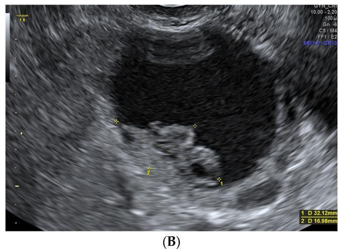
This diagnostic ultrasound image displays a transverse view of an adnexal mass, categorized as a unilocular-solid cyst. The primary feature is a large, predominantly anechoic cystic cavity containing fluid. Arising from the internal wall is a prominent, irregular papillary projection with intermediate echogenicity. Quantitative analysis is provided via electronic calipers: the solid projection measures approximately 32.12 mm in its longest axis (1 D) and 16.98 mm in height (2 D). According to the IOTA (International Ovarian Tumor Analysis) framework, the presence of a solid papillary component within a unilocular cyst increases the suspicion for borderline tumors or malignant serous carcinomas. The clinical context notes that while this specific image (B) represents a borderline serous carcinoma, such findings can visually mimic benign cystadenofibromas, necessitating detailed assessment of vascularity and papillary morphology for differential diagnosis in gynecologic oncology.

This diagnostic ultrasound image displays a transverse view of an adnexal mass, categorized as a unilocular-solid cyst. The primary feature is a large, predominantly anechoic cystic cavity containing fluid. Arising from the internal wall is a prominent, irregular papillary projection with intermediate echogenicity. Quantitative analysis is provided via electronic calipers: the solid projection measures approximately 32.12 mm in its longest axis (1 D) and 16.98 mm in height (2 D). According to the IOTA (International Ovarian Tumor Analysis) framework, the presence of a solid papillary component within a unilocular cyst increases the suspicion for borderline tumors or malignant serous carcinomas. The clinical context notes that while this specific image (B) represents a borderline serous carcinoma, such findings can visually mimic benign cystadenofibromas, necessitating detailed assessment of vascularity and papillary morphology for differential diagnosis in gynecologic oncology.

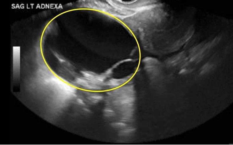
This grayscale transvaginal ultrasound image in the sagittal plane (labeled SAG LT ADNEXA) demonstrates a large, complex left adnexal mass. The mass is primarily cystic and multilocular, characterized by anechoic fluid-filled spaces separated by thin septations. A distinct, hyperechoic solid component is visible within the cyst wall (circumscribed by a yellow circle), exhibiting an irregular internal echotexture compared to the surrounding fluid. The outer margins of the cystic portion appear relatively smooth and well-defined. The findings are categorized under the Ovarian-Adnexal Imaging-Reporting and Data System (O-RADS) as score 4, indicating an intermediate risk of malignancy (10% to <50%). This imaging is clinically significant for the evaluation of potential ovarian neoplasms, such as cystadenoma or malignant tumors, in a symptomatic patient.

This grayscale transvaginal ultrasound image in the sagittal plane (labeled SAG LT ADNEXA) demonstrates a large, complex left adnexal mass. The mass is primarily cystic and multilocular, characterized by anechoic fluid-filled spaces separated by thin septations. A distinct, hyperechoic solid component is visible within the cyst wall (circumscribed by a yellow circle), exhibiting an irregular internal echotexture compared to the surrounding fluid. The outer margins of the cystic portion appear relatively smooth and well-defined. The findings are categorized under the Ovarian-Adnexal Imaging-Reporting and Data System (O-RADS) as score 4, indicating an intermediate risk of malignancy (10% to <50%). This imaging is clinically significant for the evaluation of potential ovarian neoplasms, such as cystadenoma or malignant tumors, in a symptomatic patient.

I now have rich, comprehensive material. Let me compile the full answer.

Differentials for Right Adnexal Lesion on Ultrasound

The right adnexa includes the right ovary, right fallopian tube, and surrounding structures. A lesion here has a broad differential, best organized by origin and category. Age and reproductive status are the strongest determinants of likelihood.

I. GYNECOLOGIC - OVARIAN ORIGIN

A. Non-Neoplastic (Functional) Cysts

These are the most common cause in reproductive-age women and usually resolve in 4-8 weeks.
LesionUSG AppearanceNotes
Follicular cystUnilocular, thin-walled, purely anechoic, <8 cmMost common functional cyst; incidental
Corpus luteum cystThick, irregular wall ("ring of fire" on Doppler), internal echoesRupture more common on RIGHT side; may mimic ectopic
Theca lutein cystBilateral large multilocular cystsAssociated with molar pregnancy, hCG excess, ovarian hyperstimulation
Hemorrhagic cystReticular/fishnet internal echoes, no internal Doppler flowAcute pain; self-limited
Ruptured cystCollapsed cyst + free fluid in PODMimics ectopic pregnancy

B. Benign Neoplastic Masses

LesionUSG AppearanceNotes
Dermoid cyst (mature cystic teratoma)Hyperechoic focus with posterior shadowing ("Rokitansky nodule"), fat-fluid level, "tip of iceberg" signMost common benign tumor in <50 yr; 10% bilateral; 15% torsion risk
Serous cystadenomaUnilocular, thin-walled, anechoic, may have thin septaeMost common epithelial benign tumor
Mucinous cystadenomaMultilocular, internal low-level echoes, "stained glass" pattern, large sizeCan be very large (>20 cm)
Fibroma / FibrothecomaSolid, hypoechoic with posterior shadowingAssociated with Meigs syndrome (ascites + pleural effusion)
Brenner tumorSolid, echogenic with calcificationsRare; often incidental
CystadenofibromaUnilocular or multilocular cyst with solid papillary nodulesMimics malignancy on USG

C. Endometriosis-Related

LesionUSG AppearanceNotes
Endometrioma ("chocolate cyst")Homogeneous low-level internal echoes ("ground glass"), thick wall, no internal Doppler flowClassic appearance; may be bilateral

D. Malignant / Borderline

LesionUSG AppearanceWorrying Features
Epithelial ovarian carcinoma (serous/mucinous/clear cell/endometrioid)Mixed cystic-solid, thick irregular septae (>3 mm), papillary projections, ascitesSize >9 cm, solid components, increased vascularity
Borderline tumorUnilocular-solid with papillary projection10% serous tumors have borderline potential
Germ cell tumor (dysgerminoma, immature teratoma, yolk sac tumor)Solid or complex; often in young patientsElevated AFP/hCG/LDH
Sex cord-stromal tumor (granulosa cell, Sertoli-Leydig)Solid or complex cysticMay cause hormonal symptoms
Krukenberg tumor (metastatic)Bilateral solid ovarian massesPrimary usually gastric or colorectal carcinoma
The IOTA simple rules help risk-stratify: B-features (unilocular, no solid component, acoustic shadowing, <100 mm, no blood flow) favor benign; M-features (irregular solid, ascites, ≥4 papillary projections, irregular multilocular-solid ≥100 mm, high Doppler flow) suggest malignancy. - Grainger & Allison's Diagnostic Radiology

II. GYNECOLOGIC - FALLOPIAN TUBE / PARA-ADNEXAL ORIGIN

LesionUSG AppearanceNotes
Ectopic pregnancyHeterogeneous adnexal ring ("bagel sign") ± fetal pole separate from uterusMust exclude in all reproductive-age women with pelvic pain; positive hCG
HydrosalpinxElongated, tubular, fluid-filled, folded ("cogwheel sign" on cross-section)Chronic sequela of PID/salpingitis
Pyosalpinx / Tubo-ovarian abscess (TOA)Thick-walled complex fluid collection, "cogwheel" tube + complex ovarian mass, hypervascularityFever, raised WBC, STI history
HematosalpinxEchogenic tubular fluidEctopic, endometriosis, or trauma
Paratubal / Peritubal cystSimple anechoic cyst separate from ovary, near tubeCommon incidental finding; remnant of mesonephric duct
Hydatid of MorgagniSmall simple cyst hanging from fimbrial endParamesonephric remnant
Fallopian tube carcinomaTubular solid-cystic mass, "sausage-shaped"Rare; may be confused with ovarian ca
Adnexal torsionEnlarged ovary >4 cm, peripheral follicles, absent/decreased Doppler flow (absent Doppler does NOT exclude torsion)Acute right-sided pain; ovary appears edematous

III. NON-GYNECOLOGIC / EXTRA-ADNEXAL CAUSES

These can mimic a right adnexal lesion and are important not to miss:
LesionUSG CluesNotes
Appendiceal pathology (appendicitis, appendiceal abscess, mucocele)Blind-ending tubular structure, compressible/non-compressibleRight iliac fossa lesion can track into right adnexa
Pelvic kidneyRenal parenchymal echo pattern, collecting system, Doppler renal vesselsAlways think of this if no ipsilateral kidney seen in renal fossa
Pelvic appendix of bowel / Meckel's diverticulumBowel signature (gut signature)Peristalsis helps
Iliac vessel aneurysm / enlarged iliac lymph nodeColor Doppler pulsatility; lymph nodes have hilum sign
Retroperitoneal/pelvic side wall massDisplaces rather than arises from ovary; no ovarian tissue around itLymphoma, nerve sheath tumor
Peritoneal inclusion cystMultilocular cyst conforming to peritoneal surfaces, "spider web" appearance, ovary seen floating insideHistory of prior surgery/PID
Bowel-related lesion (Crohn's mass, diverticular abscess, carcinoid)Bowel wall thickening, fistula, gas within lesion
Ureteric calculus / hydronephrosisUreteric jet on Doppler, stone shadow

IV. AGE-BASED APPROACH (Quick Reference)

Age GroupMost Likely Differentials
Pre-pubertalGerm cell tumor, functional cyst (rare), dysgerminoma, teratoma
Reproductive ageFunctional cyst, ectopic pregnancy, endometrioma, dermoid, TOA, cystadenoma, torsion
Peri-menopausalEpithelial ovarian tumors (borderline/malignant), endometrioma, fibroma
Post-menopausalOvarian carcinoma (until proven otherwise), metastatic disease, fibroma, simple cyst (may still be benign if <1 cm, purely simple)

V. KEY USG RED FLAGS for Malignancy (IOTA M-features)

  • Irregular solid tumor
  • Ascites
  • ≥4 papillary structures
  • Irregular multilocular-solid tumor ≥100 mm
  • Very strong internal Doppler flow
  • Thick irregular septae (>3 mm)
Reference: Grainger & Allison's Diagnostic Radiology, Berek & Novak's Gynecology, Goldman-Cecil Medicine

Representative USG Appearances

Here are some illustrative ultrasound examples from the medical image library:
Ovarian dermoid (struma ovarii) - complex cyst with septations and heterogeneous components:
Transvaginal USG of right adnexal dermoid cyst
Large multilocular adnexal cyst - mucinous cystadenoma vs. torsion in pregnancy:
Multicystic adnexal mass with septations
Unilocular-solid cyst with papillary projection - borderline serous carcinoma (IOTA M-feature):
Adnexal cyst with papillary projection

Bottom Line: Always combine USG morphology with clinical context (age, symptoms, hCG, CA-125, menstrual history). In reproductive age women, most right adnexal lesions are benign - functional cysts, dermoids, and endometriomas dominate. Post-menopausally, any adnexal mass must be presumed malignant until proven otherwise, and the threshold for further workup (MRI, CA-125, referral) is lower.
This is a shared conversation. Sign in to Orris to start your own chat.